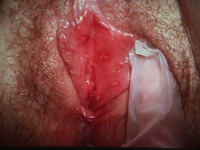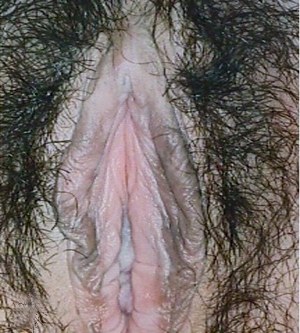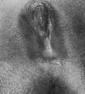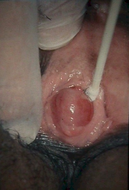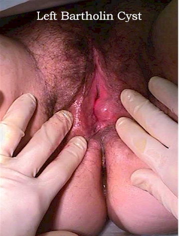
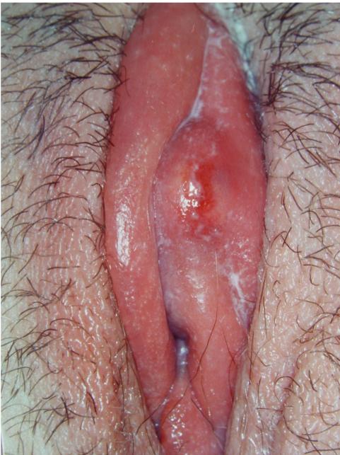
Left Bartholin Cyst (Reproduced, with permission from Michael John Hughey, MD, All rights reserved.)
|
Vulvar Disease Michael John Hughey |
|
|
Michael John Hughey, MD |
|
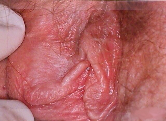
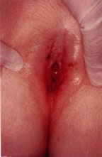
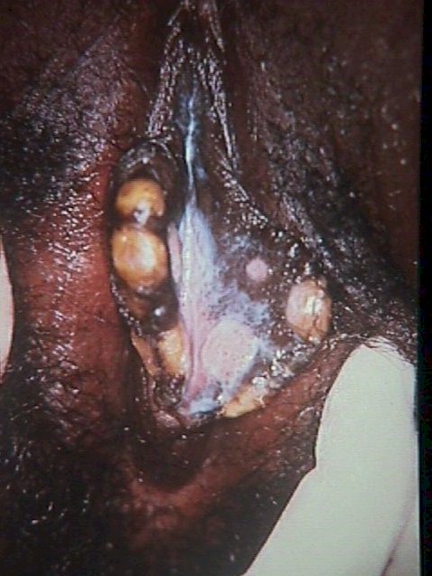
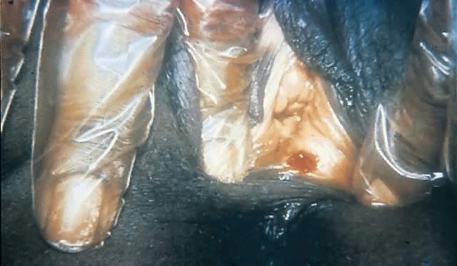
| Bartholin Cyst and Abscess |
Left Bartholin Cyst (Reproduced, with permission from Michael John Hughey, MD, All rights reserved.) |
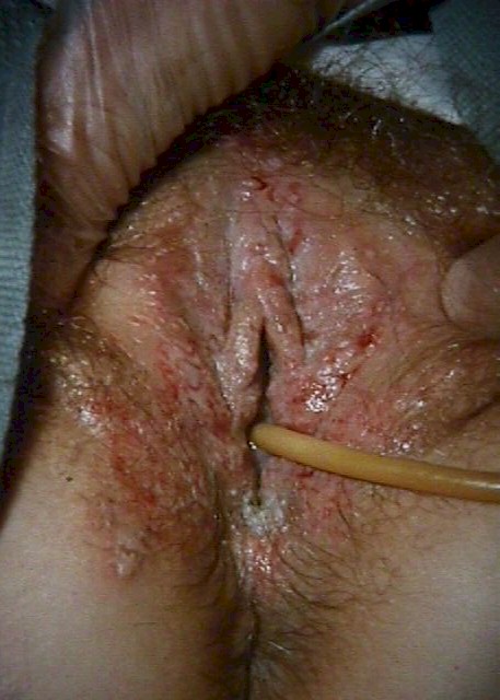
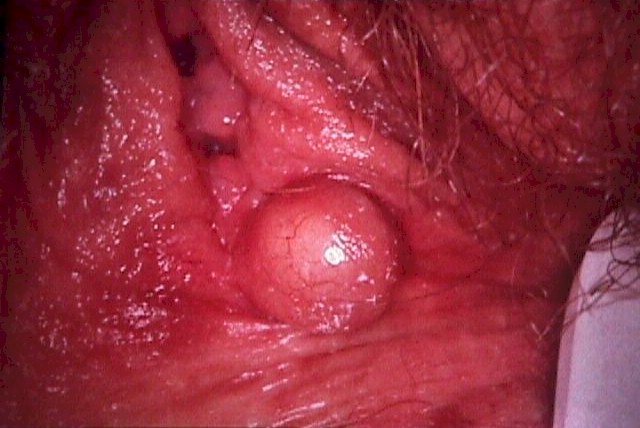
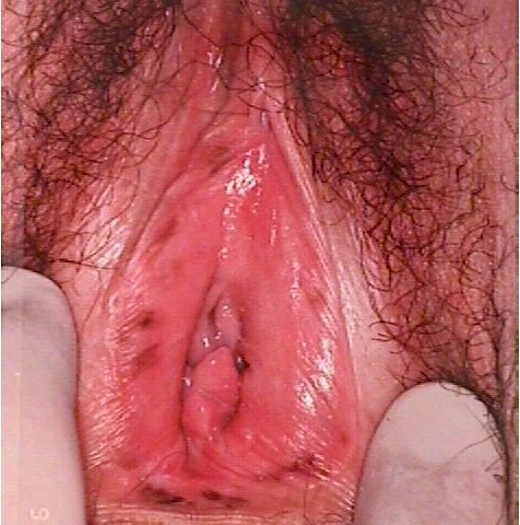
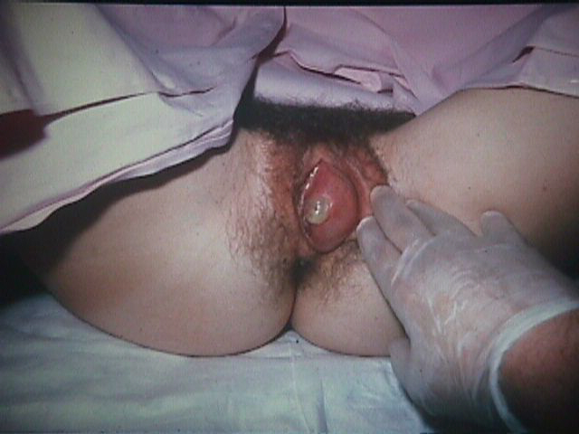
Left Bartholin Abscess (From Operational Obstetrics & Gynecology - 2nd Edition, The Health Care of Women in Military Settings, CAPT Michael John Hughey, MC, USNR, NAVMEDPUB 6300-2C, Bureau of Medicine and Surgery, Department of the Navy, 2300 E Street NW, Washington, D.C. 20372-5300, January 1, 2000. Original image courtesy CAPT Richard Stock, MC, USN) |
| Carcinoma in situ |
|
|
|
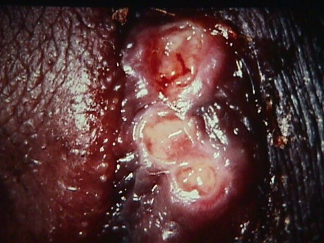
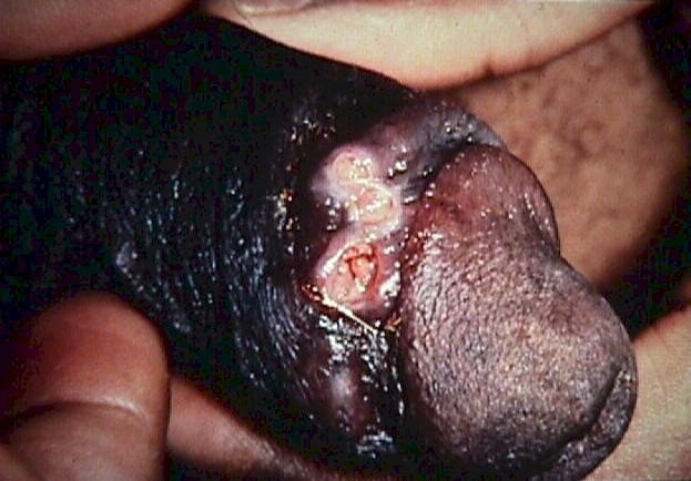
| Chancroid |
|
|
|
|
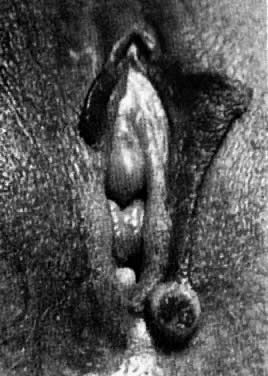
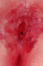
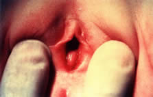
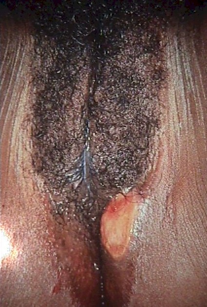
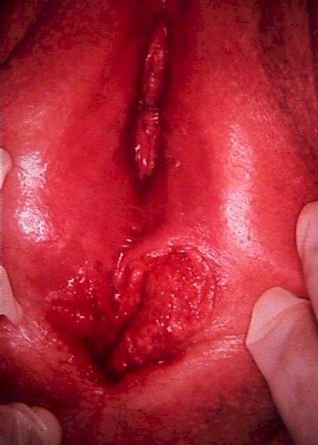
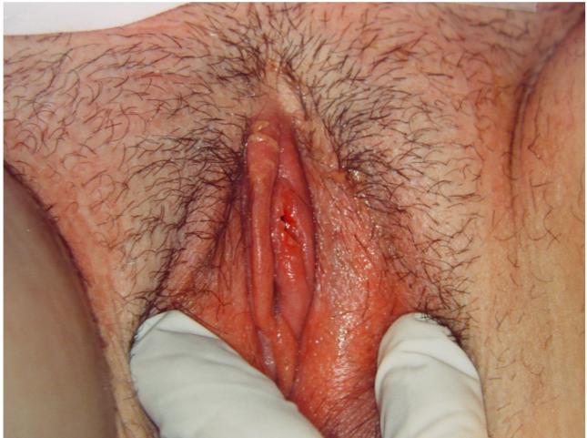
| Clitoral Abscess |
Periclitoral abscess before (left) and the next day after drainage (right) . (Reproduced, with permission from Michael John Hughey, MD, All rights reserved.) |
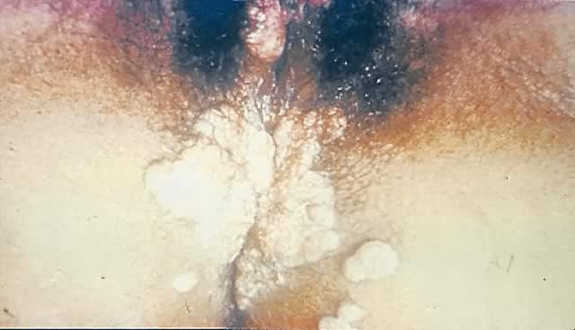
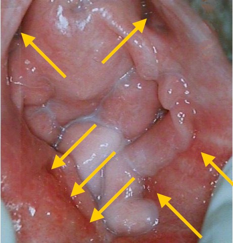
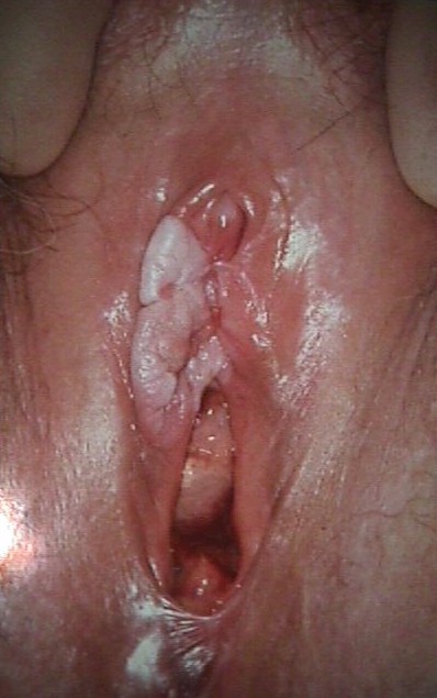
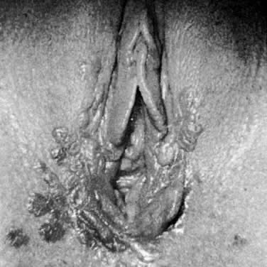
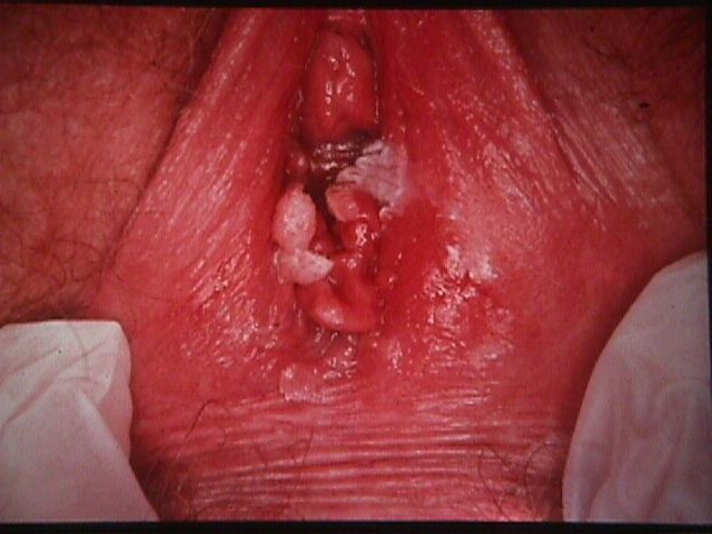
| Condyloma |
|
|
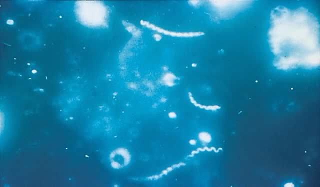
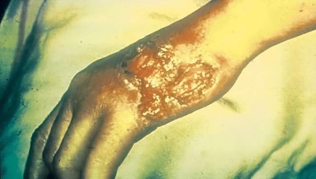
| Granuloma Inguinale |
|
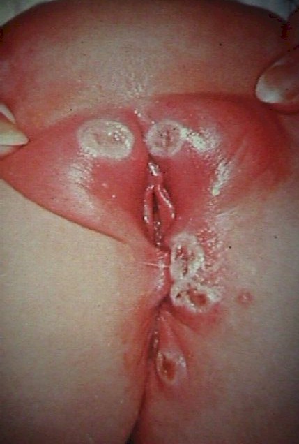
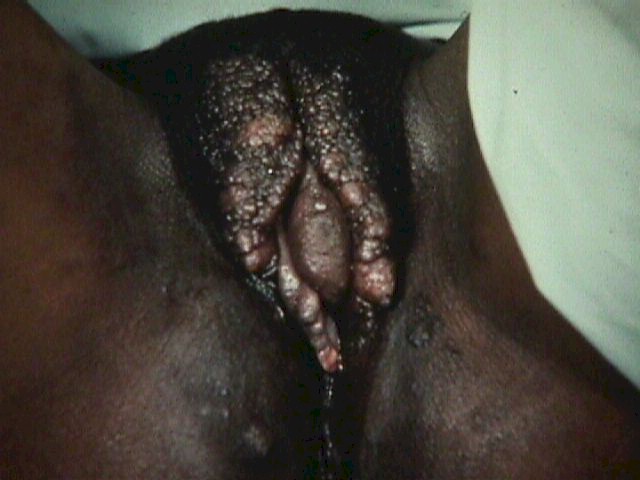
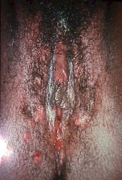
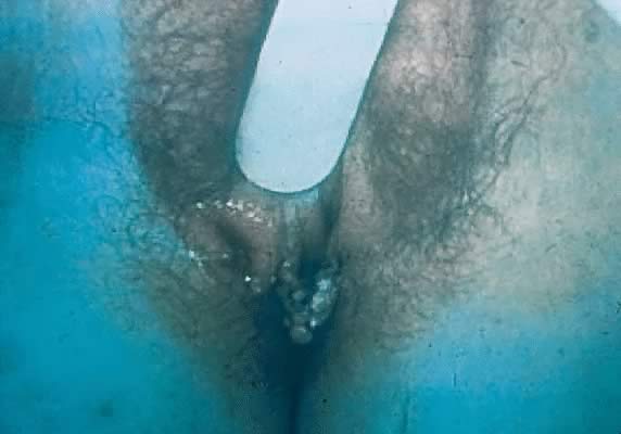
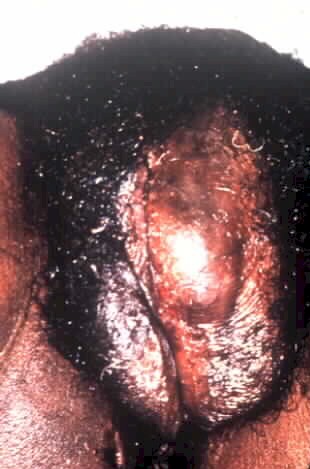
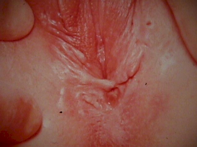
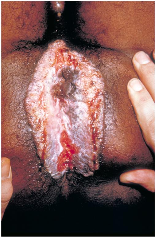
| Herpes |
Herpes Vulvitis (From Operational Obstetrics & Gynecology - 2nd Edition, The Health Care of Women in Military Settings, CAPT Michael John Hughey, MC, USNR, NAVMEDPUB 6300-2C, Bureau of Medicine and Surgery, Department of the Navy, 2300 E Street NW, Washington, D.C. 20372-5300, January 1, 2000. Original image courtesy CAPT Richard Stock, MC, USN) |
|
|
|
|
| Hidradenoma |
|
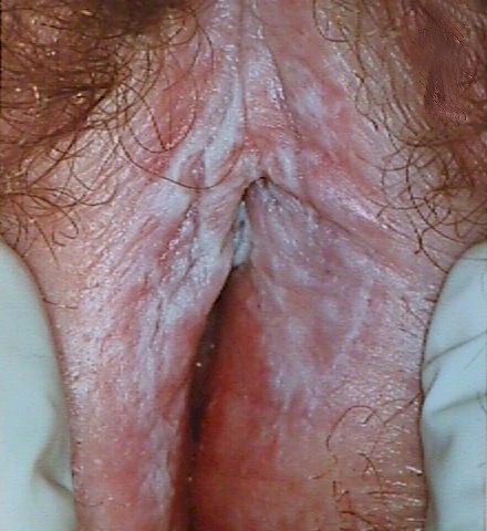
| Hypertrophic Dystrophy |
|
|
| Inclusion Cyst |
|
|
| LGV |
|
|
|
|
| Lichen Sclerosis |
|
|
|
|
|
|
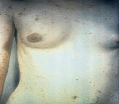
| Melanosis |
|
|
| Mixed Dystrophy |
|
|
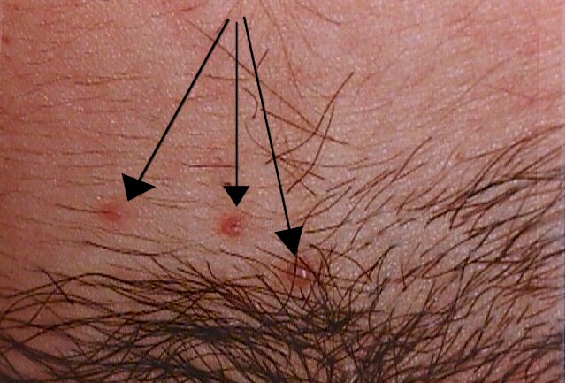
| Molluscom Contagiosum |
|
|
|
|
| Monilia |
|
|
| Pediatric Problems |
|
|
|
|
| Psoriasis |
|
|
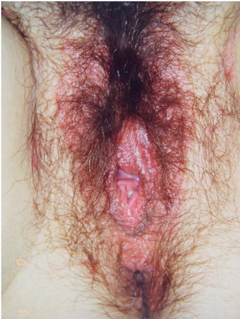
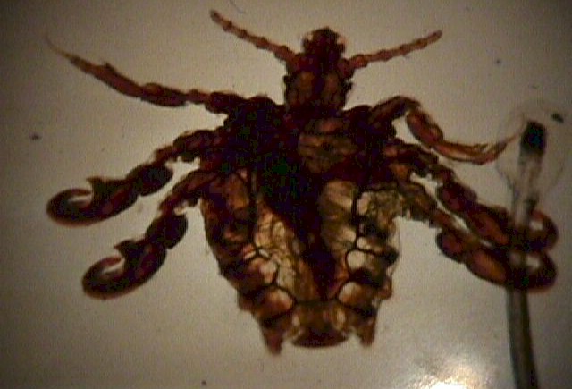
| Pubic Lice |
|
|
| Skenitis |
|
|
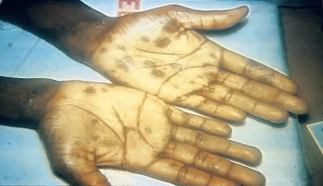
| Syphilis |
|
|
|
|
|
|
|
|
|
|
|
|
|
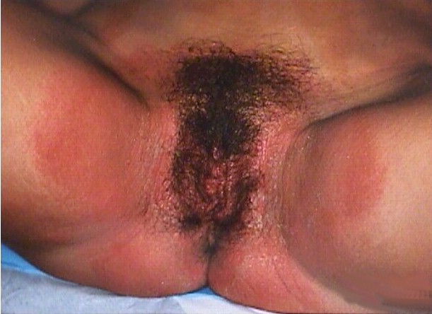
| Tinea Cruris |
|
|
| Vestibulitis |
|
|
| VIN |
|
|
| Vulvar Cancer |
|
|
| Vulvar Hematoma |
|
|
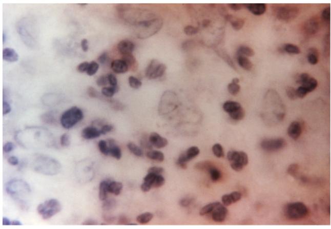 This
photomicrograph reveals "Donovan bodies" in a skin sample used to diagnose
granuloma inguinale. Diagnosis of granuloma inguinale is based on the
appearance of encapsulated bacilli called Donovan bodies. These are safety
pin-shaped microorganisms that appear inside infected tissue cells under a
microscope. (Courtesy of the Center for Disease Control, Susan Lindsley.)
This
photomicrograph reveals "Donovan bodies" in a skin sample used to diagnose
granuloma inguinale. Diagnosis of granuloma inguinale is based on the
appearance of encapsulated bacilli called Donovan bodies. These are safety
pin-shaped microorganisms that appear inside infected tissue cells under a
microscope. (Courtesy of the Center for Disease Control, Susan Lindsley.)