Chapter 31
Diagnosis and Therapy of Benign and Preinvasive Disease of the Cervix
Joanna M. Cain, Fabio Cappuccini and William Petty
Main Menu   Table Of Contents

Search

Joanna M. Cain, MD
Professor and Chair, Department of Obstetrics and Gynecology; Director, Center for Women's Health, Oregon Health Sciences University, Portland, Oregon (Vol 1, Chap 31)

Fabio Cappuccini, MD
Division of Gynecologic Oncology, Department of Obstetrics and Gynecology, Oregon Health & Science University, Portland, Oregon (Vol 1, Chap 31)

William Petty, MD
Division of Gynecologic Oncology , Department of Obstetrics and Gynecology, Oregon Health & Science University, Portland, Oregon (Vol 1, Chap 31)

 
BENIGN LESIONS OF THE CERVIX
NABOTHIAN CYSTS
CONDYLOMATA ACCUMINATA
SQUAMOUS PAPILLOMA
LEIOMYOMAS
ENDOMETRIOSIS
ADENOSIS
ENDOCERVICAL POLYPS
PREMALIGNANT SQUAMOUS LESIONS
THERMAL ABLATION: CRYOTHERAPY
LOOP ELECTROEXCISION PROCEDURE
REFERENCES

Identifying preinvasive and benign diseases of the cervix and selecting an appropriate treatment path can be challenging. Often, benign cervical disease appears malignant and malignant disease can be hidden from view. The focus of this chapter is the range of diseases from benign through preinvasive diseases of the cervix and options for management as well as guidelines that help identify those with potentially more serious or invasive diseases.

Cervical lesions range from being visible only with an instrument that augments sight such as a colposcope to gross abnormalities. Diagnosis and management requires a compilation of visual assessment (including colposcopy), tactile assessment, and laboratory assessment (Papanicolaou smears, cultures, biopsy, etc.). The details of colposcopy, one of the major tools in delineating these lesions, are covered elsewhere in the text, so this chapter covers an expanded area for management.

Back to Top
BENIGN LESIONS OF THE CERVIX

Triage of cervical lesions begins with history and symptomatology. Previous history of any malignancy, particularly those that are prone to metastases, including breast, melanoma, gestational trophoblastic disease, and high-grade malignancies of the GI tract or lung, may be a clue that this lesion represents a rare metastasis to the cervical area putting this lesion out of the benign category. Previous history of cervical surgeries, abnormal Pap smears, uterine polyps, or myoma all help guide the direction of the examination. Lesions characterized by bleeding and discharge immediately raise the possibility of infection and/or malignancy. Lesions with local pressure symptoms, for example, bladder pressure or urgency, also raise these two diagnoses. Lesions associated with cramping similar to that women associate with cervical dilation with childbirth (with or without discharge) raise the possibility of prolapsing uterine or cervical myomas, polyps, or even malignancies. Classic watery or foul discharge raises the immediate suspicion of an invasive malignancy, and a gush of fluid raises the question of tubal disease. So, eliciting a good history immediately begins to focus the differential diagnosis for the lesion.

The second element of diagnosis is the physical examination. While a full physical examination is often warranted, the elements of a pelvic examination performed before a full cervical examination are critical in continuing to delineate the diagnosis. Specifically, groin nodes should be palpated first, because enlargement may point to an unexpected infection or malignancy. The skin of the perineum and the mucosa of the vagina are inspected closely and lesions present in this area (for example condylomata) again direct the diagnosis further. Finally, the cervix itself is carefully evaluated in total. If the characteristics of the lesion are unusual, consideration of additional colposcopic assessment is appropriate to allow better assessment of the nature of the lesion and the blood supply to it.

The normal cervix varies throughout life, but understanding this transition is key to diagnosis of differences noted on evaluation of the cervix (Figs. 1A and 1B). Normal cervical epithelium should be uniformly pale pink and the epithelium thick enough that the vascular pattern is generally obscured. The cervical squamous to glandular transformation zone varies throughout life, but areas of gland openings within squamous metaplastic tissue are expected. Without estrogen, with chemotherapy, and with radiation, the epithelium tends to become atrophic with a whiter appearance and regression of the squamocolumnar junction (see Fig. 1B). If the cervix is erythematous, white in appearance, with prominent vascular patterns, friable, or if the area of transformation is beefy or edematous, these are all findings that require explication.

Fig. 1. A. Normal cervix. Note transformation zone and areas of squamous metaplasia around gland openings. B. Normal cervix/atrophy.

Back to Top
NABOTHIAN CYSTS

A common benign finding, nabothian cysts are present on the portio of the cervix and are thought to arise in areas of active metaplasia. They represent occlusion of the mucous-secreting glands of the cervix underneath a squamous covering with mucin collecting in a cystic area (Fig. 2). Usually these are small (less than .5 cm) and can be multiple; however, cysts up to 5 cm or greater in diameter have been described. They should have a smooth glistening surface with clear or slightly milky contents (Fig. 3). Deviations from this smooth surface, such as erosions or vascular anomalies, should be further investigated with colposcopy and biopsy. Clearly, very large cysts have secondary symptoms (such as pressure, heaviness, or urinary retention) unlike the absence of symptoms that characterizes the majority of nabothian cysts.

Fig. 2. Multiple small nabothian cysts of the cervix.

Fig. 3. Nabothian cysts of the cervix (×3).

The natural history of nabothian cysts may be rupture (more commonly) with a subsequent small yellow scar, stabilization, or growth. The majority of nabothian cysts, therefore, require no treatment, and the diagnosis is clinically obvious with the exception of the abnormalities noted. Large nabothian cysts may benefit from being opened with a loop electroexcision procedure (LEEP) or direct cauterization. The key is assuring that the area fully drains, which may be accomplished by assuring that the squamous covering is adequately resected or that the base is cauterized to decrease mucous production.

Back to Top
CONDYLOMATA ACCUMINATA

Cervical condyloma can take various forms (Figs. 4A and 4B), but generally they appear as one or multiple clearly delineated, elevated, white plaques on the cervical portio and often onto the vaginal apex as well. Small lesions may only be apparent through colposcopic views and acetic acid application after a cytology result of atypical cells of uncertain significance (ASCUS). The larger of these are commonly friable and can have symptoms of postcoital bleeding that would raise issues of malignancy. If a patient has external condylomata, a careful inspection of the vaginal and cervical epithelium for subclinical or clinically apparent condylomata is warranted. Clearly, full evaluation of these lesions requires cytology (preferably with liquid-based techniques to allow for human papilloma virus [HPV] testing if appropriate) and biopsy of a typical lesion with or without colposcopy at the initial evaluation. While condylomata are most commonly associated with HPV types 6, 11, 42, 43, and 44 and the potential for malignancy is low, the fact that they are present may suggest that other HPV strains could be present with higher potential for development of preinvasive disease and even malignancies.

Fig. 4. A. Condylomata. B. Condylomata, clinically unapparent, seen after acetic acid application.

Management on the cervix differs from vulvar areas where direct application of TCA or 5-fluorouracil can be accomplished with little direct absorption. Given the nature of the vaginal and cervical skin, management of these lesions tends to rely heavily on direct removal through the use of biopsy forceps, cautery, LEEP, or laser according to the size and location of the lesion as well as the availability of these modalities. All serve to eliminate the bulk of disease and are assumed to decrease the viral load by virtue of the same.

Management is problematic in women who are pregnant, who are immunosuppressed, or who have chronic recurrence of condylomata. For women who are pregnant, removal with LEEP based therapies is indicated only if there is concern about significant hemorrhage associated with cervical and/or vaginal dilatation at the time of delivery. A significant proportion of these condylomata regress after pregnancy, so therapy is preferentially targeted to the postpartum assessment if they are not thought to be significant enough to interfere with delivery. Use of topical solutions is generally prohibited in pregnancy even for external lesions. Chronic immunosuppression commonly is associated with chronic low-level cytology abnormalities of the cervix and the presence of subclinical or clinical condylomata. Rather than recurrent surgical removal, if the patient is able to continue close observation with cytology and colposcopy on a regular basis, this may well be preferable to multiple surgical interventions. Then, either a change in colposcopic appearance, gross appearance, or cytology should trigger a biopsy to ensure that any advance in the intraepithelial neoplasia associated with condylomata is identified and appropriately treated. If it remains stable at cervical intraepithelial neoplasia I or less, then close follow up may suffice. Options for removal include biopsy and/or removal, cryotherapy, laser ablation (of concern in pregnancy), and LEEP. LEEP has not been extensively investigated for this purpose in pregnancy, so the risks are as yet not fully explicated.

Back to Top
SQUAMOUS PAPILLOMA

Papillomas that are not HPV-related can occur in the cervix and usually originate from the exocervix near the squamocolumnar junction (transformation zone). These are thought to be related to local irritation or scarring and are usually less than 1 cm. Their natural history is not well described, as the usual intervention for these papillomas is excision to assure that this does not represent malignancy1 (Figs. 5 and 6).

Fig. 5. Squamous papilloma of the cervix.

Fig. 6. Benign squamous papilloma of the cervix.

Back to Top
LEIOMYOMAS

Leiomyoma is the most common tumor of the uterine body, but it is rare to find it isolated to the cervix. In the cervix, leiomyomas can occur wherever there is smooth muscle, so they can be subserosal, intramural, or submucous in analogy to those in the body of the uterus. Endocervical leiomyomas become symptomatic particularly if an endocervical polyp prolapses, often with significant cramping and bleeding or if there is endocervical obstruction with hematometra. Exocervical and endocervical leiomyomas can become large enough to obstruct urethral function as well as to prolapse into the vagina and cause bleeding. The decision to remove these masses is generally straightforward, both to adequately diagnose the condition and to alleviate the symptoms. The surgical management of these lesions is best undertaken in the operating room with adequate planning for the potential of abdominal as well as vaginal approaches. Removal generally involves a surgical procedure carefully tailored to the individual patient because of the Paracervical impingement on bladder, bowel, ureter, and even urethra. Plans for adequate control of the cervical branch of the uterine artery is always an important element. Removal of pedunculated fibroids in the office setting is generally discouraged, because the upper limits and vascular relationships of leiomyoma of the cervix are rarely straightforward.

Back to Top
ENDOMETRIOSIS

Cervical endometriosis (Fig. 7) can mimic multiple lesions, including gestational trophoblastic disease (GTD), because of its high-vascular nature. History and physical examination to evaluate the likely nature of the lesion, including a laboratory assessment of human chorionic gonadotropin are all prudent before consideration for biopsy or treatment. Biopsy of areas of trophoblastic disease are generally avoided, because the invasive neovascularization that accompanies these lesions may lead to significant hemorrhaged, unlike endometriotic implants that are less likely to have postbiopsy hemorrhage. Symptoms from a cervical lesion are generally related to bleeding or discharge and pelvic pain is more likely referred from endometriotic implants elsewhere in the pelvis. These areas should respond to general therapy for endometriosis (the use of Lupron, for example) and failure to respond should lead to biopsy to assure that this is the diagnosis if not performed previously.

Fig. 7. Cervical endometriosis.

Back to Top
ADENOSIS

Extension of the glandular epithelium onto the portio and even the vaginal apex is a classic hallmark of exposure to diethylstilbestrol (DES), but it can occur normally in some individuals. As individuals age the extent of the adenosis tends to decline toward the endocervical os, but islands can remain throughout the upper one third of the vagina. In the presence of normal cytology and, for DES patients, normal base colposcopy, no further therapy is needed. Biopsy is reserved for areas of questionable vascularity or for worrisome colposcopic findings.

Back to Top
ENDOCERVICAL POLYPS

Endocervical polyps are common and are almost always benign (Fig. 8). They are rarely seen unless they prolapse through the external cervical os, and patients often present with bleeding or spotting that is inconsistent with their menstrual cycle. Their stalk is of variable length and width. The gross appearance of endocervical polyps may vary widely and does not differentiate them from endometrial polyps nor identify them as benign or malignant by appearance alone.

Fig. 8. Cervical polyp.

Evaluation of the stalk attachment is the key to management. Often simply following the stalk to its base with a cotton swab or probe is adequate. A sonohysterogram may also be helpful if the stalk seems to progress into the uterine cavity. Colposcopy of the prolapsed portion may also give information about the likelihood of malignancy as well as the level of vascularity of the polyp. If the base is narrow and the endocervical portion easily accessed, then removal in the office can be considered. This may require only twisting the base with grasping forceps. Bleeding can occur after removal and endocervical curettage, chemocautery, or electrocautery may be required for control. Multiple polyps or broad-based polyps may require the use of excision, including LEEP and the use of laparoscopic ligature systems to control and are best managed in a more controlled environment. Recurrence can occur, although thought to be less with curettage of the base. Pathology of endocervical polyps (Fig. 9) often shows varied degrees of inflammation and edema and occasionally squamous metaplasia at the tip, accounting for the appearance of normally branching but abundant vasculature and white epithelium seen on colposcopy of these lesions.

Fig. 9. Benign endocervical polyp.

Back to Top
PREMALIGNANT SQUAMOUS LESIONS

Cytology alone, to reiterate this for its importance in management, does not make a pathology diagnosis. This is only accomplished by cervical biopsy, usually with concurrent colposcopy. A precise delineation of the size and nature of the lesion with colposcopy is required before biopsy (Figs. 10 and 11). Management of these lesions is defined by the cytologic history, the patient's risk for malignancy, and the reliability of the patient for follow-up. With the new cytologic guidelines, colposcopy and biopsy of findings for both ASCUS and HPV typing for high-risk subtypes as well as colposcopy and biopsy for ASCUS × 2, plus evaluation and biopsy of all cytologic findings higher than this level is appropriate. Biopsy may be performed with conventional biopsy forceps or with LEEP. If low-grade squamous intraepithelial lesions (CIN I) are present and this is compatible with the cytologic findings, then therapy should be individualized. Of particular concern is the use of ablative and excision techniques in young nulliparous women. For low-grade lesions with adequate evaluation, close follow-up of the young nulliparous woman with CIN I who understands the need for careful observation may be an option. The optimal interval is debatable, but an evaluation every 3 to 6 months is commonly recommended.

Fig. 10. Lesion confined to area for biopsy dear endocervix.

Fig. 11. Biopsy area in anterior cervix outlined by colposcopic evaluation.

For women who have higher-grade lesions (CIN II and III) or a suspicious benign lesion of the cervix as outlined, ablation or excision may be appropriate. Techniques include thermal ablation (cryotherapy and cautery), laser ablation, and excision with biopsy forceps, LEEP, laser, or scalpel. The choice among these options is critical and is clinically determined by whether endocervical involvement is present and whether there is an uncertainty regarding the pathology that requires accurate assessment of specific margins of resection. Of all the options, only scalpel excision affords the least chance of crush or thermal injury obscuring critical information at the margins. Because laser treatment and surgical excision are outlined elsewhere, the following discussion is devoted to thermal ablation and loop excision.

Back to Top
THERMAL ABLATION: CRYOTHERAPY

In 1968, Richart and Sciarra3 reported effective treatment of CIN diagnosed by Pap smear and biopsy with cauterization of the cervix, portio, and endocervix. In 1970, Crisp and colleagues4 reported on the effectiveness of cryosurgery in the treatment of CIN. Both methods were conservative and efficacious in the management of CIN and a significant alternative to cervical cone biopsy with hysterectomy for carcinoma in situ, which was the only other available approach. Multiple other means have been developed since that time, and cryotherapy is the primary thermal 
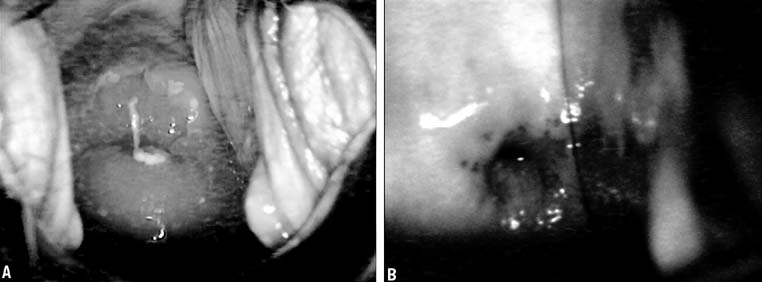
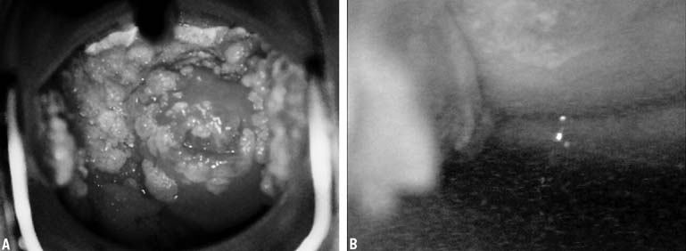
method in use at the present time. Cryotherapy may be used for treatment of condylomata or for ablation of CIN. Again, satisfactory pretreatment evaluation and individualization of therapy based on age, disease, and gravidity is important. Cryosurgery is an office procedure that usually can be performed without anesthesia or analgesia. Occasionally patients will experience discomfort, but it is seldom of a severity to require discontinuation of the treatment. Self-limiting vasomotor reactions characterized by light-headedness and flushing 
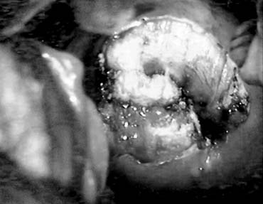
are common. After cryosurgery, patients will usually have 10 to 14 days of watery discharge requiring four or five sanitary napkins daily. Coitus and intravaginal tampons are not recommended during that time.

Technique

Most cryosurgical instruments use either nitrous oxide (freezing point of −89°) or carbon dioxide (freezing point of −65°). Proper freezing requires attention to the pressure within the tanks because a decrease in partial pressure changes the freezing rate of the probe. By changing the rate of freezing, the extent of cryonecrosis can be modified. The larger (D) tank is preferable to the narrower (E) tank for circumstances in which a number of sequential patients are treated. The pressure within the tanks must be at least 40 kg/cm2 before and at the completion of the freeze. If there is a pressure decrease during cryosurgery, the procedure should be discontinued and repeated 
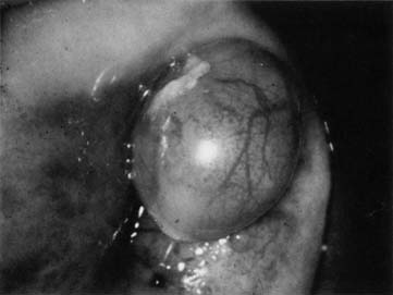
with adequate pressure levels.

Probe tips of various configurations are available and should be tailored to individual cervical anatomy. The various flat and cone tips and the 8-mm rod tip are appropriate for most cryotherapy procedures. Areas 
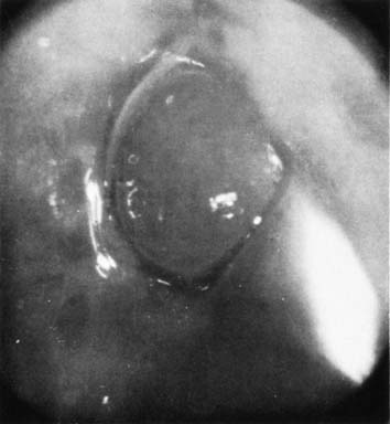
to be treated should be outlined by a visible lesion or by a colposcopic map, and direct connection of the probe tip to the area to be treated must be possible with the tip chosen.

Patients should be treated within 1 week of cessation of menstrual periods. A thin layer of water-soluble lubricant applied to the tip of the probe allows for better heat transfer between the probe and the cervix and fills any potential air gaps in the irregular surface of 
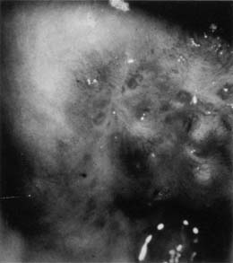
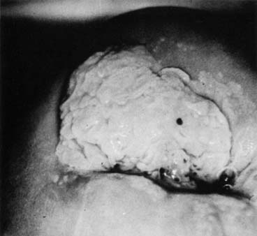
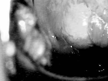
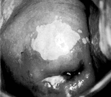
the cervix to provide a more uniform freeze. Freezing of a large ectocervical lesion should begin at the periphery and use overlapping fields of necessary. The ice ball should extend at 4 to 6 mm beyond the edge of the abnormal epithelium. The depth of cryonecrosis will be approximately 4 to 5 mm and theoretically should destroy any intraepithelial neoplastic process extending into endocervical glands on the portio. The extent of the ice ball beyond the confines of the lesion is more critical than the length of the freeze. This will usually occur within 2 minutes, so most clinicians use a freeze technique with 3 minutes on, thaw, and repeat. If constant tank pressure is maintained, tank warmers are used, and careful attention is paid to detail, then a single freeze may be effective.

Cryonecrosis and Surveillance

Cellular death occurs at a temperature of approximately −20 °C. This temperature is within 2 °C of the eutectic point of a sodium chloride solution. Cryosurgery produces severe biochemical and biophysical 
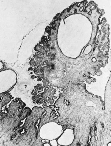
changes resulting in coagulation of the affected tissues. Rupture of the cell wall occurs with the formation of intracellular and extracellular ice crystals. Avascular necrosis is produced by circulatory 
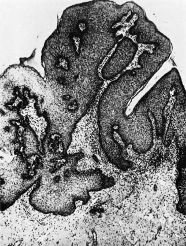
compromise because of capillary obstruction and stasis.5

Regeneration of the epithelium involves the production of initially immature squamous epithelium, which over time will mature into a stratified squamous layer that replaces the neoplastic process. The entire reparative process requires approximately 3 to 4 months. Pap smear surveillance should not be reinitiated before 3 to 4 months, and even then a Pap smear may show a high number of cells in the reparative phase. If a normal Pap smear is obtained, two additional Pap smears during the first year after cryosurgery should be planned and then should be performed every 6 months until 2 years have elapsed. Subsequent follow-up should be decided on the basis of individual risk parameters.

Efficacy

Since the introduction of thermal ablation, there has been skepticism about its efficacy in the conservative therapy of CIN. Two concerns need to be addressed. First, factors must be identified that are associated with primary failure of the technique. Second, the ability of the neoplastic process to recur must be considered along with the potential benefits or deficits of a procedure in expediting the long-term follow-up. This second concern is directly impacted by ongoing exposure to other papillomavirus infections, smoking, and the presence of other viral infections, particularly HIV. Attempts to describe the recurrence rate of any technique are heavily confounded by the action of these c-carcinogenic influences in individual patients. Concerns continue to be raised about long-term follow-up issues after thermal ablation of the cervix. In one study of a small group of patients (27), the colposcopic appearance underestimated the actual disease (11% stromal invasion) in follow-up after cryotherapy.6

Failures with cryotherapy may reflect many factors. The size of the lesion appears to be critical, with larger lesions more likely to recur.7,8 Failure rates also vary based on grade, ranging form 8.8% for CIN I to 14.5% for CIN III.9,10,11 A positive ECC before treatment is particularly worrisome, as failure rates of up to 20.8% have been reported. Inadequate freezing has also been postulated as a factor, but this is unlikely given the changes in the equipment ensuring predictable freeze levels. The majority of failures occur in the first 2 years (93%), supporting the practice of closer observation during this period.12

Immediate side effects are few.13 Long-term fertility is not appreciably altered,14 and pregnancy outcome does not appear to be affected by cryotherapy.15 An additional concern is the loss of the transformation zone, which may lead to a need for more invasive procedures in the future for young women who have a continuing need for observation and who may have additional episodes of preinvasive disease.

Back to Top
LOOP ELECTROEXCISION PROCEDURE

The addition of loop procedures to the outpatient setting has had a significant impact on office treatment of preinvasive disease. Arguments have been put forward for initial removal of the entire transformation zone with colposcopic identification of abnormality, rather than waiting for biopsy results to tailor treatment. In certain populations in which the likelihood of follow-up in tenuous, this may have appeal. For the majority of women, however, the rule that adequate colposcopy and biopsy results must be obtained before LEEP should be followed. Because this is a technique that removes tissue and leaves some level of thermal artifact at the edge, both extensive use in young nulliparous women and use when endocervical status is unknown need to be carefully considered. Again, if the histologic information required from the removal of the tissue and its margins is critical, a scalpel cone biopsy would be preferred. The technique has been proved very safe, having few complications and a stenosis rate of approximately 3.8%16

Technique

LEEP uses high-frequency, low-voltage, electrical energy produced by an electrosurgical unit to excise abnormal tissue. There are a variety of units and electrodes used for LEEP procedures, and the health care givers using the instrument must understand the various details of operation of each unit. Some variation of a grounding pad application may be required, and adequate suction ventilation with a microbial filter is required. The choice of electrodes used should be determined by careful colposcopic mapping of the disease before treatment and by the individual contour of the vagina (width) and cervix.

LEEP is best performed when patient is not menstruating. Because the procedure does involve thermal injury and tissue removal, local anesthetic with a vascular constrictor is injected into the cervical stroma before the procedure. A nonconducting speculum is inserted into the vagina, with the suction ventilation incorporated into the body of the speculum. The cervix may be treated with Lugol's solution for better definition of the lesion. Diathermy power is usually set at 50 to 60 watts cutting or at 50 watts cutting and 60 watts coagulation. An initial evaluative phantom pass of the selected loop (often approximately 1 com [10 mm] in width) identifies that the lesion will be encompassed with one or two passes and that no vaginal wall contact will be made.

Large loop excision of the transformation zone (LLETZ) uses a 15- × 7-mm loop and is intended to remove the entire transformation zone. The use of new loops and a slow steady movement will reduce thermal injury at the margin. The diathermy unit is turned on only when active movement through the cervix is ongoing. Specimens are then removed for pathologic consultation. This can be performed with one deep or shallow pass, followed by a second pass with a thinner electrode. Then the base is cauterized with a ball electrode (Fig. 12).

Fig. 12. Surgical bed after LEEP.

Efficacy

The LLETZ procedure compares favorably with laser techniques and cryotherapy in local recurrence rates.17 The additional pathologic confirmation afforded by removal of a specimen is an advantage of this technique, and coagulation artifact appears to be a lesser concern than with laser cone biopsies, although not as good as scalpel-directed cone biopsy.18,19,20,21 This artifact is directly related to the number of passes required for removal of the entire specimen.

The presence of disease at the endocervical margin and the use of LEEP for patients suspected to have endocervical disease continue to pose difficulties, and the data for loop electrosurgical cone biopsies are in its infancy. Felix and associates22 reviewed positive endocervical margins by either ECC or specimen margins in 19 of 57 women with cervical cone LEEP specimens. They found 12 of 19 patients (63%) with residual cervical cone LEEP specimens. It is unclear whether this represents the true rate of potential recurrence. In a direct comparison of laser conization versus the cold-knife cone technique,23 laser conization was found to be more costly and time-consuming; it was also found to alter the tissue the most compared to LEEP and the cold-knife technique. Between the scalpel and electroexcision, the histologic evaluation of margins was better with the scalpel, and the LEEP was technically easier and less time-consuming. This reinforces the need to make carefully individualized decisions for patients.

In this chapter, a number of alternatives for the diagnosis and management of benign and premalignant lesions have been reviewed. The number of alternatives for management on an outpatient basis is continually increasing. For premalignant lesions, however, individualized treatment according to age, diagnosis, pregnancy status, parity, cervical anatomy, and lifestyle risk factors remains the most important factor in assigning patients to different techniques. For women with HIV, particularly those with decreased immune status, the need to tailor therapy is even greater because the risk of disease recurrence is high regardless of technique.24 The role of the clinician in wisely choosing among this increasing number of options is critical for the lifelong health of their patients.

Back to Top
REFERENCES

1. Papanicolaou GN, Traut HF: Diagnosis of Uterine Cancer by the Vaginal Smear. New York, Common Fund, 1943

2. Hall FE: The use of quick freezing methods in gynecological practices. Am J Obstet Gynecol 43:105, 1942

3. Richart RM, Sciara JJ: Treatment of cervical dysplasia by out patient electrocauterization. Am J Obstet Gynecol 101:200, 1968

4. Crisp WE, Asadourian J, Romberger W: Application of cryosurgery to gynecologic malignancy. Am J Obstet Gynecol 107:737, 1970

5. Droegummueller W, Greer BE, Davis JR et al: Cryocoagulation of the endometrium at the uterine cornua. Am J Obstet Gynecol 131:1, 1978

6. Koonings PP, d'Ablaing G, Schlaerth JB et al: A clinical-pathology review of cervical intraepithelial neoplasia following cryotherapy failure. Gynecol Oncol 44:213, 1992

7. Townsend DE: Cryosurgery for CIN. Obstet Gynecol Surv 34:828, 1979

8. Ceasman WT, Hinshaw WM, Clarke-Pearson KL: Cryosurgery in the management of cervical intra-epithelial neoplasia. Obstet Gynecol 63:145, 1984

9. Creasman WT, Clarke-Pearson DL, Ahse C et al: The abnormal Pap smear: What to do next? Cancer 48:515, 1981

10. Vanheut M, Trimbos JB, Heintz AP et al: Cryosurgical treatment of CIN II in 102 patients. Gynecol Oncol 16:240, 1983

11. Kaufman RH, Irwin JF: The cryosurgical therapy of cervical intra-epithelial neoplasia. Am J Obstet Gynecol 131:381, 1978

12. Hatch KD, Shingleton HM, Austin JM et al: Cryosurgery of cervical intraepithelial neoplasia. Obstet Gynecol 57:692, 1981

13. Figge DC, Creasman WT: Cryotherapy in the treatment of cervical neoplasia. Obstet Gynecol 62:353, 1983

14. Weed JC, Cuney SL, Dunca ID et al: Fertility after cryosurgery of the cervix. Obstet Gynecol 52:245, 1978

15. Chanels EH, Savage EW: Cryosurgical treatment of CIN. Obstet Gynecol Surv 35:539, 1980

16. Bigrigg A, Haffended, Sheehan AL et al: Efficacy and safety of large-loop excision of the transformation zone. Lancet 343:32, 1994

17. Gunasekera PC, Phipps JF, Lewis BV: Large loop excision of the transformation zone (LLETZ) compared to carbon-dioxide laser in the treatment of CIN: A superior mode of treatment. Br J Obstet Gynecol 97:995, 1990

18. Naumann RW, Bell MC, Alvarez RD et al: LLETZ is an acceptable alternative to diagnostic cold-knife conization. Gynecol Oncol 55:224, 1994

19. Messing MJ, Otken L, King LA et al: Large loop excision of the transformation zone (LLETZ): A pathologic evaluation. Gynecol Oncol 52:207, 1994

20. Helkjaer PE, Schultz H, Eriksen PS et al: Thermal artifacts and inter-gynecologist variation of laser cone biopsies of the cervix. Obstet Gynecol 82:435, 1993

21. Montz FJ, Holschneider CH, Thompson LD: Large loop excision of the transformation zone: Effect on the pathologic interpretation of resection margin. Obstet Gynecol 81:976, 1993

22. Felix JC, Muderspach LI, Duggan BD et al: The significance of positive margins in loop electrosurgical cone biopsies. Obstet Gynecol 84:9960, 1994

23. Mathevet P, Dargent D, Roy M et al: A randomized prospective study comparing three techniques of conization: Cold knife, laser, and LEEP. Gynecol Oncol 54:175, 1994

24. Maiman M, Fruichter RG, Serur E et al: A recurrent cervical intraepithelial neoplasia in human immunodeficiency virus-seropositive women. Obstet Gynecol 82:170, 1993

Back to Top