Chapter 73
Surgical Management of Intractable Pelvic Hemorrhage
Louis G. Keith, Robert L. Vogelzang and Daniel L. Croteau
Main Menu   Table Of Contents

Search

Louis G. Keith, MD
Professor, Department of Obstetrics and Gynecology, Northwestern University Medical School, Chicago, Illinois (Vol 1, Chap 73; Vol 2, Chaps, 74, 81, 82; Vol 6, Chap 33)

Robert L. Vogelzang, MD
Professor of Radiology, Northwestern University Medical School; Chief, Vascular and Interventional Radiology, Northwestern Memorial Hospital, Department of Radiology, Chicago, Illinois (Vol 1, Chap 73)

Daniel L. Croteau, MD
Fellow in Vascular and Interventional Radiology, Northwestern University Medical School, Chicago, Illinois (Vol 1, Chap 73)

INTRODUCTION
ANATOMY
PHYSIOLOGY OF LIGATION
INDICATIONS FOR LIGATION
LIGATION OF THE HYPOGASTRIC ARTERY: SURGICAL TECHNIQUE
SPECIAL CONSIDERATIONS
ANEURYSMS OF THE HYPOGASTRIC ARTERY
TREATMENT OF PELVIC HEMORRHAGE WITH INTERNAL ILIAC ARTERY EMBOLIZATION EMBOLIZATION
THE NORTHWESTERN EXPERIENCE WITH PELVIC EMBOLIZATION FOR UTERINE BLEEDING UTERINE BLEEDING
CASE REPORTS
REFERENCES

INTRODUCTION

The background of ligation of the hypogastric (internal iliac) arteries for control of pelvic hemorrhage is not clear. Medical historians attribute the procedure to different surgeons in diverse specialties. In the United States, the operation was reported before 1900. Since then, diverse articles about this procedure have appeared sporadically; its usefulness has been demonstrated in many areas dealing with the pelvis and its contents.

Ligation of the hypogastric arteries can be a lifesaving procedure for patients with intractable hemorrhage from pelvic viscera. This is especially true in the field of obstetrics and gynecology, in which hemorrhage remains a major cause of mortality. Some physicians may be reluctant to perform hypogastric artery ligation for fear of injury to the pelvic viscera by interruption of the blood supply. With rare exceptions, this reluctance is unwarranted. Short- and long-term effects of hypogastric artery ligation generally are salutary. In contrast, so-called conservative procedures, such as vaginal packing, suturing of the vaginal vault, and supracervical hysterectomies, frequently fail to control hemorrhage. Before hypogastric artery ligation was considered a potential approach to the problem of pelvic hemorrhage, time, blood, and, alas, lives were lost. The technique of hypogastric artery ligation is acquired easily and should be practiced by the obstetrician and gynecologist.

Back to Top
ANATOMY

Topographic Anatomy and Surgical Landmarks

The intra-abdominal arrangement of the iliac vascular system can be projected on the abdominal surface. Beginning at a point 1/2 inch below and to the left of the umbilicus, draw a line inferolaterally so as to bisect a line running medially and inferiorly from the anterosuperior iliac spine to the middle of the symphysis pubis. The upper one third of the umbilicospinal line traces the course of the common iliac artery before its bifurcation. The distal two thirds of the same line delineates the external iliac artery; a line dropped medioinferiorly from this junction to the pelvic floor suggests the course of the hypogastric artery. The external bony landmark to the level of the bifurcation of the common iliac artery is the anterosuperior iliac spine. In most instances a line between both anterosuperior iliac spines bisects the points of bifurcation.

Internally, the aorta generally bifurcates into the common iliac arteries at the level of the fourth lumbar vertebra. The common iliac arteries, in turn, divide into the external and internal iliac (hypogastric) arteries. The external iliac artery courses along the psoas muscle laterally and ventrally to the leg, where it becomes the femoral artery. The hypogastric artery drops medioinferiorly along the border of the psoas muscle into the pelvis. An internal bony landmark for the level of the aortic bifurcation is the sacral promontory (lumbosacral articulation).

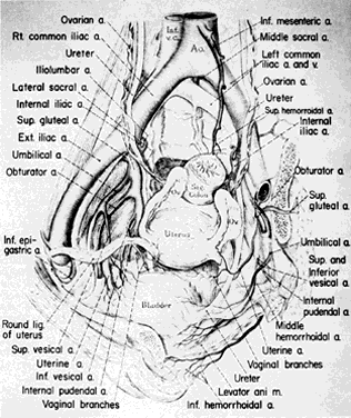
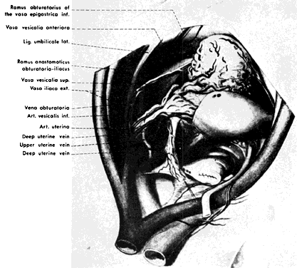
Once the hypogastric artery reaches the pelvis, it divides into the so-called anterior and posterior divisions. Those in turn divide into many branches, which are designated collectively as the hypogastric axis (Table 1). Figure 1 illustrates the gross anatomy.

TABLE 1. Branches of Internal Iliac Artery


Posterior Division

Anterior Division

Parietal

Parietal

Visceral

 Iliolumbar a.

 Obturator a.

Umbilical a. (fetal)


Superior vesical a.

 Lateral sacral a.

 Internal pudendal a.

Inferior vesical a.


Middle hemorrhoidal a.

 Superior gluteal a.

 Inferior gluteal a.

Uterine a.


Vaginal a.

Fig. 1. Surgical anatomy of pelvic circulation.(Reich WJ, Nechtow MJ: Ligation of the internal iliac [hypogastric] arteries: A life-saving procedure for uncontrollable gynecologic and obstetric hemorrhage. J Int Coll Surg 36:157, 1961)

Anatomic Relationships

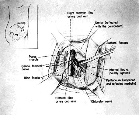
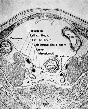
The hypogastric arteries have important relationships to neighboring anatomic structures. Knowledge of these relationships facilitates locating the arteries, as well as dissecting and ligating them. These relationships, listed below, are illustrated in Figure 2, Figure 3 and Figure 4.

Fig. 2. Cross-section of pelvis below level of bifurcation of common iliac artery, showing retroperitoneal relationship of ureter.(Reich WJ, Nechtow MJ: Ligation of the internal iliac [hypogastric] arteries: A life-saving procedure for uncontrollable gynecologic and obstetric hemorrhage. J Int Coll Surg 36:157, 1961)

Fig. 3. Lateral view of pelvic vessels, peritoneum removed.(Modified from von Peham H, Amreich J: Operative Gynecology. Ferguson LK [trans]. Philadelphia, JB Lippincott, 1934)

Fig. 4. Lateral view of pelvic vessels, peritoneum present.(Modified from von Peham H, Amreich J: Operative Gynecology. Ferguson LK [trans]. Philadelphia, JB Lippincott, 1934)

  1. Anteromedially, the hypogastric (internal iliac) artery is covered by peritoneum; it is a retroperitoneal structure. On the right side of the pelvis at this point, the terminal end of the ileum and cecum may overlie the peritoneum.
  2. The ureter lies anterior to the hypogastric artery (retroperitoneal, attached to the undersurface of the peritoneum).
  3. Posterolateral to the hypogastric artery are the external iliac vein and the obturator vein.
  4. Posteromedial to the hypogastric artery is the hypogastric vein.
  5. Lateral to the hypogastric artery are the psoas muscles, major and minor.

Collateral Circulation

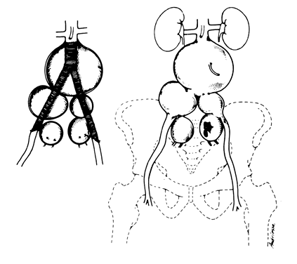
The collateral circulation of the pelvis has been the subject of discussion for at least a century. Gray's Anatomy (1870 edition) mentioned the many anastomoses. Anastomoses occur in each hemipelvis, horizontally and vertically across the pelvis.1, 2 The vertical system functions to a greater extent than the horizontal, especially after bilateral ligation. Figure 5 is a schematic representation of what might occur after unilateral ligation.

Fig. 5. Collateral circulation in pelvis.(Reich WJ, Nechtow MJ: Ligation of the internal iliac [hypogastric] arteries: A life-saving procedure for uncontrollable gynecologic and obstetric hemorrhage. J Int Coll Surg 36:157, 1961)

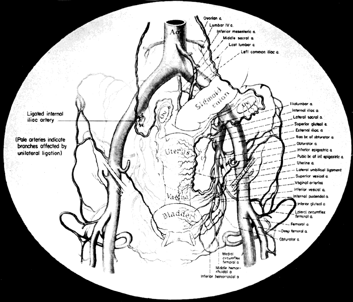
Three major vertical anastomoses exist in each hemipelvis: (1) lumbar-iliolumbar, (2) middle sacral, and (3) superior hemorrhoidal-middle hemorrhoidal. Bilateral ovarian-uterine anastomoses are another important vertical link. Anastomoses also occur between the inferior epigastric and medial circumflex femoral arteries, the circumflex and perforating branches of the deep femoral artery and the inferior gluteal artery, and the superior gluteal artery and the posterior branches of the lateral sacral artery.

The horizontal anastomoses are the branches of the vesical artery from each side and the pubic branches of the obturator artery from each side. Both systems are outlined in Table 2.

TABLE 2. Major Pelvic Anastomoses

  Vertical

  1. Ovarian artery (branch of aorta) with the uterine artery
  2. Superior hemorrhoidal artery (branch of inferior mesen-teric) with middle hemorrhoidal artery
  3. Middle hemorrhoidal artery with inferior hemorrhoidal (branch of internal pudendal from hypogastric)
  4. Obturator artery with inferior epigastric artery (branch of external iliac)
  5. Inferior gluteal artery with circumflex and perforating branches of deep femoral artery
  6. Superior gluteal artery with lateral sacral artery (posterior branches)
  7. Lumbar arteries with iliolumbar artery

  Horizontal
  1. Branches of vesical arteries from each side
  2. Pubic branches of obturator from each side

Variations in the Pelvic Circulation

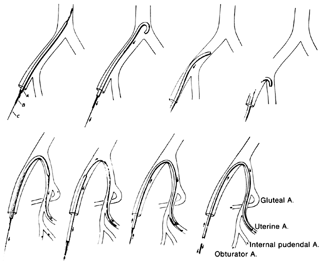
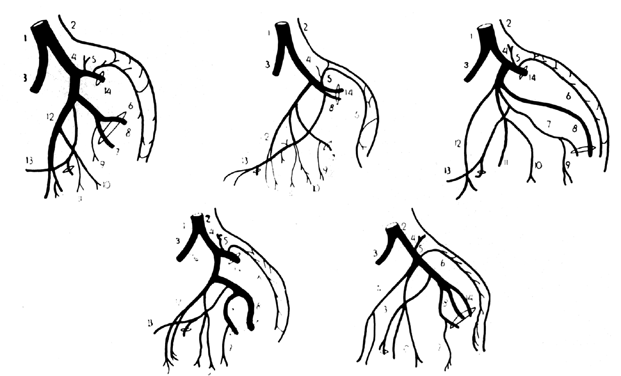
The forgoing anatomic descriptions suffice for practical purposes. Dissection of the branches of the hypogastric artery, however, frequently reveals variation in the number of branches, the relative size of the branches, and the length and diameter of the right and left hypogastric arteries.2, 3, 4 Figure 6 illustrates some of the documented variations in the branches of the hypogastric artery. Table 3 lists the collateral circulation of the branches of the hypogastric artery.

TABLE 3. Collateral Circulation of Branches of Hypogastic Arteries


Posterior Trunk

Anterior Trunk

Branch

Collaterals

Branch

Collaterals

Iliolumbar a.

Gluteal a.

Internal pudendal a.

External pudendal a.


Lumbar a.


Inferior gluteal a.


Lateral circumflex a.


Deep circumflex a.

Obturator a.

Middle circumflex a.


Spinal a.


Inferior epigastric a.


Iliolumbar a.


Inferior gluteal a.

Lateral sacral a.

Spinal a.


Superior gluteal a.


Inferior gluteal a.

Inferior gluteal a.

Lateral circumflex a.


Middle sacral a.


Middle circumflex a.


First perforating a.

Superior gluteal a.

Lateral sacral a.


Deep circumflex a.

Umbilical a.

Vesical a.


Inferior gluteal a.


Ureteric a.


Lateral circumflex a.


Uterine a.

Ovarian a.


Middle hemorrhoidal a.

Superior hemorrhoidal a.


Inferior hemorrhoidal a.

Fig. 6. Common variations in “hypogastric axis.” 1, common iliac artery; 2, middle sacral artery; 3, external iliac artery; 4, internal iliac artery; 5, iliolumbar artery; 6, lateral sacral artery; 7, internal pudendal artery; 8, inferior gluteal artery; 9, middle hemorrhoidal artery; 10, inferior vesical artery; 11, uterine or deferential artery; 12, umbilical artery; 13, obturator artery; 14, superior gluteal artery.(Shafiroff BGP, Grillo EB, Baron H: Bilateral ligation of the hypogastric arteries. Am J Surg 98:34, 1959)

The variations are relatively unimportant; however, some specific exceptions must be mentioned:

  1. Obstruction of the distal ureter by aberrant branches of the anterior division3: The obstruction usually is situated in the lower one third of the ureter at a point midway between the bladder and the brim of the pelvis. Hydroureter and dilatation of the renal calyces may occur in such cases; they can be demonstrated by an excretory urogram. Significant clinical symptoms are pain and discomfort in the loin on the affected side. Infection secondary to stasis of urine is likely to be accompanied by chills, fever, nausea, and hematuria. Therapy consists of surgical resection 
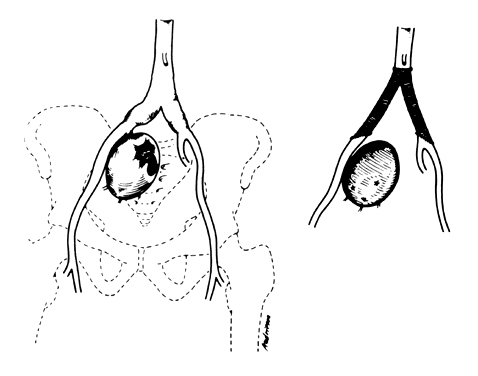
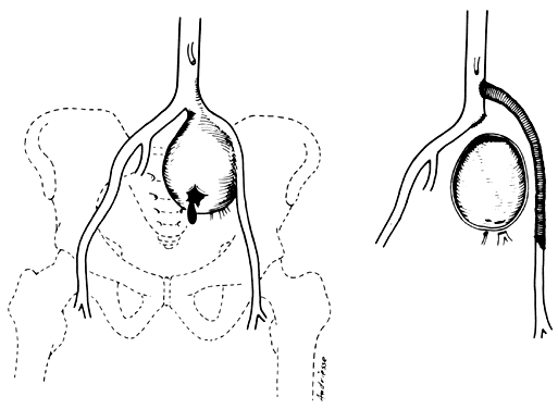
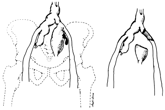
of the aberrant vessel (Fig. 7).

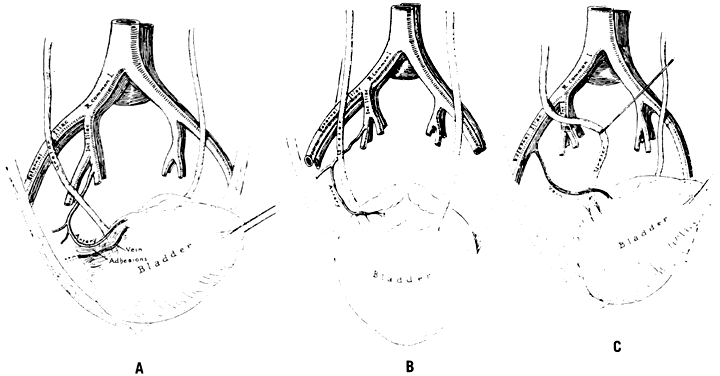
    Fig. 7. Aberrant artery causing ureteral obstruction. A. Schematic drawing from a cadaver showing bladder retracted to left and a band of fibrous tissue, artery, and vein crossing ureter at right angle. B. Bladder has been pulled anteriorly, exposing lower ureter and seminal vesicles and showing artery from hypogastric encircling ureter. C. Bladder retracted to left side, showing a vein coming from external iliac and looping about ureter.(Hyams J: Aberrant blood vessels as factors in lower ureteral obstruction: Preliminary report. Surg Gynecol Obstet 48:474, 1929)

  2. Failure of the distal hypogastric (umbilical) artery to atrophy: During fetal life, the hypogastric artery ascends from the pelvis along the bladder and onto the back of the anterior abdominal wall, where after joining its contralateral artery, it passes into the umbilicus as the umbilical artery. At the time of birth, its function normally ceases; atrophy converts the distal hypogastric artery into a fibrous cord called the lateral umbilical ligament. In the absence of atrophic changes, however, ureteral obstruction may develop.
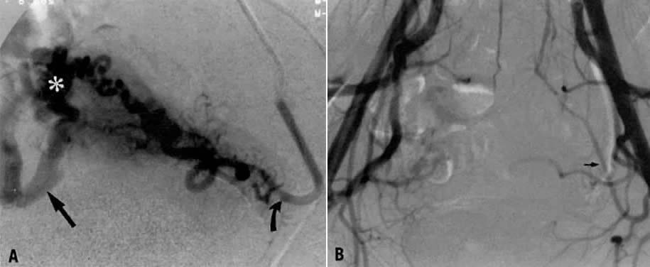
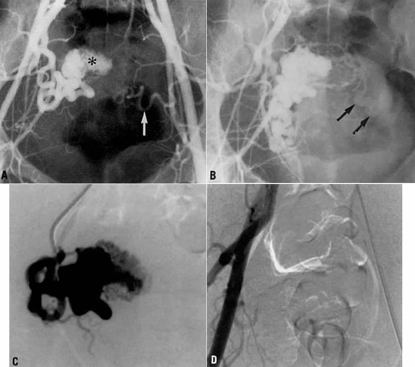
  3. Fistulous channels between branches of the hypogastric arteries and veins: Most patients with this rare condition have one or more of the following signs and symptoms:
    1. Pelvic pain in the area of the fistula radiating to the back, vagina, or down the posterior aspect of the leg on the same side
    2. A pulsatile mass with a bruit detectable vaginally or abdominally
    3. Edema of the legs
    4. Possible cardiac decompensation.

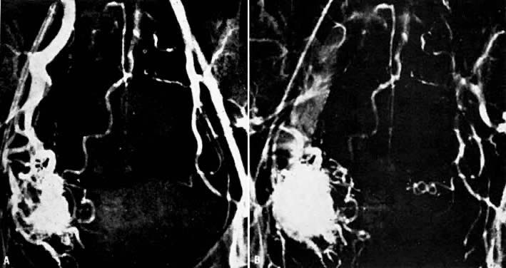
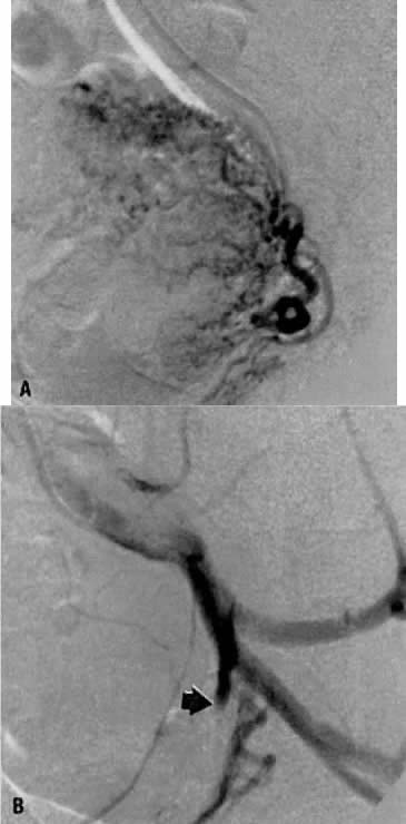
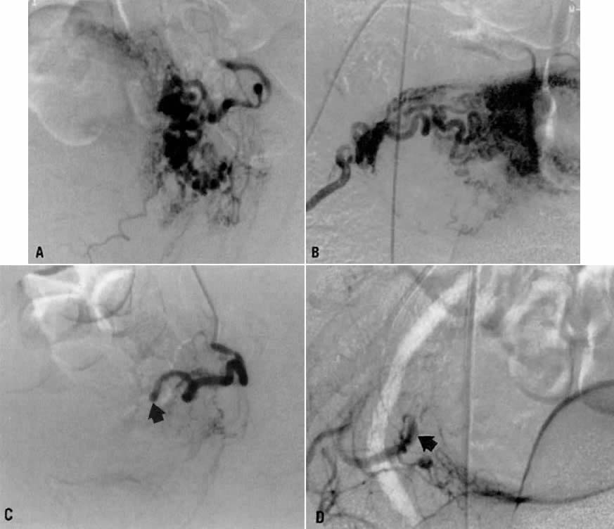
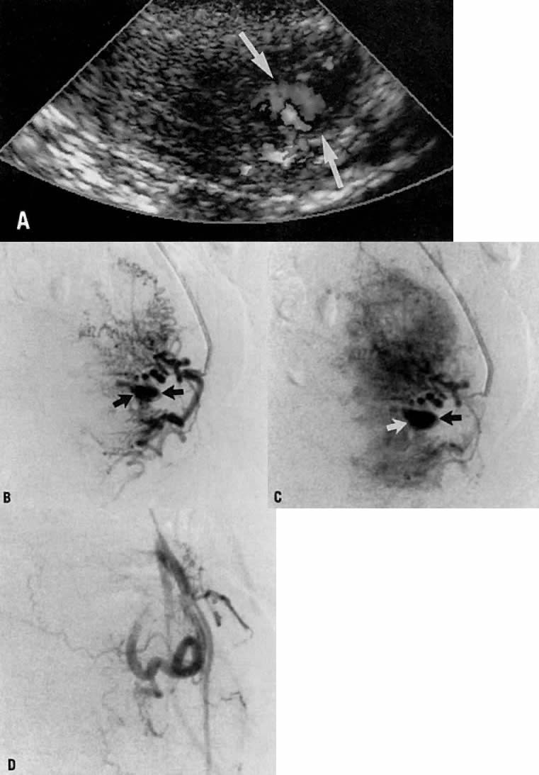
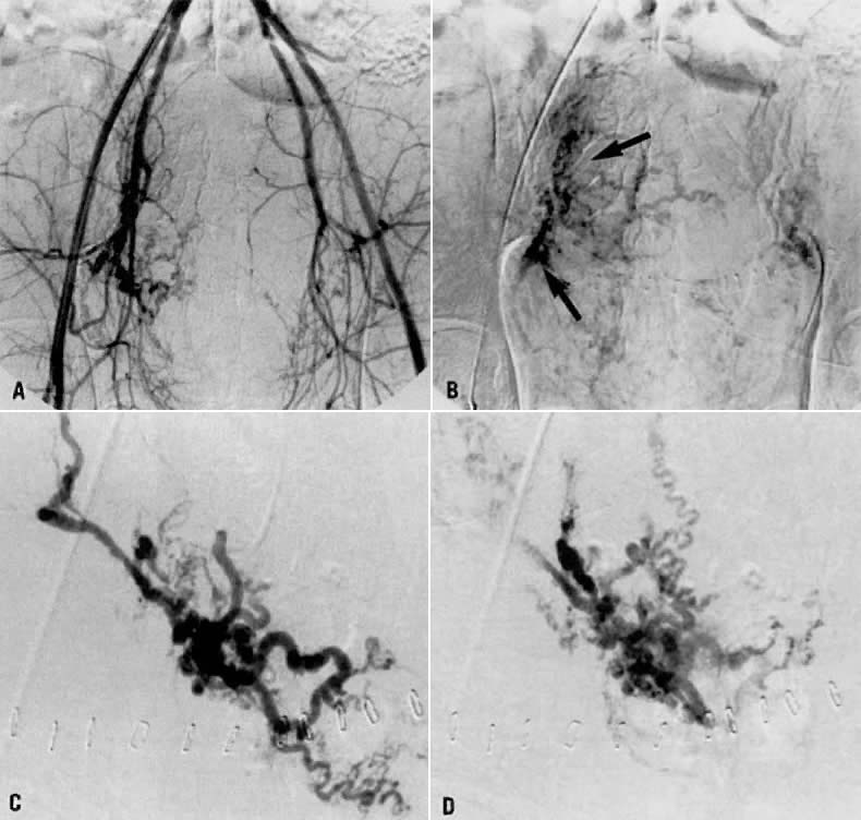
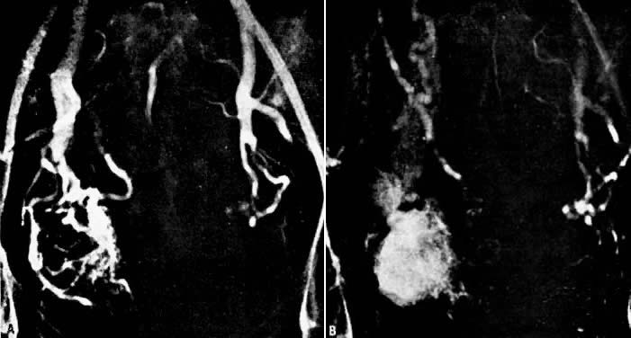
In the pregnant patient with this fistula, antepartum bleeding or abnormal enlargement of the uterus is probable. Severe, catastrophic hemorrhage has been reported in the postpartum period or after abortion. Percutaneous femoral aortography establishes the diagnosis. Therapy is achieved via surgery5 (Fig. 8 and Fig. 9)

Fig. 8. Pelvic arteriovenous fistula. Arteriographic findings during 29th week of pregnancy. A. Early arterial filling discloses nature of lesion and identifies right internal iliac artery as principal but not sole source of supply. B. Three seconds later there is filling of enlarged iliac draining veins. Enlarged uterine arteries are still opacified. Crosspelvic arterial feeder is visible, as are contributors from branches of external iliac artery. The fetal skeleton is visible.(Benson RC, Dotter CT, Peterson CG et al: Congenital arteriovenous fistula and pregnancy: Report of 3 cases. Am J Obstet Gynecol 92:672, 1965)

Fig. 9. Pelvic arteriovenous fistula 3 months postpartum. A. Early filling shows contributory sole of inferior mesenteric artery. B. Later stage of pelvic arteriogram again demonstrates intrapelvic, extrapelvic, and cross-pelvic contributing arteries. Enlarged uterine arteries have regressed normally. On the basis of this and previous arteriography it was predicted that surgical removal would be extremely difficult, perhaps even impossible.(Benson RC, Dotter CT, Peterson CG et al: Congenital arteriovenous fistula and pregnancy: Report of 3 cases. Am J Obstet Gynecol 92:672, 1965)

Back to Top
PHYSIOLOGY OF LIGATION

At one time, ligation of the hypogastric system was regarded as equivalent to shutting off all blood to the area. Fortunately, this is not true. If it were, it is likely that the procedure would not be harmless. In reality, the hypogastric artery distal to the point of ligation is never emptied of blood.6 The anastomotic system functions immediately after ligation. What does occur is the virtual abolition of the arterial pulse pressure. This is associated with reduced mean blood pressure and rate of blood flow in the collateral system. As a result, the trip-hammer effect of arterial pulsations is abolished. In effect, ligation changes the distal portion of the artery so that the net pressure is equivalent to that in a vein. Clots remain in situ and are not dislodged by arterial pulsation.

Back to Top
INDICATIONS FOR LIGATION

Active

Conventional methods of therapy for pelvic hemorrhage, such as packing, suturing, and the use of hemostatic agents frequently are ineffective. Active indications for surgical intervention include the following conditions in which rapid and life-threatening hemorrhage can occur:

  1. Intraperitoneal hemorrhage in the immediate postoperative period: Loss of a ligature around the uterine artery can result in profuse hemorrhage. Frequently, the severed artery is retracted within a hematoma. Hypogastric artery ligation stops the bleeding at a point proximal to its source.
  2. Conization of the cervix: Postoperative infection or failure to use lateral hemostatic sutures may precipitate massive bleeding. Packing and suturing may prove totally ineffective.
  3. Lacerations of the cervix, lower uterine segment, and upper vagina: It may be impossible to suture every bleeding point. The only alternative to hypogastric artery ligation would be hysterectomy and partial vaginectomy, with resultant loss of childbearing function.
  4. Uterine atony: Many patients with uterine atony are near death at the time of operation. Hypogastric artery ligation is a quick and rational alternative to hysterectomy, allowing preservation of reproductive potential.
  5. Placenta previa: Implantation of the placenta into the lower uterine segment may interfere with its ability to contract. Bleeding may be copious. Hysterectomy is the only alternative.
  6. Rupture of the uterus: If rupture occurs in the lower uterine segment and extends cephalad, the uterine artery and its branches may tear. Although hysterectomy often is necessary, it sometimes fails to control bleeding from the branches of the uterine artery, which may have retracted.
  7. Advanced endometrial carcinoma: Blood supply to the tumor will be diminished. The ovarian vessels should be ligated at the same time.
  8. Late-stage carcinoma of the cervix: Hemorrhage may be exsanguinating before or after radiation therapy. The ovarian vessels also should be ligated.
  9. Hemorrhage from vaginal angles during or after abdominal hysterectomy: With good reason, the vaginal angles are characterized as “coffin corners.” Mass sutures placed in this area frequently incorporate a ureter.
  10. Vaginal vault bleeding after vaginal hysterectomy: Postsurgical infection predisposes a patient to vaginal vault bleeding. Bleeding may occur several days after vaginal hysterectomy. Occasionally, the tissue is too necrotic to suture, and packing is useless.
  11. Placenta accreta: The uterus may be saved by hypogastric artery ligation.
  12. Abdominal pregnancy with placental implantation in broad ligament area: Disruption of the placental site can precipitate fatal hemorrhage because a mechanism does not exist for the vessels to clamp down. Hypogastric artery ligation may be crucial to patient survival.
  13. Miscellaneous: Other surgical indications are fracture of the pelvis, gunshot wounds, and spontaneous rupture of pelvic veins associated with pregnancy.

Prophylactic

Certain operative conditions are associated with hemorrhage. Operative time frequently is consumed in controlling bleeding areas. Early ligation of both hypogastric arteries allows the surgical procedure to progress in a relatively dry field and saves time.

Prophylactic surgical intervention is indicated in the following conditions and procedures:

  1. Intraligamentous pregnancy: The placental implantation is between the leaves of the broad ligament. Ligation before removal is advisable.
  2. Intraligamentous leiomyoma: An intraligamentous leiomyoma often must be removed by manually peeling it away from the broad ligament. Hypogastric ligation decreases bleeding from the base of the broad ligament.
  3. Pelvic inflammatory disease: Operation for pelvic inflammatory disease often is difficult, and bleeding is profuse. Ligation may decrease the constant ooze.
  4. Extensive endometriosis: Adhesions are frequently dense. Diminished blood supply facilitates the operative procedure.
  5. Wertheim's hysterectomy: Ligation of the hypogastric arteries should be an integral part of Wertheim's operation.
  6. Hysterectomy among Jehovah's Witnesses: Patients belonging to the sect known as Jehovah's Witnesses and to certain other religious sects refuse to receive blood transfusions. Every effort should be made to minimize blood loss.
  7. Multiple leiomyoma: The “key” fibroid may present at the corporocervical junction, distorting the branches of the uterine artery. Hypogastric artery ligation may diminish the risk in this area.
  8. Bleeding, necrotic rectovaginal fistula: Even though cure may not be possible, personal comfort and hygiene may be improved because of the decreased blood supply that sometimes follows hypogastric artery ligation.
  9. Myomectomy: Blood loss at the time of myomectomy is reduced. Future menstrual function is not impaired.
  10. Groin dissection and vulvovaginectomy: When the deep lymph nodes are removed, ligation of the hypogastric arteries reduces blood loss during the remaining procedure.
  11. Abruptio placentae with atony or Couvelaire uterus, or both: If hypofibrinogenemia is not present, uterine blood loss can be reduced effectively by ligation of the hypogastric arteries.
  12. Miscellaneous: Hemorrhage from bladder tumors before cystectomy and abdominoperineal resection are indications for prophylactic surgical intervention.

Back to Top
LIGATION OF THE HYPOGASTRIC ARTERY: SURGICAL TECHNIQUE

General Considerations

Either a midline or a transverse abdominal incision may be used. The surgeon should not use an unfamiliar incision. The transverse incision may take more time, especially when patients are obese. Visualization is better from the opposite side of the pelvis. To work on the contralateral side, the surgeon may elect to change sides during the operation.

In most situations, bilateral ligation is preferable to unilateral ligation. Not only is hemostasis more secure, but also any doubt about a possible return to the operating room is removed. Although it is possible to perform the operation by the extraperitoneal approach, the intra-abdominal approach is preferable except in cases of extreme obesity.

Some surgeons advocate complete transection of the hypogastric vessel between two ligatures. This has no practical or physiologic advantage. On the contrary, its practice may lead to injury of the underlying veins. If such an injury should occur in the course of the operation, applying pressure with a gauze sponge or suturing with an atraumatic needle and fine suture material usually suffices to repair the defect. If this should fail, however, the vein itself can be ligated above and below the defect. Incorporation of the previously tied artery into the suture in the vein adds strength and security as well as a splinting effect.

The choice of suture material depends on the preference of the surgeon. Number 1-0 chromic catgut, double-strand 2-0 black silk, and umbilical artery tape all have been used. Wet umbilical tape is particularly advisable for older patients, who are more likely to have arteriosclerosis. Two ties should be placed firmly but gently in continuity approximately 0.5 cm apart and 0.5 to 1 cm below the bifurcation.

Transabdominal Approach

The abdomen is opened and the viscera packed away in the usual manner. Identification of the bifurcation of the common iliac artery is made by the two bony landmarks: the sacral promontory and an imaginary line drawn through both anterosuperior iliac spines. A longitudinal incision into the posterior parietal peritoneum is made. If the uterine corpus is present, this incision can be started in the peritoneum on the posterior surface of the round ligament at the junction of the middle and medial thirds. The incision is extended proximally for almost 10 cm. If the uterine corpus is absent, the incision can be started over the external iliac artery and carried proximally to the level of the bifurcation. Another method is to incise into the peritoneum directly over the bifurcation. The incision then is carried distally a few inches. All these incisions have one feature in common: They result in the formation of a medial and lateral peritoneal flap. The ureter is always on the medial flap and may be visualized, reflected, and protected with ease. The ureter normally crosses the common iliac artery from lateral to medial at a point just proximal to the bifurcation.

Once the peritoneum is opened, loose areolar tissue must be wiped away with firm, gentle motions in the direction of the vessels, not across them. Small pieces of dental cotton on long, curved forceps are effective. The fingers also may be used. When the areolar tissue has been removed, the bifurcation comes into view. Correct orientation begins with digital palpation of the bounding pulse. The vein may be visualized posteriorly. The ureter should be located on the medial peritoneal flap. The bifurcation feels like an inverted Y. The branch coming off at right angles is the hypogastric (internal iliac) artery. It courses medially and inferiorly to the palpating finger. The continuing branch is the external iliac artery. It courses laterally and superiorly out over the psoas muscles to the leg, where it becomes the femoral artery.

The surgeon must accurately identify these two branches. There is no room for error. Should the external iliac artery be ligated, the leg that receives its blood supply from this vessel soon will become cold, numb, and pale. Loss of the limb can follow. If the external branch is ligated, the sutures can be cut; however, if the artery has been transected, repair often is difficult.

After identification, the hypogastric artery should be elevated from the vein by Mixter forceps or the forceps designed by Reich and Nechtow2 (Fig. 10). The artery often is firmly adherent to the underlying vein; caution is advised. The point of the instrument should be directed toward the midline and placed at the border of the artery, and the forceps tips should be spread open gently. At the same time, the forceps should be “nudged” medially. When the artery is lifted off the vein, the external branch should be reexamined and reidentified. After confirmation, ligatures should be passed beneath the artery and tied gently but firmly. The artery should not be transected.

Fig. 10. Surgical technique: transabdominal approach.(Reich WJ, Nechtow MJ: Ligation of the internal iliac [hypogastric] arteries: A life-saving procedure for uncontrollable gynecologic and obstetric hemorrhage. J Int Coll Surg 36:157, 1961)

The peritoneum should be closed with interrupted 3-0 plain catgut because a continuous suture can kink the ureter. The procedure on the left pelvic wall may be slightly more complicated because it frequently is necessary to mobilize the sigmoid flexure at the “white line” to obtain adequate exposure.

Extraperitoneal Approach

The skin incision in the inguinal area parallels the course of the external oblique muscle. It runs 6 to 8 cm in length in a line 3 to 5 cm medial to the anterosuperior iliac spine. After the fat and subcutaneous tissues are dissected away, a muscle-splitting incision bares the peritoneum. This is gently reflected medially, exposing the posterior surface; the ureter is reflected medially and the vessels laterally. Ligation is performed as previously described. Closure is the same as for a herniorrhaphy (Fig. 11).

Fig. 11. Surgical technique: extraperitoneal approach.(Reich WJ, Nechtow MJ: Ligation of the internal iliac [hypogastric] arteries: A life-saving procedure for uncontrollable gynecologic and obstetric hemorrhage. J Int Coll Surg 36:157, 1961)

Midline Extraperitoneal Approach

A midline extraperitoneal approach to the aorta is advocated by some. One authority extended its use to bilateral ligation of the hypogastric arteries. A midline abdominal incision is made. After the anterior sheath of the rectus muscle is exposed and opened below the level of the umbilicus, dissection caudal to the semilunar line of Douglas is performed, and the peritoneal and preperitoneal fat are separated. The peritoneum and its contents are reflected to the right (or left), thus exposing the retroperitoneal structures7 (Fig. 12).

Fig. 12. Extraperitoneal midline exposure. A. Sheath of anterior part of rectus muscle below semicircular line of Douglas, indicated by interrupted lines, is incised and extraperitoneal dissection begun. B. Sheath of posterior part of rectus muscle is incised proximally and dissection continued. C. Exposure of infrarenal aorta, iliac arteries, and adjacent structures has been completed. Drawings A and B are of a larger scale than that of completed dissection, C.(Shumacker HB Jr: Midline extraperitoneal exposure of the abdominal aorta and iliac arteries. Surg Gynecol Obstet 135:791, 1972)

Concomitant Ovarian Vessel Ligation

If the ovarian vessels are also to be ligated, the tube and ovary are picked up with Babcock's forceps and put on a stretch away from the origin of the infundibulopelvic ligament. The ovarian artery and vein may be palpated as cordlike structures beneath the peritoneum. Numerous varicosities may obscure the area, but selection of a free space in the peritoneum is crucial. The varicosities can be pushed away by the thumb and first finger. Once the clear spot has been found, a suture ligature (chromic catgut on an atraumatic curved noncutting needle) is passed and tied. A second, parallel ligature completes the procedure.

Pitfalls

The major pitfall associated with ligation of the hypogastric artery is delay. When hemorrhagic shock is irreversible, this operation will not overcome it. Inadequate transfusion is another pitfall in the therapy of patients with severe hemorrhage. Blood loss often is seriously underestimated.

Failure to remember that the vaginal artery is a separate branch of the hypogastric artery rather than a branch of the uterine artery may lead the surgeon into the pitfall of an unnecessary and ineffective hysterectomy for control of bleeding. Injury to the external iliac artery from retractors or mistaken ligation of this vessel can lead to loss of the entire lower limb.

Postoperative Care

Special care is not necessary. Large hematomas or collections of serosanguineous fluid can be drained through separate stab wounds. Usually this is unnecessary. Antibiotics are not indicated after ligation of the arteries. Their use is dictated only by the presence of infection. Early ambulation is advisable in all cases. Bladder atony will not develop; an indwelling catheter may be necessary to facilitate adequate assessment of urinary output.

Back to Top
SPECIAL CONSIDERATIONS

Injury to Vital Structures

Opinions endorsing the safety of the procedure are overwhelming. The available data suggest that this operation does not result in necrosis of vital pelvic structures. One report to the contrary is by Tajes,8 who cited a case of his own in which this operation resulted in necrosis of the buttocks. Tajes also reviewed two previously reported cases: In one case, the bladder mucosa sloughed; in the other, scrotal necrosis ensued.

Maintenance of Reproductive Function

It has not always been possible to follow young patients for whom this operation has been performed. More important, many patients do not understand the exact nature or extent of their operation. A patient may remember only that she was sick and bleeding, that she was operated on, and that she recovered. The following consultation case illustrated this point:

A 17-year-old para-1 hemorrhaged profusely after a Kielland's forceps assisted delivery. Examination revealed that the cervix was avulsed from the vaginal mucosa along the entire posterior aspect. Attempts to suture this were unsuccessful, and bleeding persisted. Reexamination revealed an opening in the left vaginal fornix, extending into the abdominal cavity. The patient was taken to the operating room. At laparotomy, an opening in the posterior vagina was observed to extend into the left broad ligament. The peritoneum was opened, the hypogastric artery ligated, and the laceration closed. The opposite hypogastric artery was not ligated. Eight weeks later, the vagina had healed and menses had returned. The patient recalled nothing of the event.

The incidence of postoperative amenorrhea is not known. It is common for menses to resume after the operation. There have been reports of normal pregnancy and delivery occurring after bilateral hypogastric artery ligation, although it is impossible to say how frequently this occurs. It is entirely reasonable to believe that reproductive capacity is not lost after this operation, provided that the patient has a normal uterus. It is important to remember that pituitary necrosis (Sheehan's syndrome discussed elsewhere in these volumes) can affect the ability to reproduce after postpartum hemorrhage, especially if blood replacement has been delayed or inadequate, hemorrhage has been severe, and shock profound. Fortunately, this is not a common occurrence.

Failures

Occasionally ligation of the hypogastric arteries fails to stem pelvic hemorrhage. The reason for this is not clear, but some suggestions are (1) massive necrosis after infection with destruction of the vessels; (2) the presence of large, aberrant branches feeding blood to the area; (3) dislodgment of clots when blood pressure rises; and (4) concomitant severe venous bleeding; however, these circumstances are rare.

Back to Top
ANEURYSMS OF THE HYPOGASTRIC ARTERY

Aneurysms of the hypogastric artery are uncommon. Symptoms can range from subtle to bizarre. Thus, the detection of aneurysms by physical diagnosis is difficult unless they are specifically sought, and the diagnosis of ruptured aneurysm is rarely made before surgery. Even during exploratory surgery, these deformities may be missed.

Hypogastric artery aneurysms are generally found among older individuals who develop sudden abdominal pain and vascular collapse. Other symptoms may include rectal hemorrhage, bladder-neck obstruction, and pain in the buttocks and thigh. Aneurysms of the hypogastric artery frequently are associated with aneurysms at other locations.

The majority of successful surgical procedures for this condition include some form of arterial reconstruction. Unfortunately, the use of reconstruction operations is associated with a greater risk of ischemia of the colon developing as a result of interruption of the inferior mesenteric arterial blood supply9 (Fig. 13, Fig. 14, Fig. 15 and Fig. 16).

Fig. 13. Multiple aneurysms present with rupture of a hypogastric aneurysm.(Wirthlin LS, Warshaw AL: Ruptured aneurysms of the hypogastric artery. Surgery 73:620, 1973)

Fig. 14. Rupture of a single hypogastric aneurysm repaired with aortoiliac bifurcation graft, sutured below inferior mesenteric artery. Additional suture line in right limb of graft represents an added segment of prosthesis placed after exact pathology was understood.(Wirthlin LS, Warshaw AL: Ruptured aneurysms of the hypogastric artery. Surgery 73:620, 1973)

Fig. 15. A single graft placed from aorta to external iliac artery on the involved side is feasible when terminal aorta and opposite iliac are normal.(Wirthlin LS, Warshaw AL: Ruptured aneurysms of the hypogastric artery. Surgery 73:620, 1973)

Fig. 16. Because of a previous pericolic abscess, sigmoid resection, and unstable condition of the patient, a limited correction of the vascular pathology was carried out.(Wirthlin LS, Warshaw AL: Ruptured aneurysms of the hypogastric artery. Surgery 73:620, 1973)

Arterial reconstruction is relatively easy for experienced physicians to perform; however, it becomes more difficult when the ovarian artery and its branches are not visualized, when the presence of excessive subcutaneous fat makes it difficult to locate the femoral artery, and when preexisting arteriosclerosis is present.

The major usefulness of this procedure relates to oncology. Benign lesions generally displace the principal pelvic vessels and produce a gradual reduction in the caliber of their branches, unaccompanied by infiltration of the surrounding tissue. Infiltration of adjacent tissues with accompanying new vessel formation, however, suggests malignancy; malignancy also is suggested by the presence of a rich arteriolar network of irregular distribution, the persistence of the opaque image attributable to the presence of calcium deposits within a tumor, and an early venous discharge of contrast medium as a result of arteriovenous communications.10

Back to Top
TREATMENT OF PELVIC HEMORRHAGE WITH INTERNAL ILIAC ARTERY EMBOLIZATION EMBOLIZATION

Advancements in arteriographic techniques have not been fully appreciated or widely used in gynecologic practice. The emergence of arteriography as a well-defined subspecialty of radiology has led to the development and refinement of catheterization techniques that assist in the control of pelvic hemorrhage. Even relatively small arteries, such as the uterine branches of the internal iliac vessels, can be catheterized in the presence of massive retroperitoneal hematomas that distort normal anatomic findings. As a result, therapeutic embolization is effective in arresting massive pelvic hemorrhage caused by trauma (obstetric or otherwise) or cancer. To date, however, the technique has not been widely used to control postpartum hemorrhage.

A number of investigators confirm the efficacy of embolization techniques for control of bleeding related to obstetric and gynecologic problems. Mitty and colleagues reported that nine patients with obstetric hemorrhage from uterine, vaginal, or placental tears were successfully treated with surgical gelatin sponge embolization.11 Greenwood and co-workers described nine patients (eight obstetric, one gynecologic) who also were successfully treated with embolization therapy.12 Other series or single case reports document the use of uterine artery or internal iliac embolization in the management of hemorrhage after abortion, vaginal hemorrhage, abdominal pregnancy, vascular malformations, and trophoblastic disease.13, 14, 15, 16, 17 For the most part, success rates have been high, with little or no procedurally related morbidity and no mortality. Other investigators have reported on the use of internal iliac artery embolization to treat continued hemorrhage after successful hypogastric artery ligation, pointing out the relatively high failure rate of this technique as reported in the surgical literature.18, 19

Internal Iliac Artery Embolization

For the most part, the common femoral artery approach can be used to catheterize and embolize the iliac vessels. The use of modern steerable catheters as well as hydrophilic wires and catheters permits catheterization of the anterior divisions of both internal iliac arteries by a single femoral approach (Fig. 17). Other approaches that may be necessary in particularly problematic circumstances include the bilateral femoral approach, which enables the internal iliac artery to be catheterized on the contralateral side, or the brachial artery approach, which can provide access to both internal iliac arteries and their divisions. The disadvantage of the latter approach, however, is the fairly long catheter course from the arm to the pelvis.

Fig. 17. Technique of internal iliac artery and uterine artery catheterization.

Diagnostic Arteriography

Diagnostic arteriography should be carried out before any embolization procedure. An injection at the aortic bifurcation is ideal for visualization of all pelvic vessels. This overview permits the examiner to identify the overall branching pattern of the pelvic vessels. This is particularly necessary because the arrangement of the internal iliac artery branches varies considerably. After pelvic arteriography, selective internal iliac arteriography is carried out with at least two radiographic views of the vessel. The importance of multiple views of the pelvis cannot be overstated. The complexity and overlap of multiple vessels almost always requires several projections to identify vessels and to clarify their course, position, and distribution.

Although the major angiographic sign of bleeding is extravasation of contrast material, many authors do not report this sign in all patients with pelvic bleeding. Indeed, we and others find visualization of morphologic arterial changes, such as enlargement, tortuosity, or compression of the vessel by hematoma, to be helpful signs, especially if extravasation is not present. Among the 12 patients (including a number of actively bleeding patients) in whom we have performed uterine artery embolization, we have not noted active extravasation of contrast material. Rather, our decision to embolize was based on clinical circumstances and knowledge of pelvic anatomy (see below).

In our estimation, superselective catheterization of vessels is also important for the successful performance of embolization. We have found that the uterine artery can be catheterized (in most cases bilaterally) without too much difficulty.

Embolization Agents

A number of materials can be used for embolization, including Gianturco coils, surgical gelatin pledgets, polyvinyl alcohol foam particles (200 to 1000 μm), and liquids (e.g., tissue adhesives, alcohol). For management of bleeding, we strongly prefer the use of particulate material. In general, surgical gelatin pledgets cut to appropriate sizes of 1 to 5 mm provide ideal hemostasis and allow the vessel to recanalize within 2 to 3 weeks after the threat of hemorrhage has stopped. For more permanent embolizations, such as in uterine arteriovenous malformations, small particles or liquids have been used, but these should be injected with extreme caution because they can produce undesirable side effects in adjacent branch vessels as well as significant tissue necrosis.

Position of Embolization

In most cases of uterine bleeding, superselective uterine artery embolization is the ideal approach. Superselective catheterization and embolization permits complete occlusion of uterine vessels while preserving other normal pelvic arterial structures. In nonuterine bleeding, such as bleeding from vaginal or pelvic sidewall sources, bilateral embolization of the anterior division of the hypogastric artery with surgical gelatin pledgets is the most effective therapy. Some authors prefer anterior division embolization for all circumstances. Despite the fact that specific prospective comparisons have not been made between superselective embolization and less-selective anterior division embolization, we believe that the superselective approach is superior to less-selective embolization techniques.

Embolization of the appropriate or selected branch should be performed to the point of complete vessel occlusion, with production of vascular stasis in the blind segment as judged by frequent angiographic injections of contrast material.

Complications

Complications of pelvic embolization predominantly consist of inadvertent embolization of nontarget vessels. Fortunately, the pelvis is so richly supplied by collateral vessels (see above) that the use of medium-sized (1 to 4 mm) occluding agents will rarely result in ischemia unless the (gluteal) branches of the posterior division are occluded.

Other complications have also been reported, including occasional ischemic sciatic neuropathy from overzealous embolization of pelvic artery branches with very small particles. Moreover, the use of liquids or small particles also can produce rectal ischemia (through hemorrhoidal branches) or, rarely, areas of bladder ischemia. For these reasons, all patients must be observed carefully after embolization procedures. Despite the above caveats, pelvic embolization is very well tolerated if appropriately performed by skilled interventional radiologists.

Back to Top
THE NORTHWESTERN EXPERIENCE WITH PELVIC EMBOLIZATION FOR UTERINE BLEEDING UTERINE BLEEDING

Our experience with selective transcatheter embolotherapy for uterine hemorrhage includes 12 patients treated between 1987 and 1995. They ranged in age from 16 to 42 years, and all had failed conservative therapy; most had received transfusions and one patient had von Willebrand's disease. Seven patients had postpartum uterine hemorrhage, and five had uterine vascular malformations. In the postpartum group, two had cervical artery lacerations after dilatation and curettage, one had placenta accreta after a cesarean section, one had placental subinvolution with hemorrhage 2 weeks after cesarean section, one had a uterine artery pseudoaneurysm at a cesarean section suture line, one had active bleeding from the uterine fundus after cesarean section, and one had uterine atony with coagulopathy. Among the patients with uterine vascular malformations, two had congenital arteriovenous malformations, and three had complications of gestational trophoblastic disease (choriocarcinoma, invasive mole, or hydatidiform mole).

Our technique for uterine artery embolization utilized the femoral approach. Bilateral uterine artery embolization was used in 10 patients; unilateral uterine artery embolization was used in 2 patients. All patients were embolized with either surgical gelatin pledgets or polyvinyl alcohol foam particles (300 to 700 μm). Two patients had minor complications: One had a femoral artery false aneurysm treated successfully with ultrasound-guided compression; the other patient had transient fever for 3 days after embolization, which was believed to be related to endometritis. No ischemic complications were observed. Of the 12 patients, 11 were cured of hemorrhage; the remaining patient with uterine atony had recurrent bleeding at 8 days, necessitating a hysterectomy. Of the 11 patients who retained their uterus, 1 patient became pregnant 8 months after embolization. The following six case reports illustrate the significant benefits of pelvic embolization for uterine hemorrhage.

Back to Top
CASE REPORTS

Case 1.

This 35-year-old woman (gravida 2, para 2) had brisk and unremitting hemorrhage after forceps delivery of a healthy term infant. A cervical laceration was found and sutured; however, the patient continued to bleed; 5 units of whole blood were administered. Despite uterine and vaginal packing, bleeding continued. In an effort to avoid hysterectomy, arteriography and embolization were requested. Selective left uterine arteriography (Fig. 18A), performed from the right common femoral approach, showed the typical appearance of the uterine artery and its branches postpartum. Although no extravasation was seen, the brisk hemorrhage (presumably from a descending cervical branch of the uterine artery) prompted the use of embolization with surgical gelatin pledgets. This intervention resulted in complete occlusion of the uterine artery (Fig. 18B). The right uterine artery also was embolized, resulting in prompt cessation of bleeding.

Fig. 18. Case 1. Forceps-related cervical laceration. A. Left uterine arteriogram with postpartum changes and no extravasation of contrast. B. Complete occlusion of left uterine artery ( arrow) after embolization with surgical gelatin pledgets.

Case 2.

This 26-year-old woman (gravida 2, para 1) underwent dilatation and curettage for a missed abortion and bled briskly after the procedure; the bleeding did not respond to conventional measures. To preserve reproductive function, embolization was carried out bilaterally. Figure 19 represents the left (Fig. 19A) and right (Fig. 19B) uterine artery injections, both of which were performed from a right common femoral approach. Of note is the typical spiral endometrial artery pattern of the right uterine artery. Here also, no specific bleeding was demonstrated, but bilateral selective uterine embolization with surgical gelatin produced complete occlusion of the left (Fig. 19C) and right (Fig. 19D) uterine arteries with prompt cessation of the hemorrhage.

Fig. 19. Case 2. Bleeding after dilatation and curettage. A. Left uterine artery injection. B. Right uterine artery injection. Note typical spiral endometrial pattern without contrast extravasation. C and D. Images after bilateral embolization with surgical gelatin ( arrow ).

Case 3.

This 23-year-old woman (gravida 1, para 1) had an uneventful term cesarean section 2 months earlier. Approximately 2 weeks before hospital admission, intermittent vaginal bleeding developed, with each episode producing approximately 100 mL of blood loss. Physical examination showed no abnormality, and a dilatation and curettage did not correct the problem. Ultimately, hysteroscopy revealed a small pulsatile mass in the lower uterine segment which was believed to be arterial in nature. An arteriovenous malformation was suspected.

Transvaginal ultrasound with color Doppler showed a focal arterial aneurysm with a turbulent flow pattern within it located on the left side of the uterus (Fig. 20A). Selective left uterine arteriography showed the lesion to be a false aneurysm of the uterine artery seen both in the early (Fig. 20B) and late (Fig. 20C) phases of left uterine arteriography. Embolization with surgical gelatin yielded complete occlusion of the left uterine artery (Fig. 20D). The abnormality did not fill, and the patient had an uneventful postoperative course with no further episodes of bleeding and elimination of the abnormality as evidenced by a follow-up transvaginal ultrasound taken 2 weeks later.

Fig. 20. Case 3. False aneurysm of the uterine artery after cesarean section. Doppler ultrasound showing what appeared to be focal arterial aneurysm ( A, arrows ). Early B) and late C) phases of left uterine arteriogram showing false aneurysm ( arrows ). D. Image shows uterine artery occlusion after embolization with surgical gelatin.

Case 4.

This 30-year-old woman (gravida 3, para 3) required cesarean section at 42 weeks. Despite what appeared to be complete removal of the placenta, the patient continued to bleed from the placental site after surgery, and placenta accreta was considered a diagnostic possibility. To avoid a hysterectomy and to preserve reproductive function, uterine embolization was requested. Anteroposterior pelvic arteriography (Fig. 21A) showed enlargement of the right uterine artery with persistence of contrast enhancement in the late phase (Fig. 21B). Selective right uterine arteriography (Fig. 21C) also showed enlargement of the right uterine artery with typical postpartum changes of the spiral endometrial arteries and dense staining in the late phase (Fig. 21D). Both right and left uterine arteries were embolized successfully and without complication. This intervention produced prompt cessation of bleeding.

Fig. 21. Case 4. Postpartum bleeding resulting from presumed placenta accreta. A. Anteroposterior pelvic arteriogram showing enlargement of the right uterine artery. B. Late-phase contrast enhancement of the same area ( arrows ). C. Right uterine arteriogram showing similar changes. D. Dense staining is shown in the late phase.

Case 5.

In this 36-year-old patient, bleeding occurred after successful chemotherapy for gestational trophoblastic disease (hydatidiform mole). Transfusion was required on two occasions. Figure 22A shows selective left uterine artery injection, demonstrating the uterine artery with supply of a hypervascular fundal mass with early venous drainage. The right uterine artery was normal. The left uterine artery was embolized with polyvinyl alcohol foam particles (300 to 500 μm). This intervention produced complete occlusion of the left uterine artery. A pelvic arteriogram (Fig. 22B) shows occlusion of the left uterine artery and no evidence of a hypervascular mass.

Fig. 22. Case 5. Vascular malformation after successful treatment for hydatidiform mole. A. Left uterine arteriogram showing uterine artery ( curved arrow) with supply of a hypervascular fundal mass ( asterisk) with early venous drainage ( arrow ). B. Pelvic arteriogram after embolization, resulting in occlusion of the left uterine artery ( arrow ).(Vogelzang RL, Nemcek AA, Skirtic Z et al: Uterine arteriovenous malformations: Primary treatment with therapeutic embolization. J Vasc Interv Radiol 2:517, 1991)

Case 6.

This 16-year-old patient (gravida 3, para 0) had experienced three spontaneous first-trimester abortions, all with significant hemorrhage. After the third abortion, hysteroscopy demonstrated multiple enlarged pulsatile uterine arteries in the uterine fundus; a clinical diagnosis of uterine arteriovenous malformation was made. Figure 23A is an anteroposterior view of the pelvis, demonstrating an enlarged right uterine artery supplying a hypervascular mass. Note the normal left uterine artery. Figure 23B shows the late arterial phase, in which there is early venous drainage.

Fig. 23. Case 6. Congenital uterine arteriovenous malformation. A. Anteroposterior pelvic arteriogram shows enlarged right uterine artery supplying a hypervascular mass ( asterisk ), and normal left uterine artery ( arrow ). B. Late arterial phase showing early venous drainage ( arrows ). C. Superselective right uterine arteriogram shows tortuous feeding arteries. D. Right common iliac artery injection showing no filling of arteriovenous malformation after embolization.(Vogelzang RL, Nemcek AA, Skirtic Z et al: Uterine arteriovenous malformations: Primary treatment with therapeutic embolization. J Vasc Interv Radiol 2:517, 1991)

Pelvic embolization was requested. Figure 23C shows subselective right uterine arteriography with tortuous dilated vessels supplying the mass. The malformation was embolized with polyvinyl foam particles (300 to 500 μm) and surgical gelatin pledgets. Complete occlusion of the right uterine artery was achieved. Because of the hypervascular nature of the mass, the left uterine artery also was embolized with polyvinyl alcohol particles. Figure 23D shows a right common iliac artery injection with obliteration of the arteriovenous malformation after embolization. Six months after embolization, the patient became pregnant again and had an uneventful first trimester; however, because of her persistent and overwhelming concerns about the possibility of bleeding, she insisted on and underwent an elective hysterectomy for termination of pregnancy. Pathologic examination of the uterus showed a thrombosed arteriovenous malformation without evidence of new vessels: Clearly, the embolization had been completely successful in obliterating the malformation. Unfortunately, the patient did not benefit from the therapy.

Back to Top
REFERENCES

1. Burchell RC, Olson G: Internal iliac artery ligation: Aortograms. Am J Obstet Gynecol 94: 117, 1966

2. Reich WJ, Nechtow MJ, Keith LG: Supplementary report on hypogastric artery ligation in prophylactic and active treatment of hemorrhage in pelvic surgery. Int Surg 44: 1, 1965

3. Hyams J: Aberrant blood vessels as a factor in lower ureteral obstruction. Surg Gynecol Obstet 48: 474, 1929

4. Shafiroff BGP, Grillo EB, Baron H: Bilateral ligation of hypogastric arteries. Am J Surg 98: 34, 1959

5. Benson RC, Dotter CT, Peterson CG et al: Congenital arteriovenous fistula and pregnancy. Am J Obstet Gynecol 92: 672, 1965

6. Burchell RC, Mengert WF: Internal iliac artery ligation: A series of 200 patients. J Int Fed Obstet Gynecol 7: 85, 1969

7. Shumacker HB Jr: Midline extraperitoneal exposure of the abdominal aorta and iliac arteries. Surg Gynecol Obstet 135: 791, 1972

8. Tajes RV: Ligation of the hypogastric arteries and its complications in resection of cancer of rectum. Am J Gastroenterol 26: 612, 1956

9. Wirthlin LS, Warshaw AL: Ruptured aneurysms of the hypogastric artery. Surgery 73: 620, 1973

10. Comparato M, Salgado C: Selective catheterization of the hypogastric artery. Int J Obstet Gynecol 10: 108, 1972

11. Mitty HA, Sterling KM, Alvarez M, Gendler R: Obstetric hemorrhage: Prophylactic and emergency arterial catheterization and embolotherapy. Radiology 188: 183, 1993

12. Greenwood LH, Glickman MG, Schwartz PE et al: Obstetric and nonmalignant bleeding: Treatment with angiographic embolization. Radiology 164: 155, 1987

13. Pearl ML, Braga CA: Percutaneous embolization in control of life-threatening pelvic hemorrhage from gestational trophoblastic disease. Obstet Gynecol 80: 571, 1992

14. Haseltine FP, Glickman MG, Marchesi S et al: Uterine embolization in a patient with postabortal hemorrhage. Obstet Gynecol 63: 78S, 1984

15. Vogelzang RL, Nemcek AA, Skirtic Z et al: Uterine arteriovenous malformations: Primary treatment with therapeutic embolization. J Vasc Interventional Radiol 2: 517, 1991

16. Kivikoski AI, Martin C, Weyman P et al: Angiographic arterial embolization to control hemorrhage in abdominal pregnancy: A case report. Obstet Gynecol 71: 456, 1988

17. Rosenthal DM, Colapinto R: Angiographic arterial embolization in the management of postoperative vaginal hemorrhage. Am J Obstet Gynecol 151: 227, 1985

18. Clark SL, Phelan JP, Yeh SY et al: Hypogastric artery ligation for obstetric hemorrhage. Obstet Gynecol 66: 353, 1985

19. Evans S, McShane P: The efficacy of internal iliac artery ligation in obstetric hemorrhage. Surg Gynecol Obstet 160: 250, 1985

Back to Top