Chapter 78
Urethroscopy
Kenneth J. Nelson
Main Menu   Table Of Contents

Search

Kenneth J. Nelson, MS, MD
Department of Obstetrics and Gynecology, Northwestern University Medical School and Evanston Hospital, Chicago, Illinois (Vol 1, Chap 78)
 
HISTORICAL BACKGROUND
TECHNIQUE
STATIC URETHROSCOPIC FINDINGS
DYNAMIC URETHROSCOPIC FINDINGS
INTRAOPERATIVE URETHROSCOPY
SUMMARY
REFERENCES

Urethroscopy is an essential element of the complete evaluation of patients with lower urinary tract symptomatology. This technique not only encompasses the complete visualization of the urethra, but has evolved into a dynamic assessment of the urethra in its contribution to urinary continence and lower urinary tract function in general.

Before the development of the urethroscope, evaluation of the urethra was performed during cystoscopy using a standard water cystoscope. The limitations of that instrument retarded our understanding of the contribution of the urethra to urinary continence and lower tract symptomatology. With the development of the urethroscope came a greater understanding of the processes of urinary incontinence and the role of the urethra in producing lower urinary tract symptomatology.

The varied indications for application of this technique have armed the urogynecologist with the ability to diagnose lower urinary tract pathology more precisely. With greater diagnostic precision, the most appropriate treatment modalities can be employed and greater therapeutic success can be achieved.

Back to Top
HISTORICAL BACKGROUND

Endoscopy of the lower urinary tract has been available since the introduction of the first cystoscope by Bozzini in Germany in 1804.1 It consisted of a hollow tube inserted into the bladder and used a candle as a light source. Later, Nitzke produced the first cystoscope as it is known today, using ice water as a distention medium and a platinum wire for illumination.

By the turn of the century, Kelly at Johns Hopkins was advocating the use of an open speculum to evaluate the bladder and urethra of patients with lower urinary tract symptoms. The Kelly air cystoscope proved useful in visualizing the bladder. However, the use of reflected light and the lack of a lens system made this instrument far less valuable for evaluating the urethra.

In general, before the development of the urethroscope, water cystoscopes were employed to inspect the urethra at the time of cystoscopy. Although these instruments provided excellent illumination and viewing of the bladder, the angled lenses permitted only composite viewing of the urethra.

The modern urethroscope was introduced in 1973 by Robertson.1 Finally an instrument was available that would allow panoramic viewing of the entire urethra, including the urethrovesical junction. There are major differences between the Robertson urethroscope and the conventional water cystoscope. This urethroscope is much smaller (Fig. 1A), measuring only 8 inches long and 24 inches in diameter at the tip. It has a 0° lens at the tip, unlike the cystoscope, which employs an angled lens mounted back from the tip (Fig. 1B). The system uses carbon dioxide gas as a distention medium, delivered through a single-channel gas cystometer.

Fig. 1. A. A comparison in size between the conventional water cystoscope and the Robertson gas urethroscope. B. Scope tips demonstrate the 0° lens of the urethroscope (left) and the side-mounted angled lens of the cystoscope (right).

Robertson is credited not only with the development of the instrument, but also with the technique of dynamic urethroscopy.2 For the first time a technique was available to visualize the sphincteric mechanism of the urethrovesical junction while filling the bladder, and thereby to identify correctly those patients with genuine stress incontinence.

Back to Top
TECHNIQUE

The urethroscope is fitted with an external sleeve and attached to a fiberoptic light source and a carbon dioxide gas or water cystometer. The flow rate is adjusted to 80 to 100 mL/min to provide gentle dilation of the urethra before the advancing scope. Under direct visualization, the instrument is introduced into the urethral meatus, with care being taken to avoid trauma by maintaining the lumen in the center of the visual field.

The first observation is the recording of the urethral opening pressure (Fig. 2) which is the pressure of gas required to open the urethral lumen along its length. In the normal patient this measures between 70 and 90 cm of water pressure. The urethral opening pressure roughly correlates not only with the intrinsic urethral elasticity, but also with the extrinsic proximal urethral support.

Fig. 2. The urethral opening pressures of a normal patient (A), a patient with genuine stress urinary incontinence (GSUI) (B), and a patient with an unstable bladder (C).

Therefore, patients with a loss of support at the urethrovesical junction and genuine stress incontinence will have a low opening pressure. Patients with an unstable bladder will have a high opening pressure.

Inspection of the urethral mucosa should be performed during initial insertion of the urethroscope. Any instrumentation of the urethra will create trauma. This may be misinterpreted as inflammation if seen only at the time of withdrawal of the instrument. Similarly, when carbon dioxide gas is used as a distention medium, its mixture with water will produce carbonic acid. This acid is irritating to uroepithelium and may result in a hyperemia that may be misinterpreted as chronic inflammation.

Attention should be paid to the color of the mucosa as well as the presence or absence of inflammatory exudate with polyps or fronds. Next, with the urethroscope beyond the urethrovesical junction, close inspection of the trigone is performed and both ureteral orifices are identified. The color of the trigone and the presence of inflammatory exudate should be noted, as well as the paleness of atrophic changes.

After the initial inspection, the urethroscope is withdrawn into the proximal urethra to afford viewing of the urethrovesical junction (Fig. 3). The activity of the urethrovesical junction is observed with the bladder filling that occurs with the concomitant cystometrogram. The patient is then instructed to hold urine, cough, and strain, and the response of the urethrovesical junction is noted. These responses are performed at the sensation of first urge to urinate, the sensation of fullness, and the sensation of maximum fullness (Fig. 4). The dynamic response pattern differentiates those patients with genuine stress incontinence from those with normal patterns or with an unstable bladder. Carbon dioxide gas has lost favor as a distention medium because of the effects it may have on the cystometrogram. Although it is easier to use and optically superior to water or saline, the formation of carbonic acid may produce artificial detrusor contractions that will affect the interpretation of the study.

Fig. 3. Dynamic urethroscopy is performed using a urethroscope attached to a cystometer with distention medium and a light source. Pressure readings are performed with the urethroscope in the proximal third of the urethra as the bladder is filled. The urethrovesical junction is observed in response to filling and to the instructions to cough, strain, and hold urine.

Fig. 4. Demonstrated response of the urethrovesical (UVJ) junction to filling the bladder, to coughing and straining, and to holding urine with a vesicular contraction. In the normal patient the UVJ closes with filling; remains closed with coughing and straining; and opens, but then rapidly closes with holding suppression. In the patient with genuine stress urinary incontinence (GSUI), the bladder slowly opens with filling; descends and opens with coughing and straining; and opens, but then closes slowly with holding suppression of a contraction. With an unstable bladder, the UVJ closes with filling. The UVJ remains closed initially with coughing and straining, but opens with the subsequent vesicular contraction and remains open despite attempts at suppression.

Finally, the urethroscope is removed entirely. As withdrawal occurs, a finger in the vagina palpates the urethra over the scope. Close inspection may reveal a diverticular orifice with erupting purulent exudate. On rare occasions, an ectopic ureter with emanating urine is encountered.

The urethroscope may then be replaced in the bladder and slowly withdrawn at a speed approximating the chart-paper speed of the cystometrogram. This will provide a crude urethral pressure profile that may suggest that more sophisticated testing is warranted.

Back to Top
STATIC URETHROSCOPIC FINDINGS

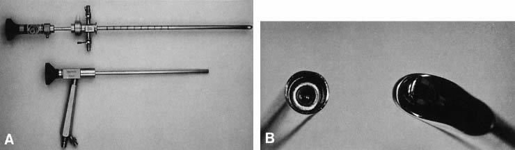
Normal Urethra

The normal urethral mucosa has epithelial folds that form a symmetric, stellate configuration. The color is lush and pink owing to the generous vascularity and thin epithelium. The urethrovesical junction is likewise pink, with more developed folds. The color and appearance are uniform throughout its length. The trigone and both ureteral orifices should be visualized with the urethroscope.

Acute Urethritis

In acute urethritis, the epithelium develops a fiery red color. Most commonly seen is an associated purulent exudate. Frequently there are friable polypoid projections or fronds from both the urethra and the urethrovesical junction. Often there is concomitant trigonitis with a shaggy, flocculent exudate.

Atrophic Urethritis/Trigonitis

Atrophic changes of the urethra and trigone are generated by a relative lack of estrogen. Estrogen maintains the uroepithelium, promotes adequate vascular supply, and stimulates the connective tissue support of the urethra and trigone. With a deficiency of estrogen comes epithelial thinning and decreased vascular perfusion, which results in a profound paleness. In addition, the urethrovesical junction fails to function well in its sphincteric capacity, resulting in urine loss. A frequent finding associated with this condition is urethral caruncles. These reddish blue projections of urethral mucosa emanate from the meatus usually at the 6-o’clock position. Caruncles are benign and require no treatment unless bleeding or pain ensues as a result of irritation. Treatment usually consists of topical estrogens, which work well. Rarely is excision required.

Urethral Diverticulum

A urethral diverticulum is a cystic outpouching of the urethral lumen usually 
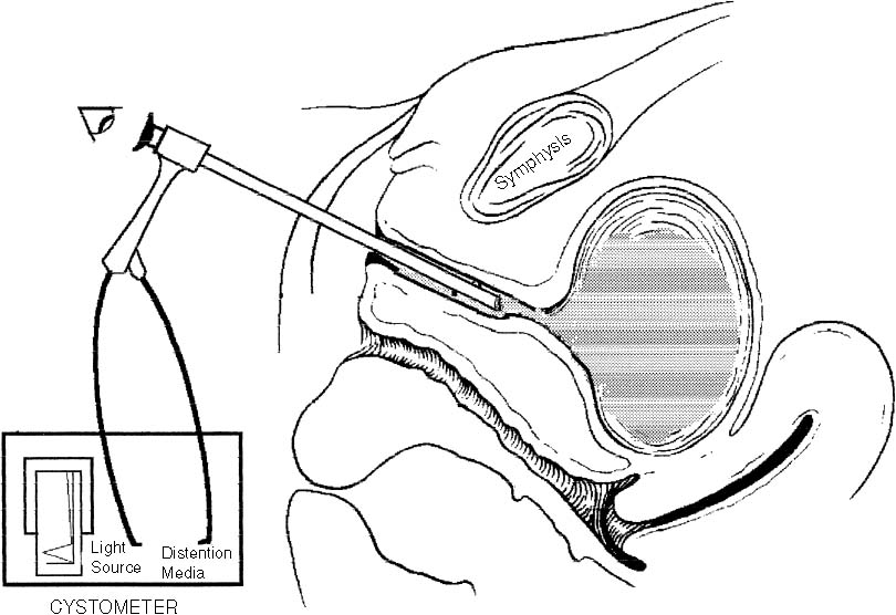
formed by chronic infection of the periurethral glands. It is best identified in conjunction with palpation. This allows easier identification of the orifice, particularly when exudate is flowing from it. Compression of the proximal urethra with the urethroscope in place will permit distention of the urethra and diverticulum, thereby increasing emanation of the exudate.3 The urethral diverticulum is most commonly seen in patients with a long history of chronic infections as well as a history of dyspareunia. It is most commonly found in the midurethra on the posterior wall. Often the diverticulum is palpable over the urethroscope with a finger inserted into the vagina. The treatment is surgical excision.

Urethral fistulas are tracts of communication between the urethra and the vagina. They often result as a complication of surgery for diverticula. Less commonly, they may develop as a result of a chronic infection of the periurethral glands. The fistula resembles a diverticular orifice when viewed with the urethroscope. It is best diagnosed radiographically.

Ectopic Ureter

Ectopic ureters are an extremely rare finding. They are identified by the presence of a ureteral orifice opening into the urethra, from which urine is seen to be emanating. The presence of an ectopic ureter should be suspected when incontinence exists in the face of an otherwise normal examination.

Urethral Condylomata

In the patient with known perineal condylomata and lower urinary tract symptoms, urethral condylomata may be seen. They appear as dense, white papillary projections into the urethral lumen. Biopsy should precede any treatment regimens. Cryotherapy has been used with good success and is generally well tolerated. Urethral condylomata are a rare finding and virtually are never seen in the absence of perineal lesions.

Urethral Neoplasms

Urethral neoplasms are rare findings and are most commonly malignant.3 Associated symptomatology includes urgency, frequency, dysuria, hematuria, urinary retention or incontinence, and occasionally purulent urethral discharge with tumor necrosis. They may be either primary or metastatic. Primary lesions are usually of the epidermoid or transitional cell type. Metastases arise from transitional cell carcinoma of the bladder, adenocarcinoma of the endometrium, and squamous cell carcinoma of the vulva and vagina. Visually they appear as fleshy growths impinging on the urethral lumen. They may be found in association with a diverticulum. Urethroscopy can identify these lesions, but it cannot differentiate between benign and malignant processes, and therefore biopsy is mandatory.

Back to Top
DYNAMIC URETHROSCOPIC FINDINGS

The dynamic evaluation of the urethrovesical junction with cystometric filling involves recognition of a response pattern to a series of direct instructions.2 As the bladder fills, the urethrovesical junction symmetrically closes (see Fig. 4). During the filling process, the patient experiences three significant sensations: (1) the feeling of filling, (2) the sensation of fullness, and (3) the point of maximum fullness. At each of these cystometric landmarks the patient is given the following instructions: “hold your urine,” “cough,” and “strain.” This is carried out with the urethroscope in the proximal urethra to afford a full view of the urethrovesical response to the instructions. The response patterns demonstrated in Figure 4 are characteristic of the normal patient, the patient with genuine stress incontinence, and the patient with an unstable bladder.

Normal

In the normal patient, the urethrovesical junction symmetrically closes with filling. It remains closed with coughing or straining and descends minimally, if at all, with those instructions. No change occurs in response to holding. Opening may occur with a vesical contraction, but closure occurs with suppression of the contraction. These responses are all consistent with good anatomic support and maintenance of continence.

Genuine Stress Incontinence

In the patient with genuine stress incontinence there is a lack of anatomic support at the urethrovesical junction. Thus, increased intra-abdominal pressure is transmitted disproportionately to the bladder and proximal urethra, resulting in descent of the bladder neck and opening of the urethrovesical junction. Indeed this is precisely what is observed during dynamic urethroscopy. With filling, coughing, or straining, the urethrovesical junction descends and opens. On the instruction to “hold your urine,” the urethrovesical junction sluggishly and incompletely closes. The response to a vesical contraction with suppression remains normal.

Unstable Bladder

The unstable bladder produces a unique response to dynamic testing. With filling, the urethrovesical junction closes and remains closed despite the increased pressure produced by coughing or straining. However, after a short delay following the provocation, a vesical contraction ensues. This produces opening of the urethrovesical junction that is not able to be overcome with voluntary suppression. Thus, the mechanism of incontinence in these patients is a strong, involuntary vesical contraction produced by an increase in intra-abdominal pressure. This vesical contraction cannot be suppressed, resulting in urine loss.

Careful observation of the urethrovesical junction in response to this dynamic testing will provide support for the etiology of incontinence in most patients.

Back to Top
INTRAOPERATIVE URETHROSCOPY

Use of the urethroscope during surgical correction of genuine stress incontinence has been advocated.4 It affords visual reassurance that supporting sutures have been properly placed and urethral injury has been avoided. Further, the technique allows visualization of the urethrovesical junction at the time tension is applied to the supporting sutures. This provides adequate correction while safeguarding against overcorrection and urinary retention. The urethroscope has been recommended not only for vaginal procedures, but for retropubic repairs as well.

Back to Top
SUMMARY

Urethroscopy has enhanced our understanding of the contribution of the urethra to female urinary incontinence. Some have criticized its development and use, claiming it adds nothing to the evaluation and treatment of the incontinent female but unnecessary discomfort and expense.5 It is my opinion, however, that any technique that provides further information about a given condition will most certainly lead to specific treatment strategies that will maximize chances for prolonged cures

Back to Top
REFERENCES

1. Robertson JR: Gynecologic Urethroscopy. Am J Obstet Gynecol 115:986, 1973

2. Robertson JR: Dynamic urethroscopy. In Ostergard DR (ed): Gynecologic Urology and Urodynamics, Theory and Practice. p 135, Baltimore, Williams & Wilkins, 1982

3. Robertson JR: Urethral lesions. In Stanton SL (ed) (ed): Clinical Gynecologic Urology. p 323, St. Louis, CV Mosby, 1984

4. Biggers RD: Intraoperative endoscopic evaluation of suprapubic urethropexy. Urology 19:268, 1987

5. Stamey TA: Urinary incontinence in the female: The Stamey endoscopic suspension of the vesical neck for stress urinary incontinence. Campbell’s Urology. p 2833, Philadelphia, WB Saunders, 1992

Back to Top