Chapter 88
The Overactive Bladder
Thomas E. Snyder
Main Menu   Table Of Contents

Search

Thomas E. Snyder, MD
Associate Professor, Department of Obstetrics and Gynecology, Wake Forest University School of Medicine, Winston Salem, North Carolina (Vol 1, Chap 88)

 
DEFINITION
EPIDEMIOLOGY
EFFECT ON QUALITY OF LIFE
PATHOPHYSIOLOGY
EVALUATION OF OVERACTIVE BLADDER
TREATMENT OF OVERACTIVE BLADDER
SUMMARY
REFERENCES

The overactive bladder and associated urinary incontinence affects 17 million Americans at a cost of $26 billion annually, mostly for pads, diapers, and other temporizing measures.1 Among the elderly, 15% to 30% of those living at home and 30% to 50% of those in assisted living situations, acute-care facilities, or nursing homes are affected.2,3 Despite a prevalence greater than other chronic diseases, such as asthma and coronary artery disease, our understanding of the problem is quite limited. Of the population with urinary incontinence, those affected by urge incontinence have their quality of life affected more severely than those with stress urinary incontinence (SUI), including sleep and/or nocturnal disturbances. Despite embarrassment, odor, and possible infection, urinary incontinence is often dismissed as a normal part of aging, and female patients rarely discuss this condition during visits with a physician. The goal of this chapter is to review the basic epidemiology, pathophysiology, and current developments in therapy of this problem.

Back to Top
DEFINITION

The Standardization Subcommittee of the International Continence Society (ICS) formally defined overactive bladder (OAB) in September 2001 as a “symptom syndrome of lower urinary tract dysfunction.” More specifically, OAB is defined as “urgency, with or without urge incontinence, usually with frequency and nocturia.”4 Synonyms include urge syndrome and urgency-frequency syndrome. Before 2001, several terms were used to refer to the syndrome. Most common was overactive detrusor function or overactive detrusor, defined as a condition of involuntary detrusor contractions during the filling phase of cystometry, either spontaneous or provoked. Previously, overactive detrusor was subdivided into detrusor hyperreflexia (caused by neurologic disease) and unstable bladder (those cases without demonstrable neurologic disease). In reviewing data, several authors5,6 believed that because of the prevalence and characteristics of the condition, many patients could be treated with conservative therapy and a minimum evaluation. In addition, problems using an urodynamically based definition of OAB include: (1) cystometry is an invasive test that requires administration and evaluation by skilled and trained specialists; (2) secondary to the worldwide prevalence of OAB, it is not economically feasible for all patients to be evaluated initially by cystometry; (3) the sensitivity of detecting involuntary detrusor contractions varies with the type of the study and 60% to 80% of patients who have negative results on supine cystometry may demonstrate involuntary contractions with ambulatory monitoring or provocative maneuvers; (4) 60% of asymptomatic volunteers show involuntary detrusor contractions; and (5) most cystometry traces that show low compliance change to phasic involuntary detrusor contractions with ambulatory monitoring. As a result of these considerations, the ICS Standardization Subcommittee changed terminology, as shown in Table 1. Detrusor hyperreflexia and detrusor instability are replaced by neurogenic detrusor overactivity and idiopathic detrusor overactivity. The commonly used terms motor urgency and sensory urgency have been eliminated.


Table 1. International Continence Society Terms


Eliminated Term Replacement Term
Detrusor hyperreflexia Neurogenic detrusor overactivity
Detrusor instability Idiopathic detrusor overactivity
Motor urgency None
Sensory urgency None
Motor urge incontinence Detrusor overactivity incontinence with urgency
Reflex incontinence Detrusor overactivity incontinence without sensation

(Wein AJ, Rovner ES: Definition and epidemiology of overactive bladder. Urology 60(Suppl 5A):7–12, 2002)


Urgency is a strong desire to void accompanied by a fear of leakage or pain. Frequency refers to the need to urinate on an abnormally frequent basis (greater than eight times per day or more than two times per night [nocturia]). Urge incontinence refers to the symptoms of urine loss preceded by a strong sense of urgency. The interested reader is referred to the ICS Committee web site, www.icsoffice.org, for the full report of the terminology subcommittee.

Back to Top
EPIDEMIOLOGY

Prevalence

There has been a wide range in the reported prevalence and incidence of OAB secondary to a variety of reported symptoms and the fact that most epidemiologic studies focused on urinary incontinence. Prevalence rates (the total number of cases at a certain point in time) are influenced by the definitions used in a particular study, as well as the methodology used for collecting data. The currently reported median prevalence of incontinence in women varies from 14% to 40.5% (by the ICS definition 23.5%).

Milsom and associates7 reported a study performed in France, Germany, Italy, Sweden, and the United Kingdom. Frequency caused by OAB was defined as more than eight micturitions in 24 hours and nocturia more than two micturitions at night. Of the 16,776 respondents, 19% reported current bladder symptoms and 17% had symptoms of OAB. Prevalence varied by country from 20% of women in Spain to 13% in France. Seventy-nine percent of patients had symptoms for more than 1 year and 49% had symptoms for more than 3 years. Frequency (85%) was the most common symptom, followed by urgency (54%) and urge incontinence (36%). The prevalence of symptoms increased with age, as shown in Table 2.

In the study by Milsom, the most widely known symptom of OAB (urge incontinence) was not the most prevalent symptom (36%). Urinary frequency (more than 8 voidings in 24 hours) was the most common symptom reported by 85% of patients. Most importantly, 65% of respondents reported that OAB adversely affected their daily life. Also surprising was the fact that despite physician consultation by 60% of respondents, only 27% were currently on medication. This fact probably reflects the side effect profile of current treatments (see section on drug therapy).

The National Overactive Bladder Evaluation (NOBLE) Program was established to provide an estimate of the prevalence and burden of illness of OAB in the United States.8 A computer-assisted telephone interview of 5204 English-speaking adults older than age 18 years standardized for race, geographic region, and age was conducted. The sensitivity and specificity of the survey were 61% and 91% for OAB symptoms. Definitions used in the survey were: (1) OAB dry, four or more episodes of urgency in the previous 4 weeks with either frequency more than eight times per day, or use of one or more coping behaviors to control bladder function; and (2) OAB wet included the same symptoms plus three or more episodes of urinary incontinence not explained by stress symptoms in the past 4 weeks. The prevalence of OAB was 16.9% in women and 16% in men, increasing with age. OAB dry and wet occurred in 7.6% and 9.3% of women interviewed, respectively. Across the United States, these figures would estimate 33.3 million adults with OAB, of whom 12.2 million would have incontinence. The prevalence was noted to be small in both sexes until approximately age 60 years in women (12%); however, it then increased to 20% at age 65 years or older. After adjusting for other disease states, patients with OAB had a significantly decreased quality of life, increased incidence of depression, and poorer quality of sleeping.


Table 2. Prevalence (%) of Overactive Bladder Symptoms, Grouped According to Age and Gender, in a Random Sample of 16,776 Subjects aged ≥40 Years From Six European Countries


Age (y) Men (n = 7048) Women (n = 9728)
40–44 3.4 8.7
45–49 6.0 10.6
50–54 9.8 11.9
55–59 13.2 16.9
60–64 18.9 16.9
65–69 23.7 17.5
70–74 22.3 22.1
≥75 41.9 31.3
All 15.6 17.4

Milsom I, et al: How widespread are the symptoms of an overactive bladder and how are they managed? A population-based prevalence study. BJU Int 87:760–766, 2001)


While the two aforementioned telephone surveys may be criticized on the basis of methodology, they present a significant advance in the understanding of OAB and its impact on daily life. Patients who have OAB may have to use coping strategies, such as decreased fluid intake, knowledge of location of the nearest toilet, and preemptive voiding, which have a significant impact on lifestyle.

Booth9 reported a study based on repeat urodynamic findings to assess OAB. Ten men and 54 women underwent repeated urodynamic studies. Of these patients, 25 were classified as having idiopathic detrusor instability and 16 enuretic instability, defined as unstable cystometry and a history of enuresis after age 7 years.

At the time of follow-up (mean: 19 months), urodynamic parameters were unchanged or similar in 73% of patients. While this is a retrospective study, it is the only study to date to consider urodynamic parameters of idiopathic detrusor overactivity over time. The study demonstrated a tendency for OAB to persist at a rate of 92% by urodynamic study despite cystodistension and drug therapy.

Racial, Gender, and Other Differences

The prevalence of incontinence is 1.5- to two-times higher in women than in men. Stress incontinence (49%) is more commonly reported than urge incontinence (22%) in women. In contrast, overactive bladder symptoms are more common in men.10 (Fig. 1) Urge incontinence was more common in black females (56%) compared with white females (28%).11 In a study of 200 women, Mattox and Bhatia12 reported that urge incontinence occurred in 18% of white women and 9% of Hispanic women matched for age, gravidity, and parity. Obesity is also associated with an increased incidence of stress incontinence and urge incontinence. Dwyer and associates13 reported a 24% incidence of detrusor instability in women more than 20% over average weight for height by age.

Fig. 1. Types of incontinence in females. (From Davila GW, Neimark M: The overactive bladder: Prevalence and effects on quality of life. Clin Obstet Gynecol 45:173–181, 2002.)

Incidence of Overactive Bladder

In contrast to prevalence, the incidence of a disease or syndrome is the number of new cases over a certain period of time. Incidence data are logistically difficult to study because of multiple factors; however, a Danish study noted the incidence and prevalence of irritative urinary tract symptoms to be 29% prevalence, 10% incidence, with a 28% spontaneous remission rate.14 An older study demonstrated the incidence of urge incontinence to increase with age from .08% to .2% between ages 45 and 59 years.15

Patients with certain conditions such as Parkinson's disease, stroke, and dementia are at risk for bladder dysfunction. Twenty percent to 30% of patients with Parkinson's and multiple sclerosis present with bladder dysfunction before diagnosis of the disease. The overall incidence of bladder dysfunction ranges from 40% to 70% in Parkinson's disease.16

Back to Top
EFFECT ON QUALITY OF LIFE

While patients with stress incontinence may be able to deal with the problem by avoiding physical activity that causes incontinence, the patient with OAB must constantly deal with symptoms. Patients may use a variety of coping skills including toilet-seeking, restriction of fluid intake, and limitation of physical and social activities to avoid embarrassment and/or odor. These behaviors may cause anxiety and distress regarding urine loss and increased urgency because of the internalized distress leading to psychological issues and social isolation.

Milsom7 reported that 65% of men and 67% of women with OAB had symptoms that had an impact on daily living and 60% of symptoms were bothersome enough to seek help from a physician. Frequency and urgency (59%) as well as urge incontinence (66%) were the most common reasons for seeking consultation. Of those who consulted physicians, only 27% were using medication at the time of the survey and 27% had been using medication that failed. Of those who had not been treated, 54% said they were likely to consult and 46% were not. Sixty-five percent of patients who had tried drugs that did not help were likely to discuss the problem with a physician and 35% were not. The findings reflect patients' frustration with the significant side effects and lack of efficacy of past and recent medical therapies.

Liberman17 used the Medical Outcomes Study Short Form (SF)-20 to study a group of patients for health-related quality of life (HRQOL). The HRQOL measured six domains: (1) physical functioning; (2) role functioning; (3) social functioning; (4) mental health; (5) health perception; and (6) bodily pain. Both OAB wet (incontinent) and OAB dry (continent but with symptoms) groups had lower HRQOL scores than controls in every domain, even after adjustment for confounders. The differences were statistically significant in five of six domains for the total group and in all six domains for the OAB wet group.

In a subset of the NOBLE study,18 Stewart assessed impact of OAB on quality of life with the 36-Item Short Form (SF-36) Health Survey, an instrument that measures HRQOB in eight domains. After adjusting for coexisting illness and other demographic factors, both men and women had significantly lower quality-of-life scores, more depression, and poorer quality of sleep than controls. Kobelt19 and Abrams20 likewise found that patients affected by OAB had significantly lower-quality lifestyles. They listed common reactions to urinary incontinence associated with OAB as embarrassment, frustration, anxiety, annoyance, depression, and fear of odor.

Brown21 found that among community-dwelling elderly women, weekly urge incontinence was associated with a 26% greater risk of falling and 34% increased risk of sustaining a fracture. More frequent incontinence was associated with an even higher risk of falling (35%) and fractures (45%). The top 10 items affecting quality of life are presented in Table 3.


Table 3. Top 10 Incontinence-Related Quality-of-Life Items


10. Urinary incontinence psychological, not physical, problem
9. Self-concept
8. Resignation
7. Loss of sleep
6. Lack of predictability
5. Fear of public embarrassment
4. Shame
3. Adaptation of daily routine
2. Lack of self-control
1. Need for pre-emptive strategies to avert urinary incontinence

Davila GW, Neimark M: The overactive bladder: Prevalence and effects on quality of life. Clin Obstet Gynecol 45:173–181, 2002. Data from DuBeau CE, et al: The impact of urge urinary incontinence on quality of life. J Am Geriatr Soc 46:683–692, 1998)


Costs of Overactive Bladder

Wagner and Hu22 noted current costs of urinary incontinence in the United States in 1995. For individuals age 65 and older, the 1995 societal cost was $26.3 billion or $3565 per individual with urinary incontinence. The authors believe that the future economic burden of OAB will be significantly greater than urinary incontinence; however, only estimates are now available.

In summary, OAB has a significant impact on the quality of life of affected individuals. In addition, a huge societal cost is incurred.

Back to Top
PATHOPHYSIOLOGY

The diagnosis of OAB suggests an involuntary increase in detrusor pressure during the storage phase of the micturition cycle. Common pathologic causes include obstruction, interstitial cystitis, spinal cord injury, denervation, Parkinson's disease, diabetes, multiple sclerosis, and aging. However, idiopathic detrusor overactivity has been identified in 29% of women and 10% of men undergoing urodynamic testing, and of all patients with detrusor overactivity, 90% are idiopathic. Our understanding of the cause of overactive bladder is still incomplete; however, through recent studies, a more detailed understanding has begun to emerge.

Sand23,24 has recently summarized possible causes of overactive bladder etiology, as shown in Table 4. While some proposed etiologies may have components of more than one theory, the following section attempts to discuss etiologies by the most appropriate theory.


Table 4. Theories Proposed to Explain OAB Etiology


MYOGENIC States that changes in the structure and innervation of the detrusor lead to increased excitability and conduction between cells, causing myogenic contractions of the whole detrusor.25
NEUROGENIC States that sensitization of peripheral afferent terminals in the bladder or damage to central inhibitory pathways unmask primitive voiding reflexes, leading to overactivity of bladder contraction reflexes.26
URETHROGENICStates that bladder muscle contraction is triggered by urine entering the urethra at inappropriate times, possibly from a decrease in urethral smooth muscle thickness.27,28 May explain stress-induced involuntary detrusor contractions, triggered by coughing, Valsalva maneuver, and other increases in intra-abdominal pressure.

(Kwon C, Sand PK: Management of Overactive Bladder and Urinary Tract Infections in Women. Ob/Gyn Special Edition, New York, McMahon Publishing Group, Spring 2003)


Normal Anatomy and Physiology

The pathophysiology of the overactive bladder is incompletely understood. However, to understand the currently proposed potential mechanisms, one must have a basic understanding of normal physiological bladder function.

The normal bladder performs three basic functions. The first is storage of a socially acceptable urine volume. To perform this, the bladder must exhibit compliance: the ability to increase in volume without a significant change in pressure. The second is that the lining urothelium must expand during filling while protecting underlying nerves and muscle from the contained urine. The third is that the smooth muscle of the bladder wall must contract in a coordinated fashion to adequately expel urine.29 In this regard, the bladder is different from other organs in the following ways: (1) normal neural function of the bladder depends on intact circuits in the brain, spinal cord, and peripheral ganglia compared with other organs that maintain a level of function even after elimination of extrinsic neural input; (2) the bladder exhibits switch-like patterns of activity compared with tonic patterns in other organs, e.g., the cardiovascular system; (3) micturition is under voluntary control and depends on learned behavior; and (4) micturition depends on integration of autonomic and somatic efferents in the lumbosacral spinal cord to coordinate urine storage and release.26 The smooth muscle of the bladder is ideally suited for this purpose. Smooth muscle consists of small, spindle-shape cells linked at specific junctions. Smooth muscle maintains a steady state of tension that may be altered by hormones, local factors, or autonomic nervous activity. Smooth muscle is able to adjust its length over a wider range than skeletal muscle.29

Neural Control

The peripheral nervous system controlling bladder function originates from three sources: parasympathetic, sympathetic, and somatic nervous system (Fig. 2). Parasympathetic preganglionic neurons originating in the sacral parasympathetic nucleus (SPN) send axons to peripheral ganglia and subsequently postganglionic neurons located in the detrusor wall and pelvic plexus where they release acetylcholine as an excitatory transmitter. Sympathetic paths emerge from the lumbar spinal cord. Through noradrenergic stimulation, they cause relaxation of the bladder body and contraction of the outlet and urethra (e.g., urine storage). Somatic pathways originate in Onuf's nucleus in the lateral ventral horn and course in the pelvic and pudendal nerves.

Fig. 2. Diagram showing the sympathetic, parasympathetic, and somatic innervation of the urogenital tract of the male cat. SCG, sympathetic chain ganglia; ISN, inferior splanchnic nerves; IMG, inferior mesenteric ganglia; HGN, hypogastric nerve; IC, ischiocavernosus; BC, bulbocavernosus; EUS, external urethral sphincter; PG, prostate gland; U, ureter; VD, vas deferens. (Chancellor MB, Yoshimura N: Physiology and pharmacology of the bladder and urethra. In Walsh PC (ed): Campbell's Urology, pp 831–886. Philadelphia, Saunders, 2002.)

Afferent pathways transmitting information from the bladder to the CNS course in the pelvic, hypogastric, and pudendal nerves. Bladder volume and amplitude of bladder contractions are monitored by pelvic nerve afferents consisting of A-delta and C axons. In the experimental animal, A-delta fibers are low-threshold mechanoreceptors and C-fibers are mechanoinsensitive except under pathologic conditions such as irritation from high potassium, low pH, and capsaicin.

Central pathways that control lower urinary tract function are organized as on–off switching circuits between the bladder and urethra (Fig. 3). Filling depends on the properties of smooth muscle and inhibition of parasympathetic pathways and stimulation of sympathetic paths to close the bladder neck and proximal urethra. The storage phase may be switched to the emptying phase either voluntarily or involuntarily, e.g., in the infant when the volume contained exceeds the micturition threshold. At this time, secondary to increased afferent input from tension receptors, the pattern of efferent outflow reverses with increasing parasympathetic and decreasing sympathetic input. This results in relaxation of sphincter and coordinated contraction of the detrusor (Fig. 4). For a very complete discussion of the normal anatomy and physiology of the bladder and mechanisms of the overactive bladder, the interested reader is referred to Chancellor and Yoshimura.29

Fig. 3. Diagram illustrating the anatomy of the lower urinary tract and the switch-like function of the micturition reflex pathway. (From Chancellor MB, Yoshimura N: Physiology and pharmacology of the bladder and urethra. In Walsh PC (ed): Campbell's Urology, pp 831–886. Philadelphia, Saunders, 2002).

Fig. 4. Mechanism of storage and voiding reflexes. A. Storage reflexes. B. Voiding reflexes. (From Chancellor MB, Yoshimura N: Physiology and pharmacology of the bladder and urethra. In Walsh PC (ed): Campbell's Urology, pp 831–886. Philadelphia, Saunders, 2002).

Neurogenic Mechanisms of Overactive Bladder

Changes in central and peripheral neurologic pathways that may result in overactive bladder include: (1) decrease in central or peripheral inhibition; (2) increase in excitatory reflex pathways; (3) increased afferent input from the lower urinary tract; and (4) development of bladder reflexes resistant to central inhibition.26

Central Lesions

Studies by deGroat30 and others31,32 using the cat lesion model noted inhibition of voiding functions by the cerebral cortex. In humans, the cerebral cortex appears to act in a similar way to suppress the micturition pathway, e.g., patients with Parkinson's disease often have overactive bladders. A monkey model simulating Parkinson-like symptoms has been developed that has demonstrated that bladder overactivity was caused by loss of dopaminergic inhibitors.33

Spinal Cord Lesions

Spinal cord injury above the sacral level disrupts normal supraspinal pathways that control urine storage and release. A period of urinary retention lasting a few weeks is then followed by automatic micturition and bladder overactivity secondary to reorganization of synaptic connections in the spinal cord.34 Normal voiding is mediated by A-delta afferents.30 C-fibers, however, in most species are relatively insensitive to bladder distension.

After spinal injury, there appears to be a reorganization of reflex correction in the spinal cord. C-fiber afferents are facilitated and A-fiber afferents are eliminated, resulting in symptoms of overactive bladder. These capsaicin-sensitive axons have been implicated in upper motoneuron diseases such as spinal cord injury, multiple sclerosis, and Parkinson's disease.35

Based on animal models, mechanisms for the emergence of overactive bladder symptoms after spinal cord injury may include: (1) elimination of bulbospinal inhibitory pathways; (2) strengthening of existing synapses or formation of new synaptic connections caused by axonal sprouting in the spinal cord; (3) changes in synthesis, release, and action of neurotransmitters; and (4) alteration in afferent input from peripheral organs.26

Myogenic Mechanisms of Overactive Bladder

It is obvious that the smooth muscle of the overactive bladder detrusor behaves in an abnormal fashion compared with the normal bladder. While the exact mechanisms of this muscular dysfunction are incompletely understood, our understanding has been enhanced by recent research. One hypothesis is that the pathology is caused by reduction in the activity of the excitatory neurons to the smooth muscle, which triggers alterations in the muscle, including excitability and increased cell coupling, leading to uncoordinated contractions.25

During filling, the normal bladder remains compliant, expanding to standard volume with minimal change in intravesical pressure. Then, it must respond to a stimulus that can smoothly and rapidly increase intravesical pressure to expel the contained urine. Smooth muscle cells in the normal bladder are spontaneously active; however, during filling, the activity does not result in a synchronous contraction, because each cell is only coupled to a few of its neighbors and the extensive coupling necessary does not exist. Synchronous activity requires dense innervation to stimulate a large number of smooth muscle cells to contract.

The bladder with overactivity appears to be both structurally and functionally altered. Within overactive bladder muscle, cell-to-cell junctions enable small foci of electronic activity to be propagated beyond their normal range, causing widespread smooth muscle contractions.24

Ultrastructural changes of overactive bladder tissue support this theory of cell-to-cell coupling of background electrical activity.24–36 Dysfunction, denervation, and neuroenervation have been shown by Saito37,38 and also in animal models.39,40 Mills41 demonstrated denervation in 35% of muscle bundles from overactive bladders compared with 1.5% of controls. Overactive bladder specimens showed increased baseline tone and more spontaneous contractions compared with controls.

Electron Microscopic Changes

In addition to the aforementioned changes, ultrastructural changes have been noted that support the concept of altered cell-to-cell communication. The ultrastructure of the usual detrusor includes polygonal to cylindrical cell profiles, like other normal smooth muscle. A continuous cell member defines each cell, and individual cells appear uniformly separated. There is a relatively small amount of collagen attached to the cell membranes between adjacent cells. Elbadwi and associates42 investigated ultrastructural changes of dysfunctional bladders with the electron microscope. Bladder tissue associated with detrusor overactivity exhibited what was termed a dysjunctional ultrastructure pattern. This pattern is characterized by: (1) marked reduction of intermediate cell junctions of the normal detrusor; (2) abundant, unique protrusion of muscle cell junctions; and (3) ultraclose cell abutments (Fig. 5). The protrusion junction is the contact between two long, finger-like processes that connect the muscle cells. Mechanical muscle coupling is essential for development of normal voiding. It is postulated that these protrusions and the close approximation of the smooth muscle cells in the unstable bladder facilitate the coupling of adjacent cells and give the entire detrusor the functional properties of a syncytium.43 The action of individual cells is easily passed to the next cell, prompting a coordinated but inappropriate contraction. Brading25 noted similar findings in the guinea pig bladder. Elbadwi and colleagues44 concluded that the close abutment of the cells and multiple cell junctions would allow the coordination of cellular activity noted in the uninhibited bladder.

Fig. 5. Dysjunction pattern in detrusor overactivity. Protrusion junctions are indicated (arrows). (From Elbadwi A: Structural basis of voiding dysjunction. In Ray S (ed): Female Urology, 2nd ed. Philadelphia, WB Saunders, 1995).

Conversely, a distinctive degenerative pattern has been noted in bladders with overactivity, but with impaired contractility.42 This pattern is characterized by widespread degeneration of muscle cells and intrinsic nerves of the detrusor.45 These cells have features of distorted myofilaments and nuclei, disrupted organelles, vacuolated sarcoplasm, and thickened or breached cell membranes.26 The degeneration inhibits the muscle cell's ability to contract, resulting in an uncoordinated, inefficient overall contraction of the bladder wall.

A myohypertrophy ultrastructural pattern has been noted in the bladder with outlet obstruction superimposed on the pattern of overactivity.25 This pattern is distinguished by hypertrophic muscle cells, wide separation of individual cells, and abundant collagen in the ultracellular spaces. According to Elbadwi and associates,44 these hypertrophic cells are characterized by inappropriate size, branching, tapering ends, and convoluted contours. They postulated that the unusual configuration of these cells may impede the ability of the detrusor to generate enough contraction to empty the bladder effectively.

It is clear that recent studies have helped to clarify the cause of inappropriate detrusor activity; however, much remains to be learned in this arena.

Bladder Outlet Obstruction

Smooth muscle of the bladder is remarkable in its adaptation to pathologic changes such as obstruction. This adaptation is referred to as plasticity. In both the experimental model and observation in humans with obstruction, the detrusor hypertrophies to accommodate obstruction. In addition, smooth muscle has been shown to become supersensitive to agonist stimulation and demonstrates increased excitability in response to denervation. Animal models of outflow obstruction and diabetic neuropathy have been extensively studied to understand these phenomena. Gosling and colleagues46 studied a rabbit model in which the bladder was obstructed for up to 70 days, and response to field stimulation and carbachol were compared with alterations in ultrastructure and innervation. He observed that chronic obstruction is accompanied by a progressive decrease in contractility of smooth muscle.

Structural changes included muscle cell hypertrophy, denervation, decrease in myofilaments, and damaged mitochondria associated with a decreased response to field stimulation and carbachol. Outlet obstruction and subsequent irritative voiding symptoms have been attributed to denervation supersensitivity.47 Brading and Turner48 proposed that smooth muscle change caused by a reduction in functional innervation of the bladder wall is a common feature of all causes of bladder instability.

Mills41 studied full-thickness specimen from 14 patients with idiopathic detrusor instability (IDI) and 14 cadaveric controls undergoing transplant retrieval. When compared with controls, he observed evidence of altered spontaneous contractile activity connected with increased electrical coupling of cells, patchy denervation of the detrusor, and potassium supersensitivity. Based on this evidence, they concluded that subsequent supersensitivity appears to be a common feature of many types of bladder instability.

Studies in the rat have demonstrated alterations in the CNS after bladder neck obstruction.49 These similar observations in humans suggest development of new spinal circuits after lower-tract obstruction. This plasticity appears to occur through a mechanisms involving nerve growth factor (NGF), which is responsible for growth and maintenance of sympathetic and sensory neurons and is responsible for neural regrowth after injury.29

Urethral Factors in Overactive Bladder

Detrusor instability is found in approximately 20% to 30% of women with urinary incontinence.50,51 Theories of causation include occult central or peripheral nerve disorders and primary dysfunction of the detrusor smooth muscle.43

The term urethrogenic detrusor instability has been applied to the finding of unstable detrusor contractions triggered by funneling of urine into a poorly supported urethra. This theory is supported by findings of lower maximal urethral closure pressure in patients with detrusor instability.52,53 Koonings and Bergman54 studied 114 women with IDI. One group demonstrated involuntary bladder contraction preceding any changes in urethral pressure. The other showed a decrease in urethral pressure preceding detrusor contraction. The latter group responded poorly to anticholinergics but responded better to alpha andrenergics. The group without urethral change responded more favorably to anticholinergics. Secondary to these changes, some authors advocate bladder neck suspension for patients with instability and stress incontinence to prevent urine entering the urethra, thus activating this reflex.55,56,57

Major and associates50 recently used intraurethral ultrasound to study 17 patients with detrusor instability and 16 patients with normal urodynamic testing. The patients with detrusor instability had decreased longitudinal smooth muscle thickness, total urethral diameter, and total urethral circumferences compared with those with normal urodynamic testing.58 Previously, the author had demonstrated an association between longitudinal smooth muscle thickness and urethral structure measured by maximal urethral closure pressure. They concluded that loss of longitudinal smooth muscle thickness could result in decreased urethral resistance, allowing urine to enter the proximal bladder neck, leading to a detrusor contraction.

In summary, neurogenic, myogenic, and urethral changes have been noted in the overactive bladder. Goldberg and Sand24 note that smooth muscle changes can induce the emergence of pathologic nerve pathways, both locally and regionally, which facilitate the spread of electrical activity. Structural alteration of the bladder wall after outflow obstruction may trigger alterations induced by nerve growth factor.59 However, neurologic changes may trigger alterations in the sensitivity and contractions of the detrusor. As Goldberg states, “Whether it is an initial neurogenic trigger leading to myogenic alterations in smooth muscle and involuntary detrusor activity or vice versa, a number of processes may converge toward a similar final common pathway at the level of the bladder wall.”24

Back to Top
EVALUATION OF OVERACTIVE BLADDER

The patient with OAB commonly presents with a combination of symptoms including frequency, urgency, nocturia, and/or incontinence. Incontinence occurs when the patient is consciously unable to control the urge to void. Frequency occurs when the urge to void occurs before complete filling of the bladder. Nocturia refers to frequency occurring at night, at times accompanied by bed-wetting. Few patients report pain associated with the symptoms. The patient will usually present to her physician when symptoms become disruptive or when there is concern of a more serious underlying problem.

History

An accurate history is essential for correct diagnosis of overactive bladder. MacLennon60 studied 1547 women age 18 to 97 years old. Twenty percent presented with stress incontinence alone, 2.9% had urge incontinence alone, and 11.6% had with mixed incontinence.

The overall initial goals of the evaluation are to identify reversible causes of incontinence, exclude other serious disease, identify proximate causes of the overactive bladder, and properly classify the type of incontinence. Reversible causes may be remembered by the mnemonic DIAPPERS (delirium, infection, atrophy, pharmaceuticals, psychological problems, endocrine problems, restricted mobility, stool impaction), most of which may be identified by history alone (Table 5).25 Medications that may affect continence are listed in Table 6.


Table 5. Causes of Transient Urinary Incontinence


Delirium (confusional state)
Infection-urinary (only symptomatic)
Atrophic urethritis, vaginitis
Pharmaceuticals
Psychological, especially severe depression (rare)
Excess urine output (e.g., CHF, hyperglycemia)
Restricted mobility
Stool impaction

CHF, congestive heart failure
(Resnick NM: Urinary incontinence in the elderly. Med Grand Rounds 3:281, 1984)


Table 6. Commonly Used Medications That May Affect Continence


Type of Medication Examples Potential Effects on Continence
Sedatives/hypnotics Long-acting benzodiazepines (e.g., diazepam, flurazepam) Sedation, delirium, immobility
Alcohol Polyuria, frequency, urgency, sedation, delirium, immobility
Anticholinergics Dicyclomine, disopyramide, antihistamines Urinary retention, overflow incontinence, delirium, fecal impaction
Antipsychotics Thioridazine, haloperidol Anticholinergic actions, sedation, rigidity, immobility
Antidepressants Amitriptyline, despiramine Anticholinergic actions, sedation
AntiParkinsonians Trihexyphenidyl, benztropine mesylate (not L-dopa/selegiline) Anticholinergic actions, sedation
Narcotic analgesics Opiates Urinary retention, fecal impaction, sedation, delirium
α-Adrenergic antagonists Prazosin, terazosin Urethral relaxation may precipitate stress incontinence in women
α-Adrenergic agonists Nasal decongestants Urinary retention in men
Calcium-channel blockers All Urinary retention: nocturnal diuresis owing to fluid retention*
Potent diuretics Furosemide, bumetanide Polyuria, frequency, urgency
Angiotensin-converting enzyme inhibitors (ACEI) Captopril, enalapril, lisinopril Drug-induced cough can precipitate stress incontinence in women and in some men with previous prostatectomy
Vincristine Urinary retention

*Dihydropyridine class of calcium-channel blockers, such as nifedipine, nicardipine, isradipine, felodipine, and nimodipine.
(Resnick NM: Geriatric incontinence. Urol Clin North Am 23:55, 1996)


The history should evaluate those factors that may be reversible, such as diet, fluid intake, and psychological and mental status. The patient should be questioned about related symptoms such as hematuria, pain, and problems with complete emptying. Many patients will have previously consulted other physicians, and the results of these evaluations should be obtained. Patients should be questioned about bowel habits and symptoms of genital prolapse.

Questionnaires may be useful to quantify symptoms. Examples of questionnaires include Incontinence Impact Questionnaire,61 Urological Distress Inventory, King's Health Questionnaire, and Bristol Female Lower Urinary Tract Symptoms (FLUTS).

Physical Examination and Laboratory Diagnosis

A careful physical examination is an often neglected part of the evaluation of OAB. The abdomen should be evaluated for a compressing mass, and a pelvic examination should be performed to assess for prolapse, urethral obstruction, estrogen status, and infection or mass. The demonstration of incontinence associated with stress phenomena has a positive predictive value of 91%, but 40% of those patients will have additional urologic problems, such as OAB.62

The neurologic examination should evaluate S2-S4 and bulbocavernosus and anal wink reflexes. Other lower extremity reflexes and strength should be evaluated as well.

Urinary diaries are often useful to assess fluid intake, time and volume of each micturition, and episodes of incontinence. The minimum length of keeping a diary should be 2 days. The diary may reveal suggestions as to the type of voiding dysfunction and excessive fluid intake, and can serve as a monitor of treatment.

Urinalysis should be performed to evaluate for bacteruria, pyuria, and hematuria. Hooton63 has shown that 103 organisms per milliliter may be used to diagnose acute cystitis instead of the traditional 105 organisms. Urine cytology may occasionally be considered to evaluate for tumors. Pad testing is a good way to confirm and estimate the volume of leakage in those patients who have incontinence. However, Versi64 has shown that the variability of the long-term test (7%) is much better than the short-term test (45%). The long-term test may be made more accurate by a concomitant voiding diary.

Residual Urine

Voiding dysfunction and OAB commonly coexist, and evaluation of a postvoid residual may help differentiate between the two and avoid therapeutic errors. Haylen65 showed that a residual urine greater than 30 milliliters by ultrasound 1 minute after voiding 200 milliliters or more occurs in only 5% of normal women and in 13% of symptomatic women. Dwyer66 noted voiding dysfunction in 13.7% of 1193 women referred for urinary symptoms. One-third of those had a residual greater than 150 milliliters and 80% had irritable bladder symptoms. Ultrasound determination of postvoid residual has the advantage of being less invasive and does not have the risk of infection, compared with transurethral catheterization.

Urodynamics

Urodynamic testing is the only way to accurately differentiate OAB from other forms of urinary tract urgency and incontinence. Women presenting with a history of OAB may be frequently shown to have stress incontinence, and women with symptoms of stress incontinence frequently have evidence of OAB.

Standard cystometry is usually performed at fill rates of 10 to 100 milliliters per minute, and note is made of volume at first sensation, any urgency, and bladder capacity. Video urodynamics, urethral pressure profiles, sphincter electromyography, and pharmacologic testing may supplant standard cystometry.

Cystourethroscopy is the gold standard to rule out bladder and urethral pathology in patients presenting with OAB symptoms. Endoscopic evaluation is recommended in patients with microscopic hematuria, previous history of pelvic surgery (e.g., previous incontinence repair), bladder pain (to rule out interstitial cystitis and other pathology), and patients who may have fistula.66 A classic example of low- and high-pressure uninhibited detrusor contractions in patients undergoing urodynamic testing is shown in Figure 6.

Fig. 6. Idiopathic detrusor overactivity. Low-pressure detrusor overactivity. High-pressure detrusor overactivity.

Back to Top
TREATMENT OF OVERACTIVE BLADDER

Therapy of OAB has recently become a popular topic both nationally and internationally for two reasons. As previously described, the number of persons affected is only now being recognized, and the economic impact is staggering. As the current population continues to age, the number of patients promises to increase in the future. New medical therapies with fewer side effects have been developed, making realistic therapy easier for the patient. Current therapies for OAB include a variety of behavioral, pharmacologic, and surgical methods, as shown in Table 7. This section concentrates on those pharmacologic therapies that are the most commonly used (Table 8).


Table 7. Overactive Bladder Treatment Modalities in Common Use


Therapy Type Specific Therapy
Behavioral Bladder drill
  Timed/prompted voiding
Biofeedback Various probes and patches
Electrical Stimulation Vaginal or anal electrical stimulation
  Transcutaneous or percutaneous stimulation
  Peripheral nerve and pelvic floor nerve stimulation
  Sacral nerve neuromodulation
Pharmacological Table 8
Surgical Autoaugmentation
  Enterocystoplasty
  Urinary diversion

(Yoshimura N, Chancellor MB: Current and future pharmacological treatment for overactive bladder. J Urology 16 8:1897–1913, 2002)


Table 8. Drugs With Bladder Action


Classification Examples Pharmacologic Action
Anticholinergic agents Atropine, glycopyrrolate oxybutynin, propantheline, tolterodine Inhibit muscarinic receptors, thus, decreasing the response to cholinergic stimulation. Used to reduce pressure during bladder filling and treat unstable bladder contractions.
Smooth muscle relaxants Dicyclomine, flavoxata Direct smooth muscle relaxation decreases intravesical pressure during filling as well as the severity and presence of unstable bladder contractions. Most agents have some degree of anticholinergic action.
Ca antagonists Diltiazem, nifedipine, verapamil Used for treating unstable bladder contractions to decrease the magnitude of spikes by reducing the entrance of calcium during an action potential.
Calcium channel openers Cromakalim, pinacidil Acts to increase membrane potential and, thus, decrease myogenic initiation of unstable bladder contractions.
Prostaglandin synthesis inhibitors Flurbiprofen Prostaglandins have been implicated in increased smooth muscle tone and the induction of spontaneous activity. Inhibition of prostaglandin synthesis could promote bladder relaxation during filling and decrease spontaneous bladder activity.
β-Adrenergic agonists Isoproterenol, terbutaline Stimulation of β-receptors induces relaxation of the bladder body, resulting in decreased intravesical pressure during filling.
Tricyclic antidepressants Amitriptyline, imipraminee These agents have anticholinergic, direct smooth muscle relaxant and norepinephrine re-uptake inhibition properties.
α-Adrenergic agonists Ephedrine, phenylpropanalamine, midodrin, pseudoephedrine Increase urethral tone and closure pressure by direct stimulation of α-adrenergic receptors.
Afferent nerve inhibitors Dimethyl sulfoxide, capsaicin, resiniferatoxin Decrease sensory input from the bladder, thereby, increasing bladder capacity and reducing bladder hyperreflexia.
Botulinum toxin Botulinum toxins A & B Direct injection into the urethral and bladder skeletal and smooth muscle to treat detrusor-sphincter dyssynergia and overactive bladder. Botulinum toxin has been demonstrated to relax the neuromuscular junction of skeletal and smooth muscle.

(Yoshimura N, Chancellor MB: Current and future pharmacological treatment for overactive bladder. J Urology 168:1897–1913, 2002)


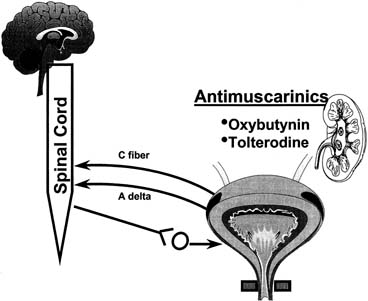
Fig. 7. Most commonly used drugs today to treat overactive bladder are anticholinergics. Despite all components of lower urinary tract and nervous system that control it, only anticholinergic drugs have been used in past 30 years to treat overactive bladder. (From Yoshimura N, Chancellor MB: Current and future pharmacological treatment for overactive bladder. J Urol 168:1897–1913, 2002).

Pharmacologic Treatment

ANTICHOLINERGIC AGENTS

Anticholinergics are competitive inhibitors of acetylcholine that block muscarinic receptors and thereby inhibit involuntary bladder contractions (Fig. 7). All drugs of this class have similar side effects that, to some degree, limit their usefulness. These include dry mouth, dry eyes, constipation, and drowsiness. The prominence of these side effects is shown in Table 9. General contraindications include closed angle glaucoma and obstructive uropathy.


Table 9. Pharmaceuticals for Overactive Bladder and Their Side Effects


Drug Dosage Side Effect
Dicyclomine 20 mg tid +++
Flavoxate 100–200 mg tid–qid ++
Hyoscyamine 0.125 mg tid–qid ++
Imipramine 10–50 mg bid +++
Oxybutynin 2.5–5.0 mg tid ++++
Oxybutynin XL 5–20 mg qd +
Propantheline 15–30 mg tid–qid ++++
Tolterodine 1–2 mg bid +
Tolterodine LA 4 mg qd +

From Cannon TW, Chancellor MB: Pharmacotherapy of the overactive bladder and advances in drug delivery. Clin Obstet Gynecol 45:205–217, 2002


OXYBUTYNIN

Oxybutynin was first approved in 1972 and is an antimuscarine with muscle relaxant and local anesthetic activity acting mainly on (M3) subtype receptors, which are responsible for the contractile properties of the bladder. Efficacy is good (more than 50% symptomatic improvement), reaching 61% to 86% at a dose of 15 milligrams per day.67 Common side effects include dry mouth (salivary glands share an M3-type receptor) and are directly proportional to the dose administered. A once-daily, controlled-release oxybutynin extended-release (XL) medicine has been released since this topic was last reviewed.68 This drug uses a push–pull osmotic system of drug release that avoids the peaks of the immediate-release formulation. Comparison studies with standard immediate-release medication showed similar efficacy but reduced side effects.69 The decrease in side effects of the XL product may be explained by absorption in the large intestine rather than stomach and small intestine, which limits the formation of oxybutynin metabolites that may be responsible for the side effects.70,71,72

TOLTERODINE

Tolterodine was approved by the FDA in 1998. This drug is more bladder-selective, which limits some side effects such as dry mouth. This is desirable, because dry mouth was responsible for the approximately 50% drug discontinuance rate in the past. In a meta-analysis of trials involving 1120 patients, moderate to severe dry mouth was reported in 6% of placebo, 4% of the 2 milligram per day tolterodine, 17% of 4 milligram per day tolterodine, and 60% of 15 milligram per day tolterodine.73 In 2001, the long-acting formation of tolterodine was approved by the FDA. This formulation has fewer side effects and similar or slightly better efficacy than the twice-per-day formulation.74

Oxybutynin Versus Tolterodine

Appell75 reported a study of 378 patients randomized to receive either 10-milligram oxybutynin XL or tolterodine immediate-release (IR) twice daily. Oxybutynin XL decreased the number of weekly episodes of urge incontinence from 25.6 to 6.1. IR tolterodine decreased the number of episodes from 24.1 to 7.8. Oxybutynin XL had better efficacy (p = .03) than IR tolterodine. There were no differences in the side-effect profiles. The Antimuscarine Clinical Effectiveness Trial (ACET) was recently reported by Sussman and Garely.76 This study consisted of two trials. Patients with overactive bladder were randomized to treatment with either 2 milligram or 4 milligram of tolterodine extended-release (TER) or oxybutynin extended-release (OER). A total of 1289 patients were enrolled. After 3 weeks of therapy, 70% of patients in the TER 4-milligram group improved compared with 60% in the OER 10-milligram group. Response to therapy was greater in patients who perceived their bladder condition as moderate to severe. Dry mouth was dose-dependent with both drugs. Patients treated with TER 4 milligram had a significantly lower severity of dry mouth compared with OER 10-milligram users. Most recently, Diokno and colleagues77 reported results of the Overactive Bladder: Performance of Extended Release Agents (OPERA) trial. The trial was a randomized, double-blind, active-control study at 71 United States centers from November 2000 to October 2001. An extended-release formulation of oxybutynin 10-milligram per day and extended-release tolterodine 4-milligram per day were administered for 2 weeks to 790 women with 21 to 60 urge incontinence episodes per week and more than 10 voids per 24 hours. Women treated with both drugs showed improvement in weekly urge incontinence episodes. Oxybutynin was more effective at reducing frequency (p = .003) and 23% of the oxybutynin group reported no episodes of incontinence versus 16.8% of the tolterodine group (p = .03). However, dry mouth was more common with oxybutynin (p = .02).

Propantheline

Propantheline bromide was an early drug with antimuscarinic activity. The usual adult dose was 7.5 to 30 milligrams, three to four times daily. The side effect profile makes this drug of minimal current use.

Future Directions in Pharmacologic Therapy

Darifenacin is a new drug currently undergoing phase 3 FDA trials. This drug has an 11-fold higher affinity for M3 than for M2 receptors. It was similar to atropine in blocking acetylcholine-induced contractions in the guinea pig bladder and had a five-fold lower affinity for parotid gland M3 receptors.78

Other Agents

TRICYCLIC ANTIDEPRESSANTS

These drugs classically act centrally and peripherally to block uptake of serotonin and norepinephrine. While the exact mode of action is unknown, tricyclics appear to have both anticholinergic and musculotropic effects. This combination of effects makes this class of drugs especially useful in patients with OAB and incontinence.

Imipramine is the most common agent in this class of drugs and is usually started at 25 milligrams per day. Dosing is increased by 25 milligrams per week until efficacy is achieved or side effects are not tolerated. Cardiac toxicity is the most important potential side effect. Additional side effects include dry mucous membranes, sleep disturbances, personality changes, weakness, and fatigue. Tricyclics should not be stopped abruptly but tapered to avoid rebound depression.

DESMOPRESSIN

Desmopressin is a synthetic vasopressin analogue with strong antidiuretic effects commonly used in children with enuresis. Desmopressin may decrease urine output for approximately 6 hours, allowing the patient more prolonged sleep.79,80 Fluid overload, hyponatremia, and subsequent seizures are possible severe side effects.

Currently, research is focused on a variety of other agents that may act on the bladder via adrenergic, purinergic various neuropeptides and tachykinins. While some of these agents show promise, they are considered research tools at the present time. For a detailed discussion of current knowledge of these agents, the interested reader is referred to a very complete review by Yashimura and Chancellor.81

Intravesical, Transdermal, and New Alternative Drugs

Several studies have established the efficacy of intravesical administration of anticholinergics as an alternative to oral therapy. However, practicality is not high, secondary to the need for intermittent self-catheterization. Agents so far studied include emperonium bromide, lidocaine, oxybutynin, and verapamil. Intravesical oxybutynin has been successful in patients who have not been helped by oral therapy.82 Symptomatic improvement was 55% to 90% and side effects were uncommon. As previously mentioned, transdermal and intravesical therapy may produce significantly fewer side effects than oral therapy secondary to lower blood levels of the oxybutynin metabolite desethyloxybutynin produced by first pass in the liver and P450 metabolism in the proximal gut.83

Transdermal oxybutynin is an exciting possibility for a new delivery system. Davila84 reported the use of transdermal versus IR oxybutynin in a randomized trial. The authors observed similar efficacy with fewer side effects of dry mouth compared with oral therapy.

Intravesical Pump

A recent novel delivery system to allow continued high intravesical levels of drug without repeated instrumentation is the intravesical pump (Fig. 8). The reservoir must obviously not be too small to be voided but not large enough to cause irritation or obstruction. The reservoir is designed to release drug at a constant rate and, when empty, to be retrieved by a flexible cystoscope.

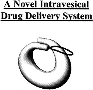
Fig. 8. Bladder pump is currently under development to deliver constant dose of oxybutynin intravesically for up to 30 days. Bladder drug delivery device is inserted using catheter-based applicator and removed using flexible cystoscope and grasper. (From Yoshimura N, Chancellor MB: Current and future pharmacological treatment for overactive bladder. J Urol 168:1897–1913, 2002).

Intravesical Peppers

The vanilloids such as capsaicin and resiniferatoxin activate nociceptive sensory nerve fibers through an ion channel known as vanilloid receptor subtype 1 (VR1).85 Intravesical capsaicin has been used recently with limited success in patients with multiple sclerosis or spinal cord injury. Chancellor and deGroat86 performed a meta-analysis of six series including 131 patients. Bladder capacity improved and symptomatic improvement occurred in 72%. Resiniferatoxin is approximately 1000-times more potent than capsaicin.87 Both agents are vanilloid receptor agonists that result in desensitization; however, resiniferatoxin causes less discomfort.

Botulinum Toxin

Botulinum toxin was first isolated in 1897.88 The toxin acts by inhibiting acetylcholine at the presynaptic cholinergic junction, resulting in decreased muscle contractility and muscle atrophy. This appears to be reversible with axon regrowth in 3 to 6 months. Recently, botulinum toxin has gained wide popularity in cosmetic surgery. Dykstra89 and others reported on use of botulinum toxin to treat patients who have detrusor dyssynergia secondary to spinal cord injury. Schurch90 reported increased bladder capacity and decreased mean maximum detrusor voiding pressure in patients treated with toxin. Phelan91 recently reported a series of patients with voiding dysfunction who were using indwelling or intermittent catheterizations. Improvement occurred in 19 of 21 patients who were able to discontinue use of catheters.

Behavior Modification and Pelvic Floor Exercises

Kegel92 first described the use of pelvic floor muscle (PFM) exercises to treat urinary incontinence in 1948. He reported a high success rate of 84% in his study. This early study did not differentiate between urge, stress, or other forms of incontinence; however, later studies have demonstrated that PFM exercises may be effective in treatment of symptoms of overactive bladder. The basis of such exercises is the observation that electrical stimulation of PFM appears to inhibit detrusor contractions. An early study by Godec93 demonstrated decreased bladder hyperactivity and increase in bladder capacity after mild electrical stimulation, and deGroat26 noted increased sympathetic firing during bladder filling, representing guarding reflexes to promote continence.

Bladder drills, bladder training, and multicomponent behavioral training are current extensions of Kegel's original work. The purpose of all these treatments is to diminish symptoms and improve bladder control through systematic changes in the patient's behavior. These therapies have been recognized as efficacious and are recommended as first-line therapy for incontinence in adults by the Agency for Health Care Policy and Research.94

The bladder drill was an intensive procedure that required patients to increase intervals between voidings. This was usually performed on an inpatient basis. Studies in the 1970s and 1980s combined anticholinergics and sedation with bladder training and reported a success rate of 82% to 86%.95 Similar success has been reported with randomized trials conducted as an outpatient procedure.96 A modification of the bladder drill, termed bladder training is performed more gradually on an outpatient basis. The rationale is that frequency and urgency are not only a result but also an initiating factor for uninhibited detrusor contractions. Increased voiding frequency leads to decreased bladder capacity and detrusor instability. Bladder training attempts to break the cycle by requiring the patient to resist urgency and postpone voiding until scheduled intervals.

Pelvic Floor Exercises and Biofeedback

Nygaard and associates97 recently reported use of pelvic floor exercises in the treatment of OAB. They report a significant decrease in mean number of incontinent episodes per day in a group of 14 women studied over 3 months. Fifty percent of the patients described excellent or good results. It is important that the patient be properly educated regarding pelvic floor anatomy and assessed by a trained examiner for performance of exercises. The typical protocol calls for 50 contractions per day in two or three divided sessions. Each contraction is sustained for five seconds followed by 10 seconds of relaxation.

In some patients, biofeedback may be added to aid in identification of appropriate muscle contractions. An intravaginal EMG probe is used to sense pelvic musculature activity and is converted to a visual signal for the patient on a computer screen or to an audio source.

Multicomponent behavioral training uses biofeedback and other techniques using the PFM in an attempt to inhibit bladder contraction. Combined feedback of bladder pressure and PFM activity allows patients to visualize detrusor contractions and respond with appropriate pelvic floor muscle contractions that increase urethral pressure to prevent urine loss.

Burgio98 conducted the first randomized trial comparing biofeedback-assisted behavioral training with standard drug therapy consisting of immediate-release oxybutynin chloride. Patients age 55 to 92 were randomized to 8 weeks of behavioral treatment, drug therapy, or placebo. Drug therapy improved 68.5% of patients and behavioral therapy improved 80.7%, which was statistically significant. In this study, 96.5% of the patients treated with behavioral therapy stated they would be willing to continue therapy indefinitely, while only 54.7% of those treated with drug therapy would continue indefinitely. Seventy-five percent of those using drug therapy stated they would like another form of therapy compared with only 14% of those on behavioral therapy.

Although both behavioral and drug therapy are useful for reducing urgency, few patients are cured by these therapies. Burgio98 showed only 23% of patients were dry after oxybutynin and 30% were dry after a trial of behavioral training. Burgio99 reported a study that demonstrated combining drug and behavioral therapy may be more effective than either therapy alone. A crossover design allowing those patients not satisfied with the initial assignment to use additional therapy was initiated. Of those initially assigned to behavioral therapy, only 8 of 65 elected to add drug to their regimen. These patients improved from 57% to 89% reduction in incontinence after the addition of oxybutynin. However, 21 of 67 agreed to add behavioral therapy after drug therapy. Of this group, 84% had reduction with combined therapy compared with 72% with drug therapy alone.

In summary, research has shown that behavioral training is effective in reducing urge-type incontinence. Addition of drug therapy to behavior modification may increase the success further; however, few patients are completely cured by these techniques.

Neuromodulation for Refractory Overactive Bladder

The scope of OAB will probably increase with the growing number of older persons in the future. Included in this number will inevitably be a group that will be refractory to usual therapy. For that group, neuromodulation techniques may offer some relief. Neuromodulation techniques are based on our current understanding of afferent and efferent neurological pathways in bladder function. The bladder and pelvic floor receive this innervation from S2, S3, and S4. Sacral parasympathetics provide excitatory pathways balanced by thoracolumbar sympathetic inhibition.

Current neuromodulation techniques are based on early experimental treatment of urinary incontinence by Caldwell, with electrical stimulation and subsequent experiments by Schmidt and Tanagho.100

Two devices are currently FDA-approved for peripheral sacral neurostimulation. The Stoller Afferent Nerve Stimulator (SANS) device was approved for use in 1999. The device uses a monopolar generator to stimulate the posterior tibial nerve via an acupuncture needle placed at the ankle. This area has been recognized as the bladder center by Chinese acupuncture and has nerve projections to S3.

Govier101 reported a study of 53 patients treated on a weekly basis with this device for 12 weeks. Based on urodynamic follow-up, voiding diaries, and quality of life surveys, 71% of patients were classified as successful. A 25% and 21% reduction in daytime and nighttime frequency were noted, and urge incontinence was reduced by 35% in previously refractory OAB patients.

Klingler102 reported 18 patients with refractory urge frequency or pelvic pain syndrome. At 10 months, pain was significantly reduced in most patients, OAB was eliminated in 77% of patients, and bladder capacity was increased from a mean of 197 to 252 milliliters. In addition, daytime voids were reduced from 16.1 to 8.3 and nighttime voids were reduced from 4.4 to 1.4. Complications are rare; however, the technique does require multiple patient visits.

The Interstim Sacral Nerve Stimulation System (Medtronics, Minneapolis, MN) was approved by the FDA for urgency/frequency and nonobstructive urinary retention in 1999. The device consists of an implantable neurostimulator attached to electrodes directed through the S3 foramen to stimulate sensory afferents. Patients initially undergo either bilateral needle test stimulation or unilateral staged implant before permanent placement of the device.

Jankneqt103 reported a group of 96 refractory patients treated with the device in 2001. The treated patients demonstrated a significant reduction in number of urge incontinence occurrences and number of pads used. Siegel104 has reported one of the largest multicenter trials of the device in 260 patients from a second group of 581 with urge incontinence, urgency/frequency, and urinary retention. At 18 months, 76% of patients with urge incontinence were deemed successful while urgency/frequency responded in 63% of patients. Patients with urinary retention responded successfully in 71% at 18 months, defined as elimination or more than 50% reduction in need of catheterization.

Kohli and Rosenblatt105 in a recent article have described a good step-by-step treatment algorithm that uses a complete history and physical examination, appropriate office testing, and treatment of patients with OAB (Fig. 9). For a complete summary of current neuromodulation techniques, the reader is referred to Kohli and Rosenblatt.105

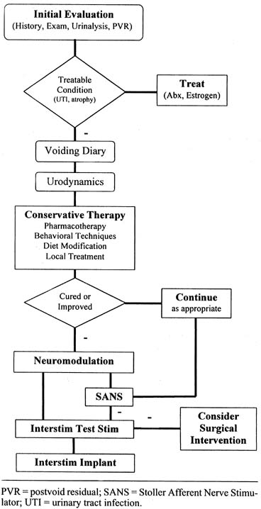
Fig. 9. Treatment algorithm for overactive bladder syndrome. PVR, postvoid residual; SANS, stoller afferent nerve stimulator; UTI, urinary tract infection. (From Kohli N, Rosenblatt PL: Neuromodulation techniques for the treatment of the overactive bladder. Clin Obstet Gynecol 45:218–132, 2002).

In summary, neuromodulation techniques hold promise as an alternative therapy for OAB cases that do not respond adequately to conventional pharmacotherapy or behavioral techniques. Future research should continue to further elucidate our understanding of the mechanisms of action and how electrical stimulation may modify somatic afferent inhibition of bladder stimulation.

Back to Top
SUMMARY

Overactive bladder is a symptom complex involving “urgency with or without urge incontinence, usually with frequency and nocturia in the absence of local or metabolic factors explaining these symptoms.” It has recently been postulated that even interstitial cystitis may be a pain variant of OAB. It is estimated that the problem affects approximately 16.5% of adults or 33 million persons in the United States. Secondary to the aging population, the scope of this problem will probably increase. Current therapy options have increased dramatically in the recent past and include pharmacotherapy, behavioral techniques, local therapy, neuromodulation, and surgery. New techniques and those still in the research arena hold promise for improved therapy of this difficult problem.

Back to Top
REFERENCES

1. Voelker R: International group seeks to dispel incontinence “taboo.” JAMA 280:951, 1998

2. Resnick RM, Ouslander SG: National Institutes of Health consensus development conference on urinary incontinence. J Am Geriatr Soc 38:263, 1990

3. Urinary Incontinence Guideline Panel: Urinary incontinence in adults: Clinical practice guidelines. Rockville, MD, Agency for Health Care Policy and Research, US Public Health Service, US Department of Health and Human Services, AHCPR publication no. 92-0038 1992

4. Abrams P, Cardozo L, Fall M et al: The standardisation of terminology of lower urinary tract function: Report from the Standardisation Subcommittee of the International Continence Society. Neurourol Urodyn 21:167, 2002

5. Abrams P, Wein AJ: The overactive bladder and incontinence: Definitions and a plea for discussion. Neurourol Urodyn 18:413, 1999

6. Wein AJ: Overactive bladder: Defining the disease. Am J Managed Care 6(S):559–564, 2000

7. Milsom I, Abrams P, Cardozo L et al: How widespread are the symptoms of an overactive bladder and how are they managed? A population-based prevalence study Br J Urol Int 87:760, 2001

8. Stewart W, Herzog AR, Wein A et al: Prevalence and impact of overactive bladder in the US: Results from the NOBLE program. Neurourol Urodyn 20:406, 2001

9. Booth CM, Whiteside CG, Turner-Warwick RT: A long-term study of the persistence of the urodynamic characteristics of the unstable bladder. Br J Urol 53:310, 1981

10. Jeffcoate TNA, Francis WJA: Urgency incontinence in the female. Am J Obstet Gynecol 94:604, 1996

11. Bump RC: Racial comparisons and contrasts in urinary incontinence and pelvic organ prolapse. Obstet Gynecol 81:421, 1993

12. Mattox TF, Bhatia NN: The prevalence of urinary incontinence or prolapse among white and Hispanic women. Am J Obstet Gynecol 174:646, 1996

13. Dwyer PL, Lee ETC, Hay DM: Obesity and urinary incontinence in women. Br J Obstet Gynecol 95:91, 1988

14. Moller LA, Lose G, Jorgensen T: Incidence and remission rates of lower urinary tract symptoms at one year in women aged 40–60: Longitudinal study. Br Med J 320:1429, 2000

15. Elving LB, Foldspang A, Lam OW et al: Descriptive epidemiology of urinary incontinence in 3100 women age 30-59. Scand J Urol Nephrol 125:37, 1989

16. Karram MM: Detrusor instability and hyperreflexia. In: Walters MD, Karram MM (eds): Clinical Urogynecology. p 297, 2nd ed. St. Louis, Mosby-Year Book, 1999

17. Liberman JN, Hunt TL, Stewart WF et al: Health related quality of life among adults with symptoms of overactive bladder: Results from a US community-based survey. Urology 57:1044, 2001

18. Stewart W, Herzog R, Wein A et al: Prevalence and impact of overactive bladder in the US: Results from the NOBLE program. Presented at the International Continence Society, Seoul, Korea September 2001

19. Kobelt G: Economic considerations and outcome measurement in urge incontinence. Urology 50:(S6):100, 1997

20. Abrams P, Kelleher CJ, Kerr LA et al: Overactive bladder significantly affects quality of life. Am J Managed Care 6:(S):S580, 2000

21. Brown JS, McGhan WF, Chokroverty SA: Comorbidities associated with overactive bladder. Am J Managed Care 6:(S):S574, 2001

22. Wagner TH, Hu TW: Economic costs of urinary incontinence in 1995. Urology 51:355, 1998

23. Kwon C, Sand P: Management of overactive bladder and urinary tract infections. OB/GYN Special Edition 25. Spring 2003

24. Goldberg RP, Sand PK: Pathophysiology of the overactive bladder. Clin Obstet Gynecol 45:182, 2002

25. Brading AF: A myogenic basis for the overactive bladder. Urology 50:(S6A):57, 1997

26. deGroat WC: A neurologic basis for the overactive bladder. Urology 50:(S6A):36, 1997

27. Hindmarsh JR, Gosling PT, Dean AM: Bladder instability: Is the primary defect in the urethra? Br J Urol 55:648, 1983

28. Major H, Culligan P, Heit M: Urethral sphincter morphology in women with detrusor instability. Obstet Gynecol 99:63, 2002

29. Chancellor MB, Yoshimura N: Physiology and pharmacology of the bladder and urethra. In: Walsh PC (ed): Campbell's Urology. pp 831-886, Philadelphia, Saunders, 2002

30. deGroat WC, Booth AM, Yoshimura N: Neurophysiology of micturition and its modification in animal models of human disease. In: Maggi CA (ed): The Autonomic Nervous System, Nervous Control of the Urogenital System. pp 227-290, Vol. 3: London, Harwood Academic Publishers, 1993

31. Torrens MJ, Morrison JFB: The Physiology of the Lower Urinary Tract. Berlin, Springer-Verlag, 1987

32. Langworthy OR, Kolb LC: The encephalic control of tone in the musculature of the urinary bladder. Brain 56:371, 1933

33. Yoshimura N, Mizuta E, Kuno S et al: The dopamine D1 receptor agonist SKF 38393 suppresses detrusor hyperreflexia in the monkey with parkinsonism induced by 1-methyl-4-phenyl-1,2,3,6-tetrahydropyridine (MPTP). Neuropharmacol 32:315, 1993

34. Araki I, de Groat WC: Synaptic modulation associated with developmental reorganization of visceral reflex pathways. J Neurosci 17:8402, 1997

35. Geirsson G, Fall M, Sullivan L: Clinical and urodynamic effects of intravesical capsaicin treatment in patients with chronic traumatic spinal detrusor hyperreflexia. J Urol 154:1825, 1995

36. Van Duyl WA, Van der Hoeven AAM, de Bakker JV: Synchronization of spontaneous contraction activity in smooth muscle or urinary bladder. Neurourol Urodyn 9:547, 1990

37. Saito M, Kondo A, Kato T et al: Response of the human neurogenic bladder to KCL, carbecho, ATP and CaC12. Br J Urol 72:298, 1993

38. Saito M, Kondo A, Kato T et al: Response of isolated human neurogenic detrusor smooth muscle to intramural nerve stimulation. Br J Urol 72:723, 1993

39. Sibley GNA: The physiological response of the detrusor muscle to experimental bladder outflow obstruction in the pig. Br J Urol 60:332, 1987

40. Sethia KK, Brading AF, Smith JC: An animal model of non-obstructive bladder instability. J Urol 143:1243, 1990

41. Mills IW, Greenland JE, McMurray G et al: Studies of the pathophysiology of idiopathic detrusor instability: The physiological properties of the detrusor smooth muscle and its pattern of innervation. J Urol 163:646, 2000

42. Elbadwi A: Functional pathology of urinary bladder muscularis: The new frontier in diagnostic uropathology. Semin Diagn Pathol 10:314, 1993

43. Elbadwi A, Yalla SV, Resnick NM: Structural basis of geriatric voiding dysfunction: III. Detrusor overactivity J Urol 150:1668, 1993

44. Elbadwi A, Meyer S, Malkowiez SB et al: Effects of short term partial outlet obstruction on the rabbit detrusor: An intrastructural study. Neurourol Urodyn 8:89, 1989

45. Elbadwi A: Pathology and pathophysiology of detrusor in incontinence. Urol Clin North Am 22:499, 1995

46. Gosling JA, Kung LS, Dixon JS et al: Correlation between the structure and function of the rabbit urinary bladder following partial outlet obstruction. J Urology 163:1349, 2000

47. Speakman MJ, Brading AF, Gilpin CJ et al: Bladder outflow obstruction-a cause of denervation supersensitivity. J Urol 138:1461, 1987

48. Brading AF, Turner WH: The unstable bladder: Towards a common mechanism. Br J Urol 73:3, 1994

49. Steers WD, deGroat W: Effect of bladder outlet obstruction on micturition reflex pathways in the rat. J Urol 140:864, 1988

50. Major H, Culligan P, Heit M: Urethral sphincter morphology in women with detrusor instability. Obstet Gynecol 99:63, 2002

51. Payne C: Epidemiology, pathophysiology, and evaluation of urinary incontinence and overactive bladder. J Urol 51:3, 1998

52. Awad S, McGinnis R: Factors that influence the incidence of detrusor instability in women. J Urol 130:114, 1983

53. Kuru M: Nervous control of micturition. Physiol Rev 45:425, 1965

54. Koonings PP, Bergman A: Urethral pressure changes in women with detrusor instability. Bladder or urethral pathologic process? Urology 37:540, 1991

55. Koonings P, Bergman A, Ballard CA: Combined detrusor instability and stress urinary incontinence: Where is the primary pathology? Gynecol Obstet Invest 26:250, 1988

56. Beck RP, Arnusch D, King C: Results in treating 210 patients with detrusor overactivity incontinence of urine. Am J Obstet Gynecol 125:593, 1976

57. McGuire EJ, Sacastana JA: Stress incontinence and detrusor instability/urge incontinence. Neurourol Urodyn 4:313, 1985

58. Heit M: Intraurethral ultrasonography: Correlation of urethral anatomy with functional urodynamic parameters in stress incontinent women. Int Urogynecol J 11:204, 2000

59. Steers WD, Kolbeck S, Creedon D et al: Nerve growth factor in the urinary bladder of the adult regulates neuronal form and function. J Clin Invest 88:1709, 1991b

60. MacLennan AH, Taylor AW, Wilson DH et al: The prevalence of pelvic floor disorders and relation to gender, age, parity and mode of delivery. Br J Obstet Gynaecol 107:1460, 2000

61. Shumaker SA, Wyman JF, Uebersax JS et al: Health-related QOL measures for women with urinary incontinence: The Incontinence Impact Questionnaire and the Urogenital Distress Inventory. Qual Life Res 3:291, 1994

62. Dwyer PL, Rosamilia A: Evaluation and diagnosis of the overactive bladder. Clin Obstet Gynecol 45:193, 2002

63. Hooton TM, Epidemiology. In: Stanton SL: Dwyer PL (eds): Urinary Tract Infection in the Female. pp 2-18, London, Martin Dunitz, 2000

64. Versi E, Orrego G, Hardy E et al: Evaluation of the home pad test in the investigation of female urinary incontinence. Br J Obstet Gynaecol 103:162, 1996

65. Haylen BT, Law MG, Frazer M et al: Urine flow rates and residual urine volumes in urogynaecology patients. Int Urogynecol J 10:378, 1999

66. Dwyer PL, Desmedt E: Impaired bladder emptying in women. Aust NZ J Obstet Gynaecol 34:73, 1994

67. Hehir M, Fitzpatrick JM: Oxybutynin and the prevention of urinary incontinence in spina bifida. Eur Urol 11:254, 1985

68. Gleason DM, Susset J, White C et al: Evaluation of a new once-daily formulation of oxybutynin for the treatment of urinary urge incontinence. Urology 54:420, 1999

69. Birns J, Lukkari E, Malone-Lee JG: A randomized controlled trial comparing the efficacy of controlled-release oxybutynin tablets (10 mg once daily) with conventional oxybutynin tablets (5 mg twice daily) in patients whose symptoms were stabilized on 5 mg twice daily of oxybutynin. Br J Urol Int 85:793, 2000

70. Versi E, Appell R, Mobley D et al: Dry mouth with conventional and controlled-release oxybutynin in urinary incontinence. The Ditropan XL Study Group Obstet Gynecol 95:718, 2000

71. Gupta SK, Sathyan G: Pharmacokinetics of an oral once-a-day controlled-release oxybutynin formulation compared with immediate release oxybutynin. J Clin Pharmacol 39:289, 1999

72. Gupta SK, Sathyan G, Lindemulder EA et al: Quantitative characterization of therapeutic index: application of mixed-effects modeling to evaluate oxybutynin dose-efficacy and dose-side effect relationships. Clin Pharmacol Ther 65:672, 1999

73. Appell RA: Clinical efficacy and safety of tolterodine in the treatment of overactive bladder: a pooled analysis. Urology 50:90, 1997

74. Van Kerrebroeck P, Kreder K, Jonas U et al: Tolterodine once-daily: Superior efficacy and tolerability in the treatment of the overactive bladder. Urology 57:414, 2001

75. Appell RA, Sand P, Dmochowski R et al: Prospective randomized controlled trial of extended-release oxybutynin chloride and tolterodine tartrate in the treatment of overactive bladder: results of the OBJECT study. Mayo Clin Proc 76:358, 2001

76. Sussman D, Garely A: Treatment of overactive bladder with once-daily extended-release tolterodine or oxybutynin: The Antimuscarinic Clinic Effectiveness Trial (ACET). Curr Med Res Opin 18:177, 2002

77. Diokno AC, Appell RA, Sand PK et al: Prospective, randomized, double-blind study of the efficacy and tolerability of the extended-release formulations of oxybutynin and tolterodine for overactive bladder: Results of the OPERA trial. Mayo Clini Proc 78:687, 2003

78. Smith CM, Wallis RM: Characterisation of [3H]-darifenacin as a novel radioligand for the study of muscarinic M3 receptors. J Recept Signal Transduct Res 17:177, 1997

79. Chancellor MB, Rivas DA, Staas WE: DDAVP in the urological management of the difficult neurogenic bladder in spinal cord injury. J Am Paraplegia Soc 17:165, 1994

80. Hill S, Khullar V, Cardozo L: Desmopressin tablets in the treatment of nocturia in women. Neurourol Urodyn 14:462, 1995

81. Yoshimura N, Chancellor MB: Current and future pharmacological treatment for overactive bladder. J Urol 168:1897, 2002

82. Brendler CB, Radebaugh LC, Mohler JL: Topical oxybutynin chloride for relaxation of dysfunctional bladders. J Urol 141:1350, 1989

83. Yaich M, Popon M, Medard Y et al: In-vitro cytochrome P450 dependent metabolism of oxybutynin to N-deethyloxybutynin in humans. Pharmacogenetics 8:449, 1998

84. Davila GW, Daugherty CA, Sanders SW: A short-term, multicenter, randomized double-blind dose titration study of the efficacy and anticholinergic side effects of transdermal compared to immediate release oral oxybutynin treatment of patients with urge urinary incontinence. J Urol 166:140, 2001

85. Cruz F, Guimaraes M, Silva C et al: Suppression of bladder hyperreflexia by intravesical resiniferatoxin. Lancet 350:640, 1997

86. Chancellor MB, de Groat WC: Intravesical capsaicin and resiniferatoxin therapy: Spicing up the ways that we treat the overactive bladder. J Urol 162:3, 1999

87. Lewis R: Why red-hot peppers are red-hot. Biophoto Int J 50:40, 1998

88. van Ermengem E: Ueber einen neuen anaeroben Bacillus and seine Beziehungen zum Botulisms. Ztsch Hyg Infekt 26:1, 1897

89. Dykstra DD, Sidi A: Treatment of detrusor-sphincter dyssynergia with botulinum A toxin: A double blind study. Arch Phys Med Rehabil 71:24, 1990

90. Schurch B, Stöhrer M, Kramer G et al: Botulinum-A toxin for treating detrusor hyperreflexia in spinal cord injured patients: A new alternative to anticholinergic drugs? Preliminary results J Urol 164:692, 2000

91. Phelan MW, Franks M, Somogyi GT et al: Botulinum toxin urethral sphincter injection to restore bladder emptying in men and women with voiding dysfunction. J Urol 165:1107, 2001

92. Kegel AH: Progressive resistance exercise in the functional restoration of the perineal muscles. Am J Obstet Gynecol 56:238, 1948

93. Godec C, Cass A, Ayala G: Bladder inhibition with functional electrical stimulation. Urology 6:663, 1975

94. Fantl JA, Newman DK, Colling J et al: Urinary incontinence in adults: Acute and chronic management. Clinical Practice Guideline, No. 2, 1996 Update. Rockville, MD: US Department of Health and Human Services, Public Health Service, Agency for Health Care Policy and Research AHCPR Publication no. 96-0682 March 1996

95. Frewen WK: A reassessment of bladder training in detrusor dysfunction in the female. Br J Urol 54:372, 1982

96. Fantl JA, Wyman JF, McClish DK et al: Efficacy of bladder training in older women with urinary incontinence. JAMA 265:609, 1991

97. Nygaard IE, Kreder KJ, Lepic MM et al: Efficacy of pelvic floor muscle exercises in woman with stress urge, and mixed urinary incontinence. Am J Obstet Gynecol 174:120, 1996

98. Burgio KL, Locher JL, Goode PS et al: Behavioral versus drug treatment for urge incontinence in older women: A randomized clinical trial. JAMA 23:1995, 1998

99. Burgio KL, Locher JL, Goode PS: Combined behavioral and drug therapy of urge incontinence in older women. J Am Geriatr Soc 48:370, 2000

100. Das AK, White MD, Longhurst PA: Sacral nerve stimulation for the management of voiding dysfunction. Urology 2:43, 2000

101. Govier FE, Litwiller S, Nitti V et al: Percutaneous afferent neuromodulation for the refractory overactive bladder: Results of a multicenter study. J Urol 165:1193, 2001

102. Klingler HC, Pycha A, Schmidbauer J et al: Use of peripheral neuromodulation of the S3 region for treatment of detrusor overactivity: A urodynamic-based study. Urology 56:766, 2000

103. Janknegt RA, Hassouna MM, Siegel SW et al: Long-term effectiveness of sacral nerve stimulation for refractory urge/incontinence. Eur Urol 39:101, 2001

104. Siegel SW, Catanzaro F, Dijkema HE et al: Long-term results of a multicenter study on sacral nerve stimulation for treatment of urinary urge incontinence, urgency-frequency, and retention. Urology 56:(S1):87, 2000

105. Kohli N, Rosenblatt PL: Neuromodulation techniques for the treatment of the overactive bladder. Clin Obstet Gynecol 45:218, 2002

Back to Top