Chapter 122
Open Laparoscopy
Harrith M. Hasson
Main Menu   Table Of Contents

Search

Harrith M. Hasson, MD
Department of Obstetrics and Gynecology, Rush University; Chairman, Weiss Memorial Hospital, Chicago, Illinois (Vol 1, Chaps 61, 122)

INTRODUCTION
SURGICAL ANATOMY
INSTRUMENTS
TECHNIQUE
MISCONCEPTIONS
ADVANTAGES OF OPEN VERSUS CLOSED LAPAROSCOPY
REFERENCES

INTRODUCTION

Laparoscopy requires distention of the abdominal cavity with a suitable gas to create a clear space between the abdominal wall and abdominal contents, as well as the introduction of a lighted telescope into the abdominal cavity to visualize various organs and to perform different procedures.

Traditionally, laparoscopy has been performed using needles to introduce the gas and sharp trocars to introduce the laparoscope into the abdominal cavity. The abdomen is first insufflated with a needle inserted blindly through the abdominal wall. After abdominal distention, a small skin incision is made, and a cannula bearing a sharp trocar is thrust forcibly against the remaining layers of the abdominal wall into the peritoneal cavity. The blind introductions of the needle and trocar into the peritoneal space are guided by tactile sensations. The sharp trocar is withdrawn and replaced by the scope, and the appropriate endoscopic procedure is performed. At the end of the procedure, the cannula and scope are removed. The skin wound is approximated, but the fascial gap is not repaired because it is not accessible. It is becoming increasingly clear, however, that the use of such sharp instruments is not necessary for performing laparoscopy, and on some occasions, it is undesirable.

Laparoscopy can be easily performed with the use of a minilaparotomy incision. This variation is called open laparoscopy. The difference between open and traditional closed laparoscopy relates specifically to the method of abdominal entry, the sequence of entry/insufflation, and the type of abdominal wall closure. All other aspects of laparoscopy remain unchanged.

In open laparoscopy, the abdomen is first entered through a minilaparotomy incision developed under visual control. A specific cannula carrying a blunt obturator then is introduced through the incision into the abdominal cavity. Gas is insufflated directly through the cannula. The obturator is removed and replaced with the laparoscope, and the appropriate endoscopic procedure is carried out. When the procedure is completed, the cannula and scope are removed and the abdominal wall is closed in layers.1-3

Back to Top
SURGICAL ANATOMY

The abdominal cavity may be entered through a minilaparotomy incision at any location on the anterior abdominal wall.4-6 For open laparoscopy, however, the lower border of the umbilicus is the preferred site of entry. In this location, the abdominal wall has the fewest layers, since it consists of skin fused with linea alba, transversalis fascia, preperitoneal adipose tissue, and peritoneum. The area is devoid of subcutaneous adipose tissue, Camper's fascia, and any muscles. It may be considered a window through which open laparoscopy can be most easily performed7 (Fig. 1). These unique anatomic relations result from developmental events that occur at the time of birth and shortly thereafter. Four ligaments resulting from closure of functional fetal structures attach to the inferior border of the umbilical ring and pull the skin against the deep fascia while the once-functional vessels and ducts retract and become fibrotic. The round ligament arises from the obliteration of the umbilical vein, the median umbilical ligament evolves from closure of the urachus, and the medial umbilical ligaments represent the obliterated umbilical arteries and allantoic duct (Fig. 2). In some subjects, the transversalis fascia is highly condensed into a strong layer called the fascia umbilicalis or Richet's fascia.8 The disposition of the umbilical ligaments about the umbilical ring, a fibrotic opening n the linea alba, and the presence and position of Richet's fascia are subject to wide individual variations,8 as shown in Figure 2. These variations influence the ease with which open laparoscopy is performed. Technical difficulty also is related to the tonal strength of the abdominal wall supports in the umbilical region. Other factors that increase technical difficulty include fibrosis and adhesions secondary to previous surgery or inflammation. Marked obesity per se has no effect on technical difficulty or cosmetic result. The larger incision (not to exceed 2.5 cm) that is needed to facilitate exposure of the linear alba can be made to disappear in one of the umbilical folds.

Fig. 1. Sagittal section of the umbilical window: (1) subcutaneous tissues, (2) linea alba, (3) skin attached to linea alba and umbilical ring ligaments, (4) round ligament (ligamentum teres), (5) urachus, (6) transversalis fascia, (7) condensed portion of transversalis fascia (Richet's fascia), and (8) peritoneum.

Fig. 2. Anatomical variations of umbilical ring ligaments and position of Richet's fascia:(1) round ligament (obliterated umbilical vein), (2) median umbilical ligament (closed urachus), (3) medial umbilical ligaments (obliterated umbilical arteries and allantoic duct), (4) umbilical ring, and (5) fascia umbilicalis (Richet's fascia).

Surgical incisions made along or parallel to the lines of Langer,9 which define natural cleavage lines of the skin, heal better than incisions that cross those lines.10 In the area of the umbilicus, the lines of Langer are aligned vertically, giving a sunburst appearance. The use of vertical laparoscopy incisions in the area of the umbilicus results in a superior cosmetic scar when compared with transverse laparoscopy incisions.11 Furthermore, the use of a vertical incision consistently exposes the position of fascial attachment to the skin (see Fig. 1). The arterial blood supply of the umbilical region comes from the inferior epigastric, superior epigastric, and superficial epigastric vessels. These arteries form a communicating arcade around the umbilicus, which is accompanied by the periumbilical venous plexus (Fig. 3).

Fig. 3. Arterial arcade around the umbilicus.

Back to Top
INSTRUMENTS

Standard instruments needed for open laparoscopy include two Allis clamps, a knife handle with a small blade, a straight scissors, two Kocher clamps, two small hemostats, a dissecting tissue forceps with teeth, and a short needle holder. S-shaped retractors formed with two retracting ends are used to expose the small operative field (Fig. 4). One of the retracting ends is acutely curved, and the other is more flattened. The curved end is used for retracting the superficial layers of the abdominal wall as it is anchored beneath the skin. The flatter end is more appropriate for retracting the deeper layers because it requires less space for insertion. The use of small, strong needles attached to sutures of adequate tensile strength is recommended to avoid breakage and to enhance the ease of manipulation.

Fig. 4. Surgical instruments used in open laparoscopy.

Standard laparoscopic equipment is used, with the exception of the primary cannula and pneumoperitoneum needles. The open laparoscopy cannula is fitted with a cone-shaped sleeve that moves freely over the shaft of the cannula. The cone is locked into position along the shaft using a metal screw. A rubber cap seals the cone. V-shaped suture holders are mounted on the instrument. A blunt obturator replaces the conventional sharp trocar (Fig. 5). Pneumoperitoneum needles are not used.

Fig. 5. The open laparoscopy cannula.

Several instrument companies offer disposable and reusable blunt-tipped open laparoscopy cannulas with suture holders mounted on the cone or with a distal balloon.

Back to Top
TECHNIQUE

Basic Tenets

Safe and effective performance of open laparoscopy is based on four tenets:

  1. Dissection of the minilaparotomy incision in layers and elevation of the fascia and deeper layers during incision: Entering the abdomen using a knife without proper dissection and fascial elevation increases the risk of complications. Bartsich and Dillon12 report cutting the superior mesenteric vein during the initial subumbilical skin incision of an intended closed laparoscopy procedure.
  2. After visual confirmation of abdominal entry, insufflation of gas into the abdomen directly through the cannula with the blunt obturator in place: This eliminates preperitoneal insufflation and shortens the time needed to establish the pneumoperitoneum. The blunt obturator should not be withdrawn until the abdomen is partially distended to avoid injury to internal structures by the sharp edge of the bare cannula.
  3. Blocking the escape of gas from the abdomen: Loss of the pneumoperitoneum through gas leaks exposes the surgeon to the risk of inadequate visualization and potential complication.
  4. Proper closure of the fascial defect at the end of the procedure: Proper fascial repair minimizes the occurrence of postoperative herniation.

Procedure

When satisfactory anesthesia has been achieved, the surgeon applies two Allis clamps on the lateral margins of the umbilicus and makes a vertical incision at the 6-o'clock position of the lower umbilical margin, extending inferiorly for a distance of 1 to 2.5 cm (Fig. 6). The Allis clamps are repositioned on the skin edges to be used for retraction. Closed scissors are introduced into the incision, and the blades are separated several times to dissect connective tissue fibers found between the skin and deep fascia. The surgeon directs a Kocher clamp toward the upper angle of the incision and grasps the inferior margin of the umbilical ring where the fascia is fused with the skin. The surgeon lifts and everts the fascia and applies a second Kocher clamp on it more inferiorly (Fig. 7). The fascia is clearly identified by separating it from surrounding fibrous, adipose, and areolar tissue with blunt or sharp dissection. The first Kocher clamp can be reapplied on the fascia to realize a more secure purchase, as indicated.

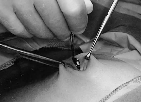
Fig. 6. Stretching the skin and making a vertical incision.

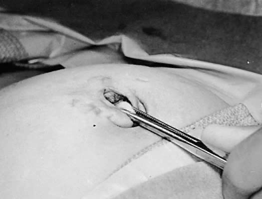
Fig. 7. Grasping the deep fascia close to its attachment to the skin and lifting it upward and superiorly.

The surgeon raises the deep fascia with the Kocher clamps and incises the fascia carefully between the clamps while maintaining fascial elevation. Raising the fascia separates the abdominal wall from the bowel and omentum and minimizes the possibility of inadvertent damage during fascial incision. The initial size of incision need not exceed 0.5 cm. The incision then is enlarged using a spreading hemostat. Usually, the peritoneum is entered by the spreading hemostat. Otherwise, the flat end of one S-shaped retractor is introduced into the incisional gap and is pressed against the peritoneum for abdominal entry (Fig. 8). The surgeon passes a suture through each fascial edge and tags it. The S-shaped retractor lodged within the incision acts as a backstop to the needle. The second S-shaped retractor then is placed through the fascial gap into the peritoneal cavity, and the abdominal wall is raised with both retractors to confirm peritoneal entry by viewing the bowel and omentum. If the surgeon discovers that the abdominal cavity had not been entered, the surgeon identifies and retracts the rectus muscle, exposes the peritoneum, and enters it with a thrust of a small hemostat. The hemostat is opened inside the peritoneal cavity, and a retractor is placed in the abdomen between the open jaws of the hemostat. The hemostat is removed, and the other retractor is applied on the opposite side. Occasionally, abdominal entry is not possible with the previously described procedures because of the presence of a well-developed Richet's fascia. In this case, the surgeon should lift Ricket's fascia and peritoneum with two hemostats and incise them carefully to avoid nicking the small bowel. In any case, the surgeon must confirm peritoneal entry through visualization of the small bowel or omentum.

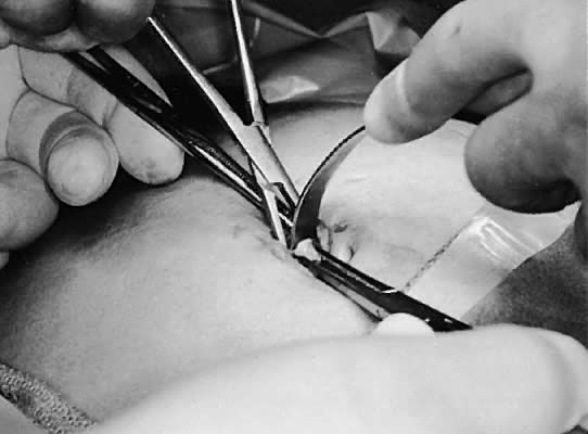
Fig. 8. Enlarging the fascial incision with the spread of a hemostat and placement of an S-shaped retractor.

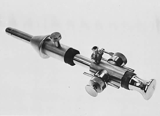
The surgeon prepares the open laparoscopy cannula for insertion by sliding the cone sleeve over the shaft and locking the cone in a position consistent with the individual thickness of the abdominal wall. The cannula then is introduced between the retractors into the abdominal cavity (Fig. 9). The fascial tag sutures are held tensely upward and threaded into the V-shaped suture holders (Fig. 10). This maneuver fixes the cannula on the abdominal wall and pulls the fascia against the cone to prevent escape of the gas (Fig. 11). Gas insufflation is initiated during threading of the fascial tag sutures into the suture holders. The blunt obturator is removed to permit a rapid flow of the gas.

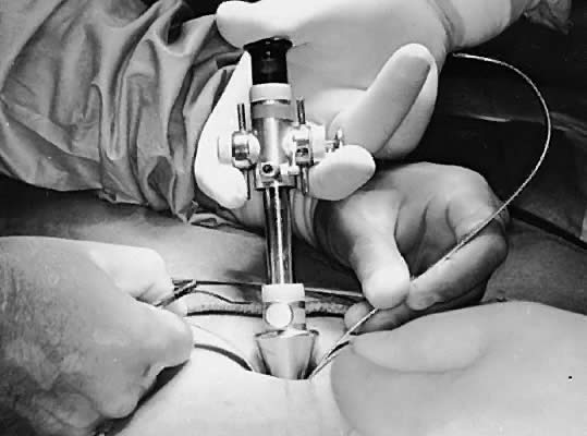
Fig. 9. Introducing the open laparoscopy cannula into the abdomen, guided by the S-shaped retractors.

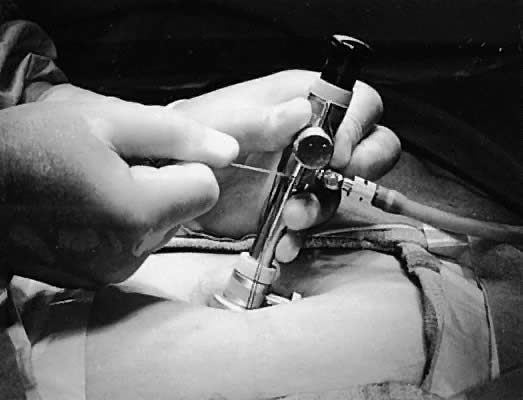
Fig. 10. Fixing the fascial tag sutures into the V-shaped suture holders.

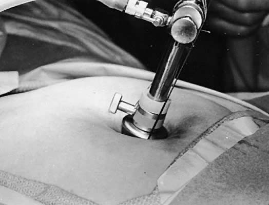
Fig. 11. Creation of an airtight seal following pulling of the sutures snugly into the holders.

The surgeon introduces the laparoscope, with or without an attached camera, through the open laparoscopy cannula and performs the intended procedure. When the endoscopic procedure is completed, the surgeon deflates the distended peritoneal cavity, withdraws the cannula, and closes the abdominal wall in two layers. The fascial tag sutures are used to approximate the fascia. The surgeon positions the sutures in parallel alignment (Fig. 12), ties a square knot on one side (Fig. 13), pulls the tied suture against the fascia, and ties a second knot on the opposite side of the fascia (Fig. 14). The skin is approximated loosely.

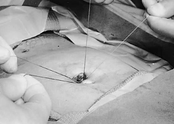
Fig. 12. Alignment of the superior and inferior tag sutures parallel to each other.

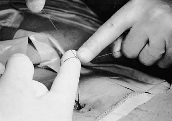
Fig. 13. Tying the first knot combining fascial tag sutures on the left side of the patient.

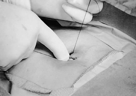
Fig. 14. Tying the second knot combining fascial tag sutures on the right side of the patient, effecting closure of the fascia.

Technical Pitfalls

Although open laparoscopy is a simple operation, there are several pitfalls that increase technical difficulty. These are outlined in Table 1.

TABLE 1. Technical Pitfalls in Open Laparoscopy


Pitfall

Reason

Remedy

Skin incision is made too small for degree of surgeon's experience or patient obesity.

Ease of operation is inversely related to size of skin incision.

To improve exposure, enlarge skin incision to maximum of 2.5 cm

Retraction is applied upward on angle greater than 60 degrees.

Such upward retraction increases distance to operative site and diminishes view.

To improve exposure, apply retraction force at projected angle of 45 degrees or less.

Failure to raise fascia during incision

Possible injury to deeper structures

To increase safety, maintain fascial elevation during incision

Fascial incision is not enlarged with hemostat.

It is difficult to pass sutures into fascial edges that are not clearly visible.

To clearly expose the fascial edges and peritoneum, enlarge the fascial gap


It is also difficult to expose or enter peritoneum through minute fascial gap.


Use of weak needles and sutures or use of large needles

Needle may break inside operative field.

To avoid breakage and to enhance ease of manipulation, use a small, strong needle attached to suture of adequate tensile strength


Sutures may break while being threaded in suture holders. Large needle is awkward to use in small operative field.


Failure to stabilize cannula while threading the fascial sutures into suture holders

Cannula may be dislodged into properitoneal space while fascial sutures are being pulled upward.

To prevent cannula from being displaced, press it gently against abdominal wall as fascial sutures are being pulled upward

Failure to pull fascial sutures tensely into suture holders

Lax fascial sutures do not pull fascial edges snugly against cone of cannula. Gas may excape.

To create an airtight seal, hold fascial sutures taut as they are wedged into suture holders

Other Procedures Using Similar Concepts

Several operations applying similar concepts of abdominal entry for laparoscopy have been proposed. These procedures use a standard laparoscopic cannula, not the open laparoscopy cannula. The modified techniques of open laparoscopy are safe and effective to the extent that they observe the basic tenets of the procedure and succeed in providing an airtight seal around the cannula. Additionally, care must be exercised while introducing the cannula into the abdomen without a protective blunt obturator to avoid internal injury by the sharp edge of the bare cannula.

Examples of methods described to block escape of the gas around a standard cannula include using a purse-string suture in the fascia (with or without a rubber tube choker)13, 14 or in the subcutaneous tissue,15 and applying towel clips,16-18 Allis clamps,19 or suture20 to the skin and subcutaneous tissue surrounding the cannula.

Back to Top
MISCONCEPTIONS

Certain misconceptions exist with regard to open laparoscopy:

  1. Open laparoscopy transforms a simple procedure into a more complex and cumbersome operation. The operative and recovery times are significantly greater for open laparoscopy than for closed laparoscopy.
    Reasonable surgeons would not contemplate doing closed laparoscopy without first acquiring proper instruments, adequate instructions, and sufficient training. One does not simply acquire a needle, a sharp trocar, and a laparoscope and begin performing conventional laparoscopy. Similarly, one should not expect to be able to perform open laparoscopy successfully without using the instruments and techniques that were developed specifically for this method. Open laparoscopy is a relatively simple operation, easily learned by any physician trained in abdominal surgery. Nevertheless, the operation does require knowledge of the anatomy and expertise related to the small size of the operative field and the need for an airtight seal.
    Given comparable circumstances, the amount of time needed to achieve competence in the procedure is less for open laparoscopy than for closed laparoscopy: surgeons can more easily master a procedure that uses a familiar standard surgical technique than one that depends on guiding a blind needle and trocar into the abdomen. Furthermore, the degree of risk to patients undergoing a procedure performed by surgeons in their initial phase of experience clearly is smaller for open laparoscopy than for closed laparoscopy.
    The belief that open laparoscopy demands a greater amount of time to perform than closed laparoscopy has been shown to be incorrect.6, 18-24 The cumulative time needed for satisfactory insertion of the pneumoperitoneum needle, establishment of an adequate pneumoperitoneum, and successful introduction of the trocar is substantially equal to if not greater than that needed for developing the open incisional technique. If a surgeon uses one of the methods routinely and the other only occasionally, the more frequently used method requires less time to perform. The recovery time is comparable for both methods.
  2. Open laparoscopy requires a relatively large incision that is cosmetically unappealing.
    In 10 studies that specified the size of the skin incision used to perform open laparoscopy, the incision was 2 cm or less in 7,4-6, 18, 20, 23, 25 2.5 cm or less in 1 study,3 3 cm or less in 1,21 and up to 4.5 cm in 1.26 The larger incisions were used in obese patients. Although many of these incisions are larger than those used with closed laparoscopy, placing the incision along the lines of Langer provides a fine, cosmetically appealing scar.
  3. Abdominal entry through an open incision increases the risk of wound infection.
    Proper surgical technique needs to be observed when either open or closed laparoscopy is performed. Existing data indicate that there is no difference between open and closed laparoscopy with regard to rates of wound infection.2,22
  4. When compared with closed laparoscopy, open laparoscopy is appreciably more difficult to perform in obese and markedly obese patients.
    Since the technical difficulty of open laparoscopy is not related to the degree of patient obesity, this procedure is easier to perform in obese patients because a larger, yet disappearing, incision can be made, and such an incision provides a more adequate field of exposure, which enhances the ease of operation. Several studies have presented excellent results with the use of open laparoscopy in markedly obese patients.3,24,27
Back to Top
ADVANTAGES OF OPEN VERSUS CLOSED LAPAROSCOPY

Safety

A consensus is emerging that most major complications of laparoscopy, defined as those leading to death or further surgery, occur as a result development of the pneumoperitoneum and blind insertion of the primary trocar, regardless of the complexity of type of laparoscopic procedure that follows.28-31 The elimination of these two potentially dangerous procedures is the main objective of open laparoscopy. Published reports of complications associated with closed laparoscopy indicate that most abdominal organs and structures have been punctured by pneumoperitoneum needles or sharp trocars.2 Misplacement of the needle and inappropriate gas insufflation has also caused emphysema of the subcutaneous tissues, the fascial compartments, and the omentum.2

For open laparoscopy, the rate of bowel injury in 26,178 patients was 0.08%, ranging from 0.03% in studies using the open method in all cases to 0.5% in those dealing exclusively with high-risk patients.2 There was one case report of a serosal bowel injury in a 2-year-old child who underwent an open laparoscopy procedure in which a standard bare 5-mm cannula and a purse-string suture were used. The suture caught the bowel. The injury was repaired, and the procedure continued as usual.32 There was one case of a major vessel injury with open laparoscopy in 26,178 patients, giving a rate of 0.004%. Occurrences of gas embolism or other visceral damage have not been reported.2 These complication rates compare favorably with those reported with closed laparoscopy.

Fewer Contraindications

Unlike closed laparoscopy, the use of open laparoscopy is not contraindicated by a history of previous abdominal surgery or by the presence of abdominal masses or marked obesity. Furthermore, because of increased safety, open laparoscopy can be used in clinical situations in which the potential risk of performing closed laparoscopy is greater than the potential benefits.

Consistency of Success

Failed attempt is a recognized problem of closed laparoscopy. Marked obesity or the development of significant abdominal wall emphysema (from inappropriate insufflation) may prevent the surgeon from completely traversing the abdominal wall with the needle or trocar. The presence of abdominal adhesions or metastatic disease also may prevent the surgeon from localizing the main compartment of the peritoneal cavity. These problems are less likely to occur with open laparoscopy because the abdomen is entered through an incision developed in a naturally occurring anatomical window, and gas is insufflated only after successful entry has been confirmed. The rate of failed attempts with open laparoscopy was 0.02% (two failures in 11,499 patients).2 Both patients had severe adhesions that prevented entry into the abdomen.

Correct Closure of the Abdominal Wall

In closed laparoscopy, the fascial tear induced by the sharp trocar is not accessible and therefore not repaired. Postoperative incisional hernias thus could develop through the unapproximated fascial gap. This complication has been reported frequently.2 Conversely, the fascial incision in open laparoscopy is clearly accessible and easily approximated. Routine closure of the fascia minimizes the possibility of postoperative herniation. The rate of umbilical hernia after open laparoscopy was 0.06% in 11,210 patients.2

Alleviation of Physician Stress

For many surgeons, the process of inserting needles and trocars into a closed abdomen is associated with a certain amount of stress. Supervising a novice while performing closed laparoscopy can produce even greater anxiety. Open laparoscopy frees the surgeon of such unnecessary stress because it uses a familiar and more predictable surgical technique.

Back to Top
REFERENCES

1. Hasson HM: Open laparoscopy: A report of 150 cases. J Reprod Med 12: 234, 1974

2. Hasson HM: Open laparoscopy as a method of access in laparoscopic surgery. Gynaecol Endosc 8: 353, 1999

3. Hasson HM, Rotman C, Rana N, Kumari NA: Open laparoscopy: 29 year experience. Obstet Gynecol 96: 763, 2000

4. Goldman LD, Cahalane MJ: The paramedian approach to open laparoscopy. J Am Coll Surg 180: 733, 1995

5. Gersin KS, Heniford BT, Arca MJ et al: Alternative site entry for laparoscopy in patients with previous abdominal surgery. J Laparosc Endosc Adv Surg Techn 8: 125, 1998

6. Lafullarde T, VanHee R, Gyst T: A safe and simple method for routine open access in laparoscopic procedures. Surg Endosc 8: 769, 1999

7. Hasson HM: Window for open laparoscopy [letter]. Am J Obstet Gynecol 137: 869, 1980

8. Orda R, Nathan H: Surgical anatomy of the umbilical structures. Int Surg 58: 458, 1973

9. Langer K: Zur anatomie und physiologie der haut: Uber die spaltbarkeit der cutis. Sitzungsberichte KaiserlichenAkad Wissenschaften 44: 20, 1862

10. Mantagna W: The Structure and Function of Skin, pp 112–114. New York, Academic Press, 1962

11. East MC, Steele PRM: Laparoscopic incisions at the lower umbilical verge. Br Med J 296: 753, 1988

12. Bartsich EG, Dillon TF: Injury of superior mesenteric vein: Laparoscopic procedure with unusual complication. NY State J Med 81: 933, 1981

13. Horattas MC, Rosser JC: A new and simple approach to open laparoscopy. Surg Gynecol Obstet 176: 287, 1993

14. Ahmad A, Quayle AR: New technique of open laparoscopy. Br J Surg 80: 1184, 1993

15. Grimes EM: Open laparoscopy with conventional instrumentation. Obstet Gynecol 57: 375, 1981

16. Treat M, Forde KA, White J: A technique for open peritoneoscopy. Surgery 92: 544, 1982

17. Grundsell H, Larsson G: A modified laparoscopic entry technique using a finger. Obstet Gynecol 59: 509, 1982

18. Hurd WW, Randolph JF, Holmberg RA et al: Open laparoscopy without special instruments or sutures: Comparison with a closed technique. J Reprod Med 39: 393, 1994

19. Perone N: Laparoscopy using a simplified open technique: A review of 585 cases. J Reprod Med 37: 921, 1992

20. Wallace DH, O'Dwyer PJ: Clinical experience with open laparoscopy. J Laparosc Endosc Adv Surg Techn 7: 285, 1997

21. Nuzzo G, Giuliante F, Tebala G et al: Routine use of open technique in laparoscopic operations. J Am Coll Surg 184: 58, 1997

22. Sigman HH, Fried GM, Garzon J et al: Risks of blind versus open approach to celiotomy for laparoscopic surgery. Surg Laparosc Endosc 3: 296, 1993

23. Ballem RV, Rudomanski J: Techniques of pneumopertioneum. Surg Laparosc Endosc 3: 42, 1993

24. Pelosi MA III, Pelosi MA: Alignment of the umbilical axis: An effective maneuver for laparoscopic entry into the obese patient. Obstet Gynecol 92: 869, 1998

25. Zaraca F, Catarci M, Gossetti F et al: Routine use of open laparoscopy: 1006 consecutive cases. J Laparosc Endosc Adv Surg Techn 9: 75, 1999

26. Adana JC, Jover JM, Lopez J et al: Advantages of the Hasson trocar in laparoscopic surgery [abstract]. Br J Surg 81 (Suppl): 81, 1994

27. Eltabbakh GH, Piver S, Hempling RE et al: Laparoscopic surgery in obese women. Obstet Gynecol 94: 704, 1999

28. Hill DJ: Complications of the laparoscopic approach. Bailliers Clin Obstet Gynaecol 8: 865, 1994

29. Capelouto CC, Kavoussi LR: Complications of laparoscopic surgery. Urology 42: 2, 1993

30. Jansen FW, Kapiteyn K, Trimbos-Kemper T et al: Complications of laparoscopy: A prospective multicenter observational study. Br J Obstet Gynaecol 104: 595, 1997

31. Chapron CM, Pierre F, Laccroix S et al: Major vascular injuries during gynecologic laparoscopy. J Am Coll Surg 185: 461, 1991

32. Sadeghi-Nejad H, Kavoussi LR, Peters CA: Bowel injury in open technique laparoscopic cannula placement. Urology 43: 559, 1996

Back to Top