Chapter 4
Diagnostic Ultrasound in the First Trimester of Pregnancy
Leeber Cohen
Main Menu   Table Of Contents

Search

Leeber S. Cohen, MD
Associate Professor, Obstetrics and Gynecology, Northwestern University Medical School, Chicago, Illinois (Vol 1, Chaps 25, 124; Vol 2, Chap 4)

INTRODUCTION
FIRST-TRIMESTER ULTRASOUND: NORMAL LANDMARKS
THREATENED ABORTION AND COMPLETED ABORTION
ECTOPIC PREGNANCY
DATING
MULTIPLE GESTATION
TRISOMY SCREENING
DIAGNOSIS OF FETAL ABNORMALITIES
MAJOR UTERINE ANOMALIES
MOLAR PREGNANCY
REFERENCES

INTRODUCTION

Diagnostic ultrasound is a powerful and frequently used tool in the assessment of first-trimester pregnancy. Today's obstetrician gynecologist is frequently called upon to interpret and in many cases perform ultrasound scans in the first trimester. In fact, certification of residency programs requires documentation of adequate exposure to and training in the evaluation of first-trimester ultrasound. Failure to understand the limitations of diagnostic ultrasound or inadequate training of physicians in this technique can result in grave complications for the patient and liability for health-care providers.

Any health-care provider performing diagnostic ultrasound must understand the physics of diagnostic ultrasound and have thorough, supervised training. This includes but is not limited to power settings, basic orientation, and proper cleaning of ultrasound probes between uses. The reader is referred to the American Institute of Ultrasound In Medicine for its extensive publications, workshops, and meetings (American Institute of Ultrasound In Medicine, 14570 Sweitzer Lane, Suite 100, Laurel, MD 20707–5906).

Back to Top
FIRST-TRIMESTER ULTRASOUND: NORMAL LANDMARKS

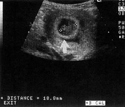
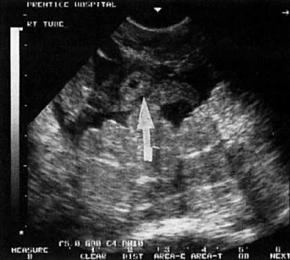
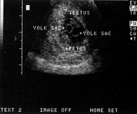
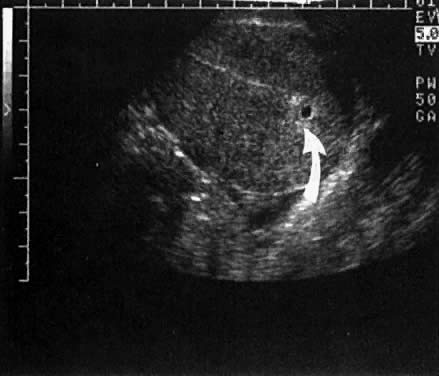
Warren and associates1 described the orderly appearance of gestational sac, yolk sac, and embryo with heartbeat at a given number of days from the onset of the last menstrual period (Table 1). With a transvaginal probe, a 3- to 4-mm gestational sac can usually be seen by 5 weeks from the last menstrual period (Fig. 1). A yolk sac or small fetal pole is usually seen by 6 menstrual weeks, when the mean diameter of the sac has reached 10 mm. As shown by Fossum and colleagues,2 the appearance of these structures can be correlated with β-human chorionic gonadotropin β-hCG) levels (Table 2). The literature regarding the correlation between quantitative β-hCG titers and early intrauterine gestational sacs and embryonic structures has been made somewhat confusing by the array of reference standards used to quantify β-hCG. Suffice it to say that the Third International Standard used by most companies marketing β-hCG kits corresponds roughly to the First International Reference Preparation.

TABLE 1 The Appearance of Early Gestational Structures


Days from LMP

28–35

35–42

42–49

49–56

Gestational Sac

100%


Yolk Sac

0%

91%

100%


Embryo with + FHTs

0%

0%

86%

100%


LMP = last menstrual period; +FHTs = positive fetal heart tones
(Warren WB, Timnor-Trisch I, Peisner DB et al: Dating the early pregnancy by sequential appearance of embryonic structures. Am J Obstet Gynecol 161:747, 1989)

TABLE 2 Correlation Between β-Human Chorionic Gonadotropin (β-hCG) Levels and Appearance Of Early Gestational Structures


First IRP

Second IS


Days from

β-hCG

β-hCG

Structure

LMP

(mIU/ml)

mIU/ml

Sac

35

1400

914

Fetal pole

40

5100

3800

Heart motion

47

17,200

13,200


LMP = last menstrual period; IRP = International Reference Preparation; IS = International Standard
(Fossum GT, Davajan V, Kletzky OA: Early detection of pregnancy with transvaginal ultrasound. Fertil Steril 49:788, 1988)

Fig. 1. A very early, 3-mm mean diameter intrauterine gestational sac at 5 weeks postmenstruation ( arrow ).

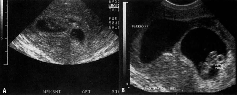
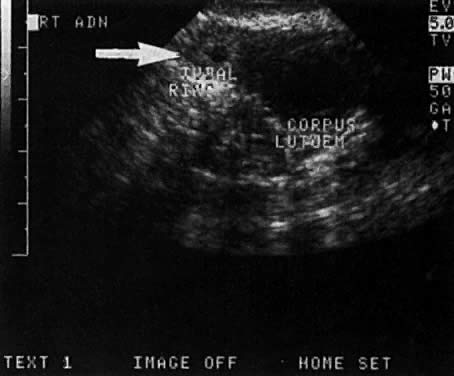
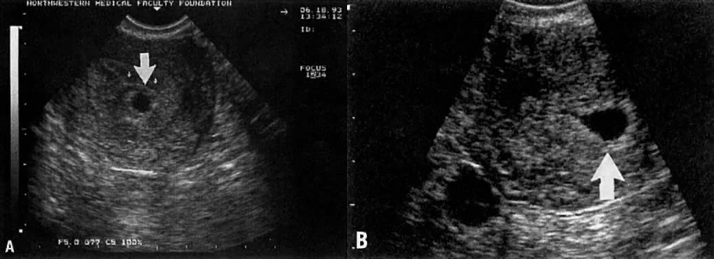
Considerable caution must be exercised not to confuse collections of fluid within a decidualized endometrium with early gestational sacs. These “pseudogestational sacs” can lead to a missed diagnosis of ectopic pregnancy. Normal early gestational sacs are seen eccentrically placed, adjacent to the echogenic central stripe (Fig. 2A and Fig. 2B). Even in experienced hands, pseudosacs and early gestational sacs can be confused.

Fig. 2. A. An eccentrically placed intrauterine gestational sac 6 weeks postmenstruation ( arrow ). B. In contrast, a pseudosac ( arrow) in a patient with ectopic pregnancy representing a collection of blood or fluid collected within the endometrial cavity.

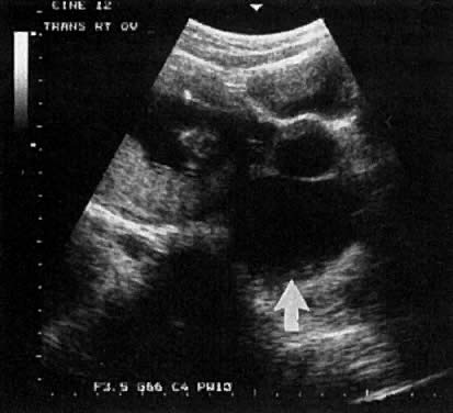
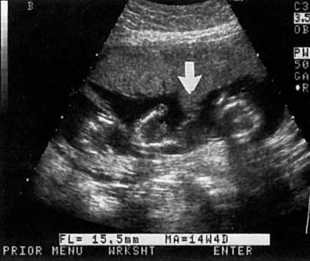
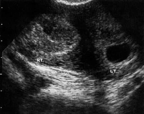
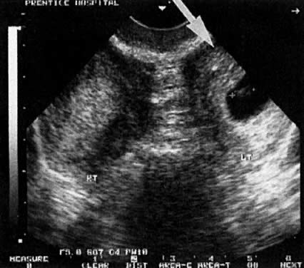
In the first 8 weeks of pregnancy, the corpus luteum is often identified as a cystic mass measuring 1 to 3 cm in diameter (Fig. 3), although they may reach as large as 8 cm.3 These masses usually resolve spontaneously by the onset of the second trimester. They can contain areas of complex echogenicity that may masquerade as a neoplasm or an ectopic pregnancy. Consultation with specialists should be obtained if an adnexal mass persists into the second trimester. The two most common benign neoplasms of the ovary during pregnancy are serous cystadenoma and benign cystic teratoma. The risk of a persistent adnexal mass during pregnancy subsequently diagnosed as malignant has probably been overestimated: it is significantly less than 1%.4 The place for expectant management of persistent adnexal masses thought to be benign by ultrasound criteria is controversial and currently being investigated.

Fig. 3. A 7 × 5 cm septated ovarian cyst found at 10 weeks postmenstruation ( arrow ). This mass resolved by the beginning of the second trimester and most likely represented a resolving corpus luteum.

Back to Top
THREATENED ABORTION AND COMPLETED ABORTION

Approximately 15% to 20% of women have a spontaneous, clinically recognized pregnancy loss in the first trimester.5 Ball and co-workers6 found that a subchorionic bleed (identified on ultrasound) is associated with an increased risk of miscarriage, stillbirth, abruptio placentae, and preterm labor (Fig. 4A and Fig. 4B). Their data suggested that the risk of spontaneous abortion increased in proportion to an increase in the size of the subchorionic bleeds; however, a larger sample size was needed to determine statistical significance. Bradycardic fetal heart rates, small sac size, abnormal yolk sacs (Fig. 5), and large subchorionic bleeds have all been associated with an increased risk of first-trimester pregnancy loss.7

Fig. 4A. A 7-week intrauterine gestational sac with a large subchorionic bleed and clot. B. Ten days later, the clot has resolved but a residual subchorionic bleed is noted.

Fig. 5. An 8+ week fetal pole crowded into the gestational sac. A subchorionic bleed is noted ( arrow ).

Missed Abortion

  1. Pennell and associates,8 using transvaginal scanning (TVS), found that a 12-mm mean diameter sac is seen at approximately 6+ menstrual weeks. Failure to see a yolk sac or small fetal pole when the sac size reaches this diameter should heighten concern of a loss. If a TVS repeated 7 to 10 days later fails to reveal embryonic structures, the diagnosis of missed abortion can be made unequivocally.
  2. By TVS, fetal heart motion should be seen 100% of the time when the fetal pole reaches 5 mm8; absence of fetal heart motion at this stage is a strong indication of missed abortion. By transabdominal scan (TAS), fetal heart motion should be seen when the fetal pole reaches 12 mm. The reliability of TAS can be compromised by maternal obesity, obscuring leiomyomas, and retroversion.
  3. Goldstein and colleagues,9 using TVS, found that fetal heart motion should be seen when the mean sac diameter reaches 20 mm. Absence of fetal heart motion at this stage is consistent with a missed abortion. By TAS, fetal heart motion is usually seen at a diameter of 25 mm. Again, diagnosis of missed abortion via TAS may be unreliable in the presence of maternal obesity, leiomyomas, or retroversion.
  4. For patients who appear not to believe the diagnosis of pregnancy loss, a repeat scan at an appropriate interval may be indicated.
  5. Many patients expect that TVS will be performed. Both they and their physicians may be uncomfortable if the diagnosis of early pregnancy loss is not confirmed by this technique. In addition a full bladder is not required for TVS. The majority of patients are more comfortable being scanned transvaginally with an empty bladder. Furthermore, anesthesiologists prefer that patients do not have full stomachs from drinking large volumes of fluid.

Completed Abortion

TVS can be used to evaluate women thought to have completed abortions. In a study by Rulin and co-workers,10 48 of 49 women determined to have an empty uterus via TVS were spared dilatation and curettage.

The practitioner must not forget that it is quite common for a patient to pass a decidual cast and falsely think they have a spontaneous abortion of an intrauterine pregnancy, when they actually have an ectopic pregnancy. For the practitioner to be satisfied with an ultrasound diagnosis of completed abortion, one of three conditions must be met:

  1. A previous ultrasound documented an intrauterine pregnancy, and the endometrial cavity is now empty.
  2. Products of conception have been identified pathologically, and the uterine cavity appears empty.
  3. Quantitative titers are heading toward zero at an appropriate rate, and the uterine cavity appears empty. This may also be consistent with a nonviable ectopic or tubal abortion.

Both sonologist and practitioner must also entertain the possibility of a heterotopic pregnancy, which is a simultaneous intrauterine and extrauterine twin pregnancy. Particularly with the rise of patients undergoing assisted reproduction, this entity is being encountered more frequently.11

Back to Top
ECTOPIC PREGNANCY

The incidence of ectopic pregnancy has now reached nearly 2%. Any patient with a history of ectopic pregnancy, tubal ligation or tubal surgery, or pelvic inflammatory disease should undergo TVS by 6 weeks from the last menstrual period (LMP). For patients who are not at high risk for ectopic pregnancy, the two most common presenting symptoms are bleeding and pelvic pain. The pain is typically lateralized over the adnexa.

In a 1981 study, Kadar and associates12 found that a “discriminatory” β-hCG value of 6000 mIU/ml could be used as a cutoff for when an intrauterine gestational sac should be be seen via TAS. With TVS, an early gestational sac should be seen at a β-hCG level of 1500 to 2000 mIU/ml (Third International Standard). For patients with lower titers, the β-hCG should double within 48 hours. In the presence of slowly rising or plateauing β-hCG titers and nonvisualization of an intrauterine gestational sac, the patient can be diagnosed with an early intrauterine pregnancy loss or ectopic pregnancy. In clinically stable patients, a dilatation and curettage may help differentiate between a failed early intrauterine pregnancy and an ectopic (Fig. 6).

Fig. 6. Algorithm for the diagnosis of unruptured pregnancy without laparoscopy. Progesterone measurements increase the sensitivity of the algorithm by inexpensively screening large numbers of patients during the first trimester of pregnancy. The definitive diagnosis is made by transvaginal ultrasound or uterine curettage and does not depend on the serum progesterone concentrations obtained during screening. To convert values for progesterone to nanomoles per liter, multiply by 3.18. D&C denotes dilation and curettage.(Carson SA, Buster JE: Ectopic pregnancy. N Engl J Med 329:1174, 1993)

The sensitivity of TVS in detecting actual ectopic adnexal masses is probably dependent on both β-hCG levels and the skill of the sonographers. Stika and colleagues13 detected an ectopic adnexal mass only 13% of the time in patients with a mean pretreatment β-hCG level of 1900 mIU/ml. This compares with the findings of Stovall and co-workers,14 who visualized 94% of the ectopic adnexal masses in patients with a mean pretreatment β-hCG level of 3950 mIU/ml.

Stovall and associates14 noted a fetal heartbeat in 12% of the ectopic pregnancies, compared to 23% in the study by Timor-Tritsch and colleagues.15 (Fig. 7) A large percentage of the time, identification of an ectopic adnexal mass is based on the findings of a tubal ring or complex adnexal mass (Fig. 8 and Fig. 9). The corpus luteum itself and hemoperitoneum secondary to it can lead to a false-positive diagnosis of ectopic pregnancy. In addition, the clinician should be aware that very small ectopic pregnancies identified on ultrasound may be difficult to identify laparoscopically.

Fig. 7. A tubal pregnancy with fetus and yolk sac.

Fig. 8. An ectopic pregnancy identified by a small tubal ring ( arrow ). The fallopian tube is outlined by fluid collection, which was found to represent hemoperitoneum at laparoscopy.

Fig. 9. A 7-mm ectopic tubal ring ( arrow) identified adjacent to a larger cystic corpus luteum.

Although the pros and cons of the medical management of ectopic pregnancy with methotrexate are beyond the scope of this chapter, two points are worth making. First, LMP dating is off by at least 1 week 15% of the time. Failure to check serial titers can result in improper administration of methotrexate to patients with healthy pregnancies. Litigation has occurred in cases where methotrexate was inadvertently given to patients subsequently found to have an early intrauterine pregnancy. Second, methotrexate therapy for proven ectopic pregnancies appears to work best if the ectopic adnexal mass is less than 4 cm and the β-hCG titer less than 5000 mIU/ml. Pregnancies that do not meet these criteria are more likely either to require multiple doses of methotrexate or to be unresponsive to treatment.

Back to Top
DATING

Crown-rump measurements at 6 to 10 weeks are accurate in assigning gestational age (95% confidence interval [CI] ± 3 to 5 days). This compares to BPD assessment at 16 to 24 weeks, which has a 95% CI accuracy of plus or minus 7 to 10 days. In contrast, as noted by Gardosi,16 LMP dating is less accurate, with a 95% CI of -9 to +27 days. The inaccuracy of LMP dating can lead to errors in assessing both preterm and post-term pregnancy rates, as well as false-positive PANAFP screens. A recent, well-referenced editorial by Gardosi16 discusses the inaccuracy of LMP dating and advocates routine ultrasound confirmation of dates.

Robinson and Fleming17 published the first crown-rump length tables. More recent studies with timed ovulation have shown that their table underestimated gestational age by about 1 week (Table 3).18,19 A simple rule at early gestation is that a 7-mm embryo is about 7 menstrual weeks and grows about 1 mm/day for the next 3 weeks. Crown rump lengths at gestational ages greater than 10 weeks are less accurate.

Scroll Right to see more columns.

TABLE 3 Gestational Age (Menstrual Age) Estimates Relative to Crown-Rump Length


Crown Rump

Gestational Age

Crown-Rump

Gestational Age

Crown-Rump

Gestational Age

Length (cm)

(Weeks + Days)

Length (cm)

(Weeks + Days)

Length (cm)

(Weeks + Days)

1

7 + 5

3

9 + 6

5

11 + 6

1.1

7 + 6

3.1

10 + 0

5.1

12 + 0

1.2

8 + 0

3.2

10 + 1

5.2

12 + 0

1.3

8 + 1

3.3

10 + 2

5.3

12 + 1

1.4

8 + 1

3.4

10 + 2

5.4

12 + 1

1.5

8 + 2

3.5

10 + 3

5.5

12 + 2

1.6

8 + 3

3.6

10 + 4

5.6

12 + 3

1.7

8 + 4

3.7

10 + 4

5.7

12 + 3

1.8

8 + 5

3.8

10 + 5

5.8

12 + 4

1.9

8 + 5

3.9

10 + 6

5.9

12 + 4

2

8 + 6

4

10 + 6

6

12 + 5

2.1

9 + 0

4.1

11 + 0

6.1

12 + 6

2.2

9 + 1

4.2

11 + 1

6.2

12 + 6

2.3

9 + 1

4.3

11 + 1

6.3

13 + 0

2.4

9 + 2

4.4

11 + 2

6.4

13 + 0

2.5

9 + 3

4.5

11 + 3

6.5

13 + 1

2.6

9 + 4

4.6

11 + 3

6.6

13 + 2

2.7

9 + 4

4.7

11 + 4


2.8

9 + 5

4.8

11 + 5


2.9

9 + 6

4.9

11 + 5

  
(Adapted from MacGregor SN, Tamura RK, Sabbagha RE et al: Underestimation of gestational age by conventional crown-rump length dating curves. Obstet Gynecol 70:344, 1987)
Back to Top
MULTIPLE GESTATION

The determination of chorionicity of multiple gestations is of obvious interest to the obstetrician because of the greatly increased morbidity and mortality in monochorionic pregnancies and in particular monoamniotic-monochorionic twin pregnancies. In a well-illustrated study, Monteagudo and co-workers20 demonstrated the extreme reliability of first-trimester ultrasound in predicting chorionic and amniotic type (Fig. 10). Sepulveda and associates,21 in a series of 288 twins, correctly identified all 63 monochorionic twins at 10 to 14 weeks using the lambda sign, which is a triangular projection of placenta where dichorionic placentas meet (Fig. 11). Sepulveda and colleagues22 also described the ipsilon zone, where the chorionic membranes converge centrally, which is useful in identifying the chorionicity of most triplet pregnancies (Fig. 12).

Fig. 10. A monochorionic twin pregnancy. A separating membrane was noted on a later ultrasound.

Fig. 11. A lambda configuration ( arrow) of the chorion identified between two triplets.

Fig. 12. An ipsilon configuration of the chorion in a quadruplet pregnancy.

Back to Top
TRISOMY SCREENING

Recently there has been a great deal of interest in screening for chromosomal defects using first-trimester nuchal translucency measurements. Pandya and co-workers,23 in a study of 20,804 English women scanned at 10 to 14 weeks' gestation, achieved an 80% detection rate for trisomy with 5% of the population being identified at risk. In another recent study of 1303 Italian women less than 35 years of age, Orlandi and associates24 found the combination of first-trimester nuchal translucency thickness measurement at 10 to 13 weeks and biochemical markers (free β-hCG and PAPP-A) yielded an 87% sensitivity for trisomy 21, with a false-positive rate of 5%. In the same group, the detection rate for trisomy 18 was 76%, with a 1% false-positive rate. The issue of training sonographers and sonologists in obtaining nuchal translucency measurements was reviewed by Braithwaite and colleagues.25 A large multicenter trial is underway in the United States.

Back to Top
DIAGNOSIS OF FETAL ABNORMALITIES

The literature is full of reports of anomalies and syndromes identified during the 10- to 14-week ultrasound examination; the reader is referred to the excellent review by Souka and Nicolaides.26 It is very interesting to note that chromosomally normal fetuses with increased nuchal translucency are at significantly increased risk for cardiac abnormalities.27 Screening for congenital anomalies at this gestational age requires great expertise; currently, general screening of the population at this gestational age is rarely indicated. The reader is referred to the review by Yagel and co-workers,28 who outlined the limitations of early pregnancy scanning for fetal anomalies.

Back to Top
MAJOR UTERINE ANOMALIES

Major uterine anomalies are not infrequently diagnosed during the first-trimester ultrasound examination (Fig. 13 and Fig. 14). The bicornuate uterus is characterized by its widened transverse diameter and a notched fundus. The septated uterus has a normal uterine contour but a septated endometrial cavity.

Fig. 13. A bicornuate uterus with a pregnancy in the left horn.

Fig. 14. A uterus didelphys with a pregnancy in the left horn ( arrow ).

Back to Top
MOLAR PREGNANCY

The characteristic “grapelike clusters” or vesicular pattern seen in molar pregnancy is easily identified on TVS (Fig. 15). In rare cases, a molar pregnancy will be noted concomitant with a normal twin pregnancy. For a discussion of the management of these cases, the reader is referred to the article by Fishman and associates30 (Fig. 16).

Fig. 15. Typical ultrasound appearance of a complete molar pregnancy.

Fig. 16. A normal twin (placenta not in view) shown adjacent to a molar pregnancy.

The author gratefully acknowledges the intellectual contribution of Rudy E. Sabbagha in the preparation of this chapter.

Back to Top
REFERENCES

1. Warren WB, Timor-Tritsch I, Peisner DB et al: Dating the early pregnancy by sequential appearance of embryonic structures. Am J Obstet Gynecol 161, 1989

2. Fossum GT, Davajan V, Kletzy OA: Early detection of pregnancy with transvaginal ultrasound. Fertil Steril 49: 788, 1988

3. Perkins KY, Johnson JL, Kay Helen HK: Simple ovarian cysts: Clinical features on first trimester ultrasound. J Reprod Med 42: 440, 1997

4. Bromley B, Benacerraf B: Adnexal masses during pregnancy: Accuracy of sonographic diagnosis. J Ultrasound Med 16: 447, 1997

5. Chervenak FA: Fetal testing in the first trimester of pregnancy. Female Patient 22: 15, 1997

6. Ball RH, Ade CM, Schoenborn JA, Crane J: The clinical significance of ultrasonographically detected subchorionic hemorrhages. Am J Obstet Gynecol 174: 996, 1996

7. Falco P, Milano V, Pilu G et al: Sonography of pregnancies in with first trimester bleeding and a viable embryo: A study of prognostic indicators by logistic regression. Ultrasound Obstet Gynecol 7: 165, 1996

8. Pennell RG, Needleman L, Pajak T et al: Prospective comparison of vaginal and abdominal ultrasound in normal early pregnancy. J Ultrasound Med 10: 63, 1991

9. Goldstein I, Zimmer EA, Tamir A et al: Evaluation of normal gestational sac growth: Appearance of embryonic heartbeat and embryo movements using transvaginal technique. Obstet Gynecol 77: 885, 1991

10. Rulin MC, Bornstein SG, Campbell JD: The reliability of ultrasonography in the management of spontaneous abortion thought to be complete: A prospective study. Am J Obstet Gynecol 168: 12, 1993

11. Tal J, Haddad S, Gordon N, Timor-Tritsch I: Heterotopic pregnancy after ovulation induction and assisted reproduction and assisted reproductive technology: A literature review from 1971–1993. Fertil Steril 66: 1, 1996

12. Kadar N, Devore G, Romero R: Discriminatory B-hCG zone: Its use in the sonographic evaluation of ectopic pregnancy. Obstet Gynecol 58: 156, 1981

13. Stika CS, Anderson L, Frederiksen M: Single dose methotrexate for the treatment of ectopic pregnancy: Northwestern Memorial Hospital three-year experience. Am J Obstet Gynecol 174: 1840, 1996

14. Stovall TG, Ling FW, Gray LA: Single dose methotrexate for the treatment of ectopic pregnancy. Obstet Gynecol 77: 754, 1991

15. Timor-Tritsch IE, Yeh MN, Peisner DB et al: The use of transvaginal ultrasound in the diagnosis of ectopic pregnancy. Am J Obstet Gynecol 161: 157, 1989

16. Gardosi J: Dating of pregnancy: Time to forget the last menstrual period. Ultrasound Obstet Gynecol 9: 367, 1997

17. Robinson HP, Fleming JEE: A critical evaluation of sonar “crown-rump length” measurements. Br J Obstet Gynaecol 82: 702, 1975

18. MacGregor SN, Tamura SK, Sabbagha RE et al: Underestimation of gestational age crown-rump length dating curves. Obstet Gynecol 70: 344, 1987

19. Rossavik IK, Torjusen GO, Gibbons WE: Conceptual age and ultrasound measurements of gestational sac and crown-rump length in vitro fertilization pregnancies. Fertil Steril 49: 1012, 1988

20. Monteagudo A, Timor-Tritsch IE, Sharma S: Early and simple determination of chorionic and amniotic type in multifetal pregnancy in the first fourteen weeks of pregnancy by high frequency transvaginal ultrasound. Am J Obstet Gynecol 170: 824, 1994

21. Sepulveda W, Sebire NJ, Hughes K et al: The lambda sign at 10–14 weeks gestation as a predictor of chorionicity in twin pregnancies. Ultrasound Obstet Gynecol 7: 421, 1996

22. Sepulveda W, Sebire NJ, Psarra A et al: Prenatal detection of chorionicity of triplet pregnancy by ultrasonographic examination of the ipsilon zone. Obstet Gynecol 88: 855, 1996

23. Pandya PP, Snidjers RJM, Johnson SJ et al: Screening for fetal trisomies by maternal age and fetal nuchal translucency at 10 to 14 weeks gestation. Br J Obstet Gynaecol 102: 957, 1995

24. Orlandi F, Damiani G, Hallahan TW et al: First trimester screening for fetal aneuploidy: Biochemistry and nuchal translucency. Ultrasound Obstet Gynecol 10: 381, 1997

25. Braithwaite JM, Kadir RA, Pepera TA et al: Nuchal translucency measurements: Training of potential examiners. Ultrasound Obstet Gynecol 8: 192, 1996

26. Souka AP, Nicolaides: Diagnosis of fetal abnormalities at the 10–14 week scan. Ultrasound Obstet Gynecol 10: 429, 1997

27. Hyett J, Moscoso G, Papapangiotou G et al: Abnormalities of the heart and great vessels in chromosomally normal fetuses with increased nuchal translucency thickness at 11–13 weeks of gestation. Ultrasound Obstet Gynecol 7: 245, 1996

28. Yagel S, Achiron R, Ron M et al: Transvaginal ultrasonography at early pregnancy cannot be used alone for targeted organ ultrasonographic examination in a high risk population. Am J Obstet Gynecol 172: 971, 1995

29. Jauniaux E, Nicolaides KH: Early ultrasound diagnosis and followup of molar pregnancies. Ultrasound Obstet Gynecol 9: 17, 1997

30. Fishman DA, Padilla L, Keh P et al: Management of twin pregnancies consisting of a complete hydatidiform mole and normal fetus. Obstet Gynecol 91: 546, 1998

Back to Top