Chapter 74
Cesarean Birth: Surgical Techniques
Grainger S. Lanneau, Patrick Muffley and Everett F. Magann
Main Menu   Table Of Contents

Search

Grainger S. Lanneau, Jr, MD
Clinical Instructor, Departments of Family Practice/Ob/Gyn, Puget Sound Family Practice Residency, Bremerton, Washington, Ob/Gyn Residency, Madigan Army Medical Center, Tacoma, Washington, Staff, Department of Ob/Gyn, Naval Hospital Bremerton, Bremerton Washington (Vol 2, Chaps 74, 75)

Patrick Muffley, MD
(Vol 2, Chaps 74, 75)

Everett F. Magann, MD
Professor, Department of Obstetrics & Gynecology, University of Western Australia, Perth Australia, Head, Department of Labor & Delivery, King Edward Memorial Hospital, Perth, Australia (Vol 2, Chaps 74, 75, 97)

 
HISTORY
EPIDEMIOLOGY
INDICATIONS
TECHNIQUE
SPECIAL SITUATIONS
COMPLICATIONS
CONCLUSION
REFERENCES

A cesarean section is the delivery of a fetus through an abdominal and uterine incision; technically, it is a laparotomy followed by a hysterotomy.1 This definition considers only the location of the fetus and not whether the fetus is delivered alive or dead. Over the past two decades, cesarean delivery has become more commonly used in the United States, and this increase has generated a number of controversial issues, including what constitutes a suitable indication, what is proper technique, and what is the correct terminology to describe the operation known as cesarean section.

Legends and myths about the abdominal delivery of an infant appear in many cultures. One of the earliest Greek myths include the birth of Aesculapius, who according to legend, was cut from his mother's abdomen by Apollo, Bacchus, and Jupiter.2 Legend holds that Julius Caesar was also delivered abdominally. Because very few neonates would have survived an open laparotomy in those times, his mother's survival well into adult life makes the story highly unlikely. It is the birth of Caesar that some authors have attributed to the origin of the term cesarean delivery.1,3 Another possible source for the term is the Latin verb caedare, meaning to cut, or the term for the children who were born by postmortem cesarean sections, who were called caesones. The Roman law Lex Regis, which dates from 600 BC, required that infants be delivered abdominally after maternal death to facilitate separate burial; this has also been proposed as the origin of the term. The specific law in question was called the Lex Cesare.4,5

Back to Top
HISTORY

Historic records that elude to the performance of cesarean section date back as far as the second and fifth centuries BC and seem to imply that the outcome for both mother and child were favorable.1 The earliest authenticated report of a child who survived cesarean birth is a document describing the birth of Gorgias in Sicily in approximately 508 BC.4 There are no other accurate descriptions of the performance of a cesarean section or the immediate outcome of the mother or the neonate until 1610.1

Gabert and Bey assessed the evolution of the cesarean section by dividing its development into three eras: before 1500, between 1500 and 1877, and from 1878 until the present.1 Before 1500, references to cesarean section are often clouded in mystery and misinformation, although some religious texts lead us to believe that cesarean sections were performed with the survival of both the mother and the infant.

After 1500, the available literature describing delivery by cesarean section and the success of the operation is more plentiful. In 1500, Nufer is reported to have performed the first successful modern cesarean section, with both the mother and infant surviving. The authenticity of this report is doubtful, because it was not documented until 82 years after the operation was performed. In his book Treatise on Caesarean Section published in 1581, Roussett advised that the cesarean operation be performed on a living woman; as such, he was the first physician to do so.4 In 1610, Trautmann performed a well-documented cesarean section in Wittenberg. Unfortunately, the patient died from infectious complications on postoperative day 25. In 1692, a patient who had died 14 years after delivering a child by cesarean section underwent autopsy. The accuracy of the claimed cesarean section was validated by finding a well-healed scar on her uterus.

During this time period, the cesarean operation remained crude at best. The abdominal incision was made lateral to the rectus muscles, and the uterus was incised at whichever portion was accessible through the laparotomy incision. The uterine musculature was not reapproximated, and the patient had to be physically restrained during the procedure because anesthesia was not available.1

By the modern era of cesarean section (1878 to present), several modifications were being made in the cesarean operation. The Porro operation was instituted and became popular in the United States and England as it became evident that this procedure was associated with decreased maternal mortality. The operation consisted of a laparotomy and hysterotomy followed by supracervical hysterectomy and bilateral salpingo-oophorectomy. The rationale behind this radical cesarean section was that with removal of the uterus and adnexa, the rates of uterine infection, sepsis, and hemorrhage would decrease.1,4 Sterility and premature menopause were unfortunate side effects of the Porro procedure.

The first step toward the cesarean operation as it is currently performed was described by Sanger.6,6A He proposed a procedure that was much less radical and designed to conserve fertility. His operation did not involve hysterectomy and adnexectomy but instead consisted of removing the peritoneum from a portion of the anterior uterine wall and performing a 2-cm-wide wedge resection of the anterior uterine wall. The wedge was cut so that a thick edge of myometrium was adjacent to the peritoneum and a thin edge was adjacent to the endometrial cavity. These modifications allowed the serosal edges to be incorporated into the closure with interrupted silk sutures.1,4 The technique was further improved on by Garrigues, who did not resect the myometrium but instead simply closed the uterine incision. Other modifications included not dissecting the uterine serosa from the uterus and the introduction of silver wire to approximate the myometrium in addition to the interrupted silk sutures on the serosal surface.1

Operative techniques continued to improve during the 18th and 19th centuries. Cesarean section became a safer operation that could be used at an earlier stage in difficult labors, and a number of perioperative and intraoperative modifications emerged. The bladder and the rectum were emptied preoperatively, with catheters and enemas, respectively, to decrease the volume of these organs in the operative field, thereby reducing the risk of injury during the surgical procedure. Preoperative antimicrobial preparation was introduced by Lister in 1876 and included shaving the operative area and applying antiseptic solutions to the operative field. Vaginal douching was also introduced and routinely performed before performing cesarean deliveries.1

The technique of laparotomy and site of hysterotomy incision were vigorously debated and modified. Abdominal incisions were made to the right or left of the rectus muscles or in the midline along the linea nigra. The uterine incision was made vertically in the midline, obliquely, transversely through the contractile myometrium, laterally 7.2 to 10 cm from the fundus, or on the posterior aspect of the uterus.1

The first report of uterine closure was not until 1769. A number of suture materials were used to close the uterus, including silver wire, silk, and catgut. Uterine closure was associated with decreased perioperative blood loss. Early surgeons often sutured the uterine incision to the anterior abdominal wall to encourage adhesion formation to reinforce the uterus and allow it to tolerate future gestations.1

Closure of the abdominal incision slowly evolved from choosing to leave the wound open and apply only bandages to allow healing by secondary intention to full closure of the abdominal wall. However, the proponents of abdominal closure were in disagreement regarding which layers required reapproximation. Many closed only the skin, whereas others closed all layers. Drainage of the surgical site was also introduced.

Johnson first described a lower segment uterine incision in 1786.1 In 1908, Selheim suggested that a uterine incision made in the lower uterine segment rather than the contractile segment of the myometrium would decrease blood loss at surgery and decrease blood loss in the event of uterine dehiscence.1,4

The development of the modern cesarean operation has not been a recent accomplishment but instead represents a series of innovations over many centuries of trial and error. Many aspects of the operation as it is commonly performed today are not based on randomized trial or techniques that have been proven to be superior by rigorous study, but instead are the culmination of many years of trial and error.

Back to Top
EPIDEMIOLOGY

In 1965, the total cesarean rate in the United State was 4.5%.7 By 1988, the cesarean rate had increased to an all-time high of 24.7% and accounted for more than 965,000 live births that year. The cesarean rate continues to fluctuate and was 24.49% in 2001.8 The high incidence of cesarean delivery has caused a great deal of debate over the past several years among health care providers, third-party payers, and health policy developers. The questions debated include what is an appropriate cesarean rate, are cesareans necessary for optimal patient care, and how should the cost of abdominal delivery be addressed (Fig. 1)?

Fig. 1. Total and primary cesarean rate and VBAC rate in the United States from 1989 to 2001. (Macdormon MF: Annual Summary of Vital Statistics, Pediatrics 110:1037–1052, 2002.)

The vaginal birth after cesarean (VBAC) rate in 1985 rate was 6.6 per 100 deliveries by women with a previous cesarean section. The VBAC rate increased 33% between 1991 and 1996 (from 21.3 to 28.3 per 100 births to women with a previous cesarean) but then decreased 17% between 1996 and 1999 (to 23.4 per 100 births). The dramatic increase in VBAC rates between 1991 and 1996, followed by its subsequent decrease, was experienced by women of all ages and for each major race/ethnicity group. The VBAC rate in 1993 had increased to 25.4 per 100 deliveries by previous cesarean patients.9 These increases in the cesarean section rate were the impetus for a number of investigators to assess the causes of these trends and to search for possible methods to decrease the cesarean birth rate.8 The Healthy People 2000 Work Group recommended a target national cesarean delivery rate of 15% for the year 2000 (including the specific targets of 12% for primary [first time] cesarean deliveries and 65% for repeat cesarean deliveries among women who had a previous cesarean delivery); it is evident that this goal was not met. A Department of Health and Human Services expert working group on cesarean delivery rates, which included ACOG representatives, discussed the Healthy People 2010 objectives and developed evidence-based cesarean delivery rate goals for the year 2010. The National Center for Health Statistics (NCHS) has determined the cesarean delivery rates for two major categories of patients from 1996 birth certificate data. Target cesarean delivery rates determined by the expert working group were based on the 25th and 75th percentiles of state ranking for these two categories. Targets for the two major categories should be set at the 25th percentile for primary cesarean delivery rates and the 75th percentile for VBAC rates. The task force has adopted 1996 rates to be consistent with NCHS and the expert working group rates, but uses 1997 national vital statistics elsewhere in this report.

The expert working group proposes the following cesarean delivery rate benchmarks: nulliparous women at 37 weeks of gestation or greater with single fetuses with vertex presentations, the national 1996 cesarean delivery rate for this group was 17.9%; the expert working group goal at the 25th percentile for this group is 15.5%.

Multiparous women with one previous low-transverse cesarean delivery at 37 weeks of gestation or greater with singleton fetuses with vertex presentations: The national 1996 VBAC rate for this group was 30.3%; the expert working group goal at the 75th percentile is 37%.

Cesarean section and VBAC rates differ among geographic regions, demographic populations, and third-party payer sources. For example the cesarean section rate continues to be highest in the southern and northeastern United States compared with the West and the Midwest. Conversely, the VBAC rates tend to be lowest in the southern and northeastern regions of the United States8,9 (Figs. 2A and 2B).

Fig. 2. A. State ranking of cesarean delivery rates in the United States in 1997 (Ventura SJ, Martin JA, Curtin SC et al: Births: Final data for 1997. Natl Vital Stat Rep 47:1–84, 1999). B. State ranking of VBAC rates in the United States in 1997. VBAC indicates vaginal birth after cesarean delivery. (Ventura SJ, Martin JA, Curtin SC et al: Births: Final data for 1997. Natl Vital Stat Rep 47:1–84, 1999.)

Fig. 2. Continued.

The rate of cesarean delivery was also increased in women older than age 35 years, in hospitals with more than 500 beds, in for-profit hospitals, and in patients with private insurance.10 The cesarean section rate in the United States, when compared with that in other developed countries, is the among the highest in the world.10,11

Several authors have examined the incidence of cesarean section by indication. An analysis of cesarean deliveries at the University of Vermont by Pollard and Capeless revealed the that the total cesarean section rate was 14.4% and the primary cesarean section rate was 11.4%.12

Abnormal presentation and dystocia were the major indications for cesarean delivery. A much smaller proportion was performed for nonreassuring fetal surveillance, failed induction, and 25% for other indications.

Repeat cesarean deliveries account for a large percentage (37%) of the cesarean sections in the United States.9 It is believed that through both patient and health care provider education, the number of repeat cesarean sections can be reduced. VBAC success rates in excess of 70% have been reported in diverse populations with the implementation of an aggressive VBAC policy. Strategies to improve VBAC rates might include educating women about the risks for complications and benefits of VBAC, ensuring careful selection of VBAC candidates, developing guidelines for management of labor, and educating health care providers about reducing VBAC risks.5 Further work is required to encourage patients and physicians to accept this alternative to repeat cesarean delivery. The indications, contraindications, and predictors of success for VBAC are discussed later in this chapter.

The diagnosis of dystocia is another important contributor to the high rate of cesarean delivery. In 1995, Pollard and Capeless found that more than 35% of cesarean sections in their institution were performed for either arrest of dilation or arrest of descent.12 Diagnoses such as dystocia, cephalopelvic disproportion, and failure to progress are unacceptably vague and do not reflect the true reason why the labor is not progressing as anticipated. An accurate recording of the reason that labor has not progressed should include the adequacy of the maternal pelvis, the fetal size and position, and strength and frequency of the uterine contractions. To more thoroughly evaluate the cause of the dystocia, an attempt to achieve active-phase labor should be undertaken. It is important to note that a primary cesarean section for dystocia occurs primarily in first labors, it is a relatively uncommon event in parous patients,13 and also that the majority of repeat cesarean operations result from primary cesarean deliveries for dystocia.13 Therefore, the thorough evaluation of patients with evidence of dystocia in labor and the exhaustive search for the underlying cause, with reasonable attempts to alleviate correctable problems, could significantly impact the cesarean section rate.

Fetal malpresentation currently accounts for approximately 3% to 4% of cesarean sections in the United States.13 Because of the International Term Breech Trial, cesarean section is routinely offered to nonvertex infants. Because of these changes, external cephalic version has emerged as a realistic means of correcting fetal malpresentations prior to labor and therefore avoiding an unnecessary abdominal delivery.14,15,16 The ACOG defines dystocia as difficult labor or childbirth resulting from abnormalities involving the cervix and uterus, the fetus, the maternal pelvis, or combinations of these factors.17

Fetal intolerance of labor contributes minimally to the overall cesarean rate. However, the cesarean section rate has increased with the widespread use of electronic fetal monitoring.18,19,20,21,22 The use of centralized fetal monitoring increases the cesarean rate even more.23 Because of the well-documented low specificity of a nonreassuring fetal heart rate pattern, further assessment by other diagnostic means should be undertaken in all but the most pressing cases. Fetal heart rate acceleration with scalp stimulation or a normal scalp pH in the fetus with repetitive late decelerations is reassuring of fetal well being. Scalp pH, which is time consuming and may be difficult to obtain, may be replaced by a newer technology. Fetal pulse-oximetry is currently used in Europe but has failed to become a standard of care in this country pending a more thorough evaluation of its benefit.24

A number of other programs have been implemented at various institutions in an attempt to reduce the cesarean section rate. A labor-adjusted cesarean rate has been proposed as a more accurate indicator of the appropriateness of the rate of cesarean section than raw numbers and rates.25 This labor-adjusted rate excludes patients who are determined not to be candidates for vaginal delivery by a reasonable physician standard. For example, excluded patients would include women with a history of classical cesarean section, proven pelvic inadequacy, invasive cervical malignancy, suspected ruptured uterus before labor, maternal disease that may be life-threatening because of the physiologic changes involved in labor, macrosomia, macrocephaly, funic presentation, monoamniotic twins, and nonreassuring fetal heart rate on antenatal surveillance. In one population in which the labor-adjusted rate was studied, the adjusted cesarean rate was almost one third of the raw cesarean rate.

Peer review committees have also been established in many hospitals to evaluate cesarean section indications and rates.25 Although the institution of a peer review process for cesarean deliveries has not been shown to significantly decrease the cesarean section rate, it remains an excellent method of evaluating and adjusting medical practice within a hospital community.

Back to Top
INDICATIONS

Indications for cesarean delivery vary depending on the clinical situation, resources available for patient care, and individual physician management techniques. There are no definitive algorithms available to the practicing obstetrician to direct when an abdominal delivery will benefit the mother and/or the fetus in every clinical situation. The decision to perform an abdominal delivery remains a joint judgment between the physician and patient after carefully weighing the pros and cons of a cesarean delivery versus continued labor and/or operative or spontaneous vaginal delivery.

Primary Cesarean Section and Repeat Cesarean Section

Primary cesarean section is the delivery of the fetus through the maternal abdomen in a gravida who has not previously undergone a cesarean delivery. Repeat cesarean section refers to the gravid who refuses trial of labor or who is not a candidate for vaginal delivery. Common indications for repeat cesarean deliveries include complete placenta previa, breech presentation, fetal lie other than longitudinal after failed external cephalic version, nonreassuring fetal assessment that cannot be further evaluated, clinically proven inadequate maternal pelvis, and severe preeclampsia hemolysis, elevated liver enzymes, and thrombocytopenia (HELLP) syndrome that is clinically worsening and is remote from delivery, cervical cancer, obstructing fibroid, and acquired immune deficiency. Indications for cesarean delivery can be divided into indications that are of benefit to the mother, the fetus, or both.

Indications for cesarean delivery for maternal benefit include any situation in which it is inadvisable to continue to strive for a vaginal delivery out of concern for maternal outcome. In these situations, the gravida undergoes a major abdominal operation for indications that are likely to decrease her risk for morbidity and/or mortality.

In contrast, when a cesarean section is performed for fetal indications, the mother is undergoing major abdominal surgery when there is no immediate benefit to her but there is potential benefit to the neonate. In these situations, fetal health would be compromised if further efforts toward vaginal delivery are pursued. Indications for abdominal delivery that fall into this classification include delivery of a nonvertex infant or nonreassuring fetal assessment. When counseling the patient before cesarean section regarding the risks and benefits of abdominal delivery, the possibility of morbidity and mortality must be discussed. This point is no less important when counseling for a repeat cesarean delivery.

Before performing an elective repeat cesarean delivery, several considerations must be addressed. In 1995, the ACOG Committee on Quality Assessment published a criteria set outlining these considerations. The committee suggested that the type of previous uterine incision should be documented from the previous operative notes, the risks and benefits of VBAC should be thoroughly discussed with the patient and documented in the chart, the risks and benefits of repeat cesarean delivery should be thoroughly discussed with the patient and documented in the chart, and fetal maturity should be verified and documented.26 Fetal lung maturity may be assumed if any of the following criteria are met: fetal heart tones have been documented for 20 weeks by a DeLee fetoscope or for 30 weeks by Doppler, it has been 36 weeks since a documented positive pregnancy test (serum or urine) by a reliable laboratory, ultrasound measurement of fetal crown–rump length performed between 6 and 11 weeks is consistent with a gestational age of 39 weeks or more, or ultrasonographic fetal biometry obtained between 12 and 20 weeks gestation agrees with historic and physical examination assessment and supports a gestational age of 39 weeks or more.27 If these criteria are not met, an elective cesarean section may not be performed without documentation of fetal lung maturity by amniocentesis or awaiting the onset of spontaneous labor. Cesarean section for maternal or fetal indications or in the event of spontaneous labor when tocolysis is no longer indicated may be performed without meeting these criteria.

Back to Top
TECHNIQUE

As noted in the historical review at the beginning of this chapter, the cesarean operation has undergone a number of technical changes as the procedure has evolved. Many different practitioners extol the benefits of various techniques of skin incision, uterine incision, uterine closure, and many other technical aspects of the operation. However, there are relatively few randomized trials to support many of the commonly used techniques in performing a cesarean section.28

Preoperative Evaluation

The preoperative assessment should include a full history and physical examination, past medical and surgical history, current medications, drug allergies, and indication for cesarean section. In the uncomplicated patient, no preoperative laboratory investigation is needed except for the routine labor and delivery admission laboratory values. Rarely will chest x-ray films and electrocardiograms be indicated unless there is a history of significant maternal medical disease. In instances in which these studies are indicated, preoperative consultation with an anesthesiologist, cardiologist, or both should be considered.

Abdominal Preparation

Although abdominal preparation and shaving the maternal abdomen the night before the procedure have been the norm in the past, there are little data to support the use of night-before preparations. There is evidence that any abdominal shave performed should be performed in the operating room just before applying the antibacterial preparations and not the night before. Shaving the patient the night before surgery actually increases the bacterial count on the maternal abdomen.29 Shaving should be performed only to remove the hair that will physically interfere with the operation itself. There is no reason to shave most patients. Placing the patient in the left lateral tilt position using either a hip wedge or an operative table with lateral tilt capability will help avoid uterine compression of the inferior vena cava, which can cause fetal bradycardia during preparation for and performance of the cesarean section. Before the abdominal preparation and draping of the patient, a Foley catheter should be placed to allow the bladder to drain during the operation so that urinary output can be evaluated intraoperatively and the presence of the bladder in the operative field can be minimized.

Skin Incision

A number of skin incisions have been used in abdominal deliveries. The most frequently used type of skin incision in the United States is the Pfannenstiel incision; the midline vertical incision is the next most common (Fig. 3). Other skin incisions used include the Maylard, Cherney, right paramedian, and the low transverse. In general, the skin incision should be determined by the physician based on maternal body habitus, clinical situation, time available to deliver the infant, and skill of the surgeon. Midline vertical incisions are generally more hemostatic and require less dissection; therefore, less time from incision to birth than transverse incisions. Transverse incisions fall along the lines of expression of the anterior abdominal wall and therefore should create less pronounced scarring and risk of dehiscence. Transverse incisions have also been associated with less postoperative pain.

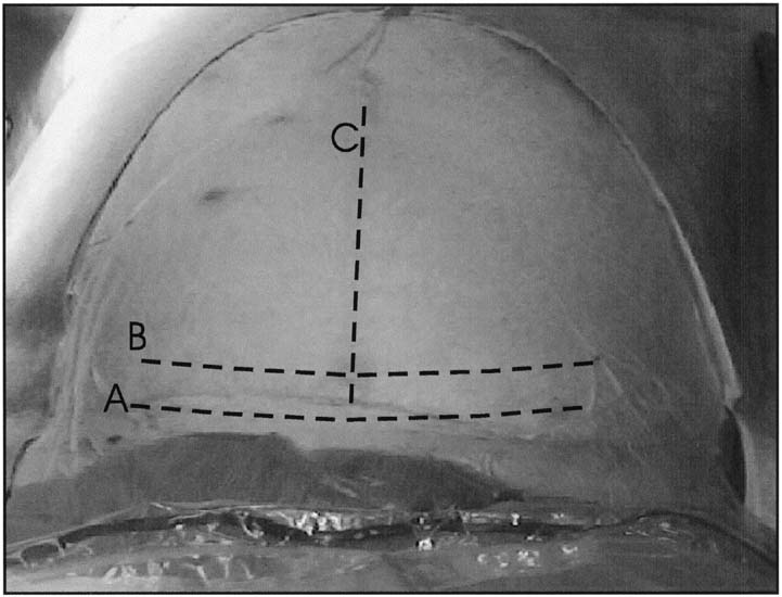
Fig. 3. Abdominal incisions. A. Pfannenstiel incision should be made in a curvilinear fashion approximately 2 cm above the pubic symphysis. B. Joel-Cohen incision should be made in a linear fashion approximately 2 to 3 cm above the traditional placement of the Pfannenstiel incision. C. Midline vertical incision should be made in the midline and extend from just below the umbilicus to just above the symphysis pubis and may be continued around the umbilicus if more exposure is necessary. D. Wiser (right paramedian) incision should be made several cm to the right side of the umbilicus and somewhat higher than the normal midline vertical incision.

TRANSVERSE INCISIONS

Although transverse incisions are commonly performed because of the widely held belief that there is a decreased incidence of wound dehiscence and incisional hernia and greater cosmetic appeal, more recent studies have not supported this belief and point to infection as the greatest risk for dehiscence regardless of incision type.30,31,32 The Pfannenstiel incision is made transversely in the maternal abdomen approximately 3 cm above the symphysis pubis and is curvilinear, with the lateral apices of the incision smiling up toward the anterior superior iliac spines (see Fig. 3). This incision is performed sharply to the level of the anterior rectus fascia. The anterior rectus fascia is then sharply incised with the scalpel in a transverse manner in the midline to expose the belly of the rectus muscle on either side of the midline. At this time, the incision in the anterior rectus fascia may be extended laterally using either the scalpel or the Mayo scissors.

Care must be taken not to cut the underlying rectus muscles. This may be accomplished by placing the Mayo scissors, with the tip up, underneath the fascia, and then sliding the scissors laterally along the length of the proposed fascial incision, opening the blades of the scissors at the proposed apex of the incision and withdrawing the scissors before closing the blades. At this point, the Mayo scissors can be used to extend the fascial incision laterally by merely pushing the blades of the scissors against the fascia. Care should be taken to avoid the transverse oblique muscle when incising the fascia. After the fascia is incised, the anterior rectus fascia can then be dissected from the underlying rectus muscles in both the cephalad and caudad directions. This is accomplished by grasping the cut edges of the fascia with a pair of strait Kocher clamps and using a combination of blunt and sharp dissection to free the muscle from the overlying fascia. This dissection allows the rectus muscles to be retracted laterally without being cut. During this dissection, care must be taken to identify and ligate or electrocoagulate the perforating vessels between the rectus muscles and the anterior fascia; this can be performed at entry, or in the event of an emergency cesarean delivery, at the time of closure. The posterior sheath consists of the fascia of the transversalis muscle and is closely opposed to the peritoneum. These tissues may be incised in either a longitudinal and transverse manner. Regardless of which manner is chosen, the entry point should be high in the operative field to avoid injury to the maternal bladder. Sharpening of the peritoneum may be performed by elevating the peritoneal membrane between two hemostats, palpating the opposing pieces of membrane for evidence of entrapped bowel, and making circumcision with a scalpel or by bluntly introducing a finger through the peritoneum at the level of the umbilicus. Once the peritoneal cavity is entered, the peritoneal incision is extended using Metzenbaum scissors to maximize surgical exposure, with care being taken to avoid inadvertent damage to the bladder or to any bowel or omentum that may be adherent to the anterior abdominal wall.

The Maylard and Cherney incisions differ from the Pfannenstiel incision in the manner in which the anterior rectus sheath and the rectus muscles are approached. With the Maylard incision, once the anterior rectus sheath is incised in a transverse fashion, the fascia is not dissected free from the underlying rectus muscles; instead, the inferior epigastric arteries are identified and ligated, and the rectus muscles are incised, usually with electrocautery to minimize bleeding. The posterior rectus sheath and the peritoneum are then incised in a transverse fashion.

The Cherney incision is performed in the same manner as the Pfannenstiel and the Maylard incisions except that the rectus fascia is not entered; instead, the rectus muscles are cut free from the symphysis pubis at their tendinous insertion and reflected superiorly. There are few if any indications for the use of this type of incision for an abdominal delivery.

The Joel-Cohen incision is performed in a transverse manner several cm above the location of a Pfannenstiel incision and is linear, not curvilinear. The fascia is not dissected off of the rectus muscles, and the peritoneum is entered transversely, as in the Maylard incision. An advantages of this type of incision include decreased operating time, however there are no maternal or fetal advantages other than speed.33,34

In the moderately obese patient, a variation of the Pfannenstiel incision is performed several cm higher than the true Pfannenstiel to avoid placing the incision in the fold created by the abdominal pannus and thereby decreasing the rate of wound complications.

VERTICAL INCISIONS

Historically, the midline vertical skin incision has been the preferred incision for cesarean section because of the speed and ease of entry into the peritoneal cavity. The decreased dissection that is required reduces intraoperative blood loss. Vertical incisions remain useful in situations such as cesarean section for fetal bradycardia and in the morbidly obese patient in whom a transverse incision may not allow for adequate exposure of the operative field. The incision is performed vertically from just below the umbilicus and extended to just above the symphysis pubis and can easily be extended around the umbilicus if exposure of the upper abdomen is required. When making a midline vertical incision, it is important to remember that the linea nigra may not represent the true midline. The incision is carried sharply down to the level of the rectus sheath, which is then incised sharply with the scalpel in a vertical direction. This incision may be completed with the scalpel or by using the Mayo scissors. The fascial edge closest to the midline is then grasped with a pair of Kocher clamps, and sharp and blunt dissections are used to separate the rectus muscles from the overlying fascia. The rectus muscles are then separated in the midline, and the peritoneum is entered vertically as described previously.

A right paramedian incision is useful in the morbidly obese patient in whom the abdominal pannus is grossly displaced when the patient is placed in the left lateral tilt position. Advantages of this incision are that once the skin has been incised, an incision that is continued perpendicular toward the floor of the operating room will incise the fascia approximately in the midline of the patient, resulting in better exposure for the delivery of the infant.

Repeat cesarean delivery account for the majority of cesareans. In patients undergoing repeat cesarean delivery, the abdominal scar may be revised at the time of repeat operation. In the case of an emergency cesarean section, any scar revision should be performed at the time of abdominal closure and not at entry. It is also important to remember that the choice of skin incision should be that which the primary surgeon believes will be most beneficial for the present operation and should not be dictated by the location of a previous scar.

Uterine Incisions

There are three standard uterine incisions that can be performed for delivery of the fetus: low transverse, low vertical, and classical (Fig. 4). The specific type of uterine incision should be determined by the primary surgeon at the time of the operation based on gestational age and lie of the fetus and any uterine anomalies.

Fig. 4. Uterine incisions. A. Low-transverse uterine incision should be made through the thin, noncontractile portion of the lower uterine segment in a curvilinear fashion. Also pictured is a low-vertical incision, which is made through the noncontractile lower uterine segment in a vertical fashion. B. J-extension of the low-transverse incision. When additional exposure to the uterine cavity is required to deliver the fetus, the low-transverse incision can be extended laterally and cephalad to increase the length of the incision without endangering the uterine arteries. C. Another option in this situation is to use a T-extension in the midline. D. The classical uterine incision is made through the contractile portion of the myometrium above the bladder reflection.

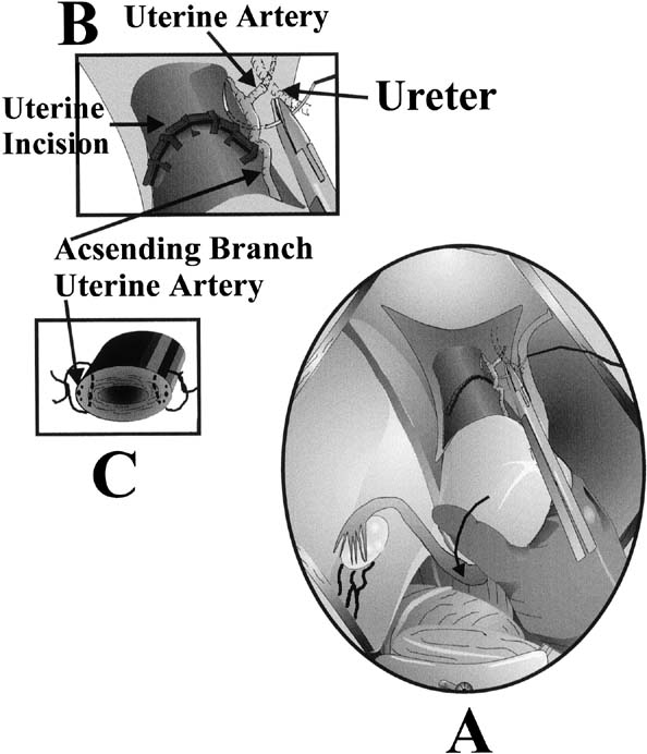
Historically, the creation of a bladder flap was advocated before making any uterine incisions. More recently, randomized controlled trials have noted that the omission of the bladder flap provides short term advantages such as reduction of operating time and incision-delivery interval, reduced blood loss and need for analgesics. Long-term effects remain to be evaluated.35 When developing a bladder flap, a segment of loose areolar peritoneum can be visualized at the area where the bladder is adjacent to the lower uterine segment. The peritoneum is grasped with a pair of forceps, elevated, and then incised transversely with Metzenbaum scissors, with care being taken not to extend the incision laterally into the vascular broad ligament. Next, the inferior portion of the incision is elevated, and careful blunt and sharp dissection is used to separate the posterior wall of the bladder from the lower uterine segment. This dissection serves two purposes: it allows better access to the lower uterine segment and it allows the bladder to be retracted out of the operative field. Before making the uterine incision, the surgeon should also identify the round ligaments to properly orient the degree of dextrorotation of the uterus and to evaluate for the presence of any myomas or other malformations that might affect the choice and/or placement of the incision.

The standard low-segment transverse incision accounts for 90% of all uterine incisions.28 It is initiated sharply in the lower uterine segment, perpendicular to the long axis of the uterus. This incision is made sharply with the scalpel in the midline and performed down to the level of the fetal membranes, with care being made not to incise the membranes. This incision is then extended laterally using either blunt dissection with the fingers or bandage scissors (Fig. 5). There was thought to be no difference between the two methods in amount of blood lost or in the rate of extension of the incision into the lateral uterine vessels when they were compared and correlated by the stage of labor.36 However, a recent investigation revealed a greater risk of subsequent blood transfusion in woman whose incision was extended sharply compared to those extended bluntly.37 When blunt dissection is used, an upward curve of the incision may be created by the surgeons placing their thumbs on the patient's anterior superior iliac spines and index fingers in the uterine incision. By keeping the hand in this position, the incision is pulled open in an arc.

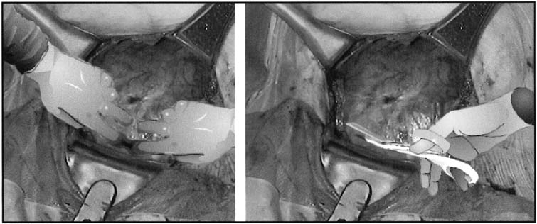
Fig. 5. Extension of the lower uterine incision may be accomplished either by inserting fingers into the uterine cavity and bluntly stretching the myometrial incision in a curvilinear fashion or by sharply cutting the lower uterine segment with bandage scissors. When the uterus has a poorly developed lower uterine segment, using bandage scissors is often preferable.

Intentional extension of the low-transverse incision is necessary in 1% to 2% of cases.38 Typically, the extension of the low transverse incision is performed by creating a low vertical incision in the midline, T-ing the uterine incision, or creating a vertical incision at the lateral aspect of the uterine incision, a J-extension. These extensions are commonly performed for malpresentations, poorly developed lower uterine segment, or deep transverse arrest.38 When performed, extensions of the low-transverse incision are associated with increased incidence of maternal blood loss, broad ligament hematoma, and uterine artery laceration compared with low-segment transverse incisions that do not require extension.

The low-vertical uterine incision is made parallel to the longitudinal axis of the uterus in the midline, with care being taken to remain below the contractile portion of the uterus and within the thin lower uterine segment. Other than the direction of the incision, technical aspects are carried out as described for the low-transverse uterine incision. Studies have shown that there is no increased risk of uterine rupture in patients with this type of incision compared with the low-segment transverse incision as long as the incision remains primarily in the thin lower uterine segment.39

A classical uterine incision is made by incising the uterus parallel to the longitudinal axis of the uterus through the contractile portion of the myometrium. Indications for classical uterine incision include situations in which the lower uterine segment is not adequately developed to accommodate a low-transverse or a low vertical incision; cases of abnormal fetal lie such as back-down transverse lie, in which the low-transverse or low-vertical incision will not allow the operator adequate access to the fetus for manipulation and delivery, or when myomas or uterine abnormalities distort the uterus in such a way as to make a low transverse incision inadvisable.

Delivery of the Fetus

After the uterine incision has been made, the fetal membranes, if still intact, are ruptured with an Allis clamp. If the fetus is in a noncephalic presentation, leaving the membranes intact until the fetal feet or head can be moved into the uterine incision will increase the ease of delivery. When the fetus is in a cephalic presentation, delivery is performed by the surgeons placing their dominant hand into the uterine cavity and elevating the fetal head into the uterine incision (Fig. 6). If the fetus is not in an occiput anterior position, rotating the head into this position will allow the fetal neck to extend around the upper portion of the incised myometrium and more closely mimic the cardinal movements of vaginal delivery. When the fetal head is impacted in the maternal pelvis, such as in deep transverse arrest, there are a number of options to assist with delivery of the fetal head. The surgeon can place a hand in the lower uterine segment in the standard fashion to cup and then disengage the fetal head. Care must be taken by the surgeon not to flex the wrist, because this often causes extension of the uterine incision caudally toward the bladder and vagina. If this does not work, an assistant can place a sterile, gloved hand into the vagina from the introitus and disengage the fetal head from below (Fig. 7). Another option is for the surgeon to the dominant hand between the lower uterine segment and the reflected bladder and attempt to disengage the head by cupping it through the lower uterine segment. It is our experience that care must be taken with this maneuver to ensure that the bladder is not damaged by inadvertent blunt cystotomy.

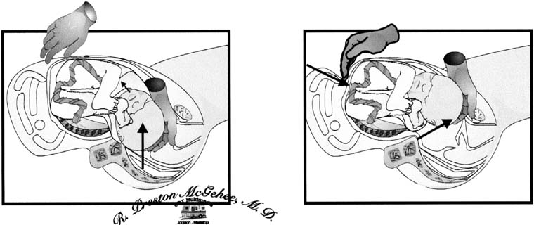
Fig. 6. Extraction of the fetal head. The surgeon's dominant hand is placed into the uterine incision so that the back of the hand is against the inside of the lower uterine segment and the fingers cup the fetal head. Firm, gentle traction is used to elevate the fetal head toward the incision. The fetal head may then be rotated to an occiput anterior position and delivered through the uterine incision with the assistance of fundal pressure.

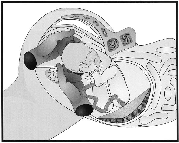
Fig. 7. Disimpaction of the fetal head. When the fetal head has descended so far into the vagina that extraction of the fetal head is difficult, having an assistant place a gloved hand into the vagina and elevate the fetal head from below can increase the ease of delivery and decrease the trauma to the lower uterine segment and vagina.

Once the fetal head is at the uterine incision, mild fundal pressure by the first assistant will encourage the expulsion of the fetal head from the uterus. At this point, the nares and mouth of the fetus should be suctioned and, after checking for and reducing any nuchal cord, the fetal body is delivered by standard maneuvers as in a vaginal delivery. After the infant is delivered, the cord is doubly clamped and cut, and the infant is handed to the personnel who have been assigned to care for the newborn. It is important to remember that if the newborn requires resuscitation, the obstetrician is responsible for its care as well as the care of the mother. Qualified personnel should be available to assume care of the newborn.

Attention is now turned to the delivery of the placenta. The delivery of the placenta may be accomplished either by manual extraction or by awaiting spontaneous delivery. Spontaneous delivery of the placenta, when assisted with uterine massage and gentle traction on the umbilical cord, is associated with a lower rate of postpartum endomyometritis and maternal blood loss compared with manual extraction.40,41,42 Once the placenta has been delivered, the uterus may be either exteriorized or left in situ to be repaired. Blood loss is not significantly different with either method.42 Exteriorization of the uterus does allow for better visualization of the adnexal structures and increases the ease with which tubal ligation can be performed.

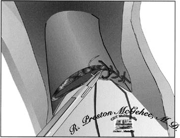
Fig. 8. Single-layer repair of the low-transverse uterine incision. To obtain optimal hemostasis of the incision in a single layer, the surgeon should be careful to include all layers of incised myometrium while taking care to avoid including excess decidua and serosa.

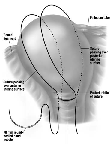
Uterine closure may be performed with either a single- or double-layer closure technique. Single-layer closure using a running locking stitch (Fig. 8) has been shown to be associated with decreased operative time and fewer additional hemostatic sutures. A large Canadian study found a four-fold increase in the risk of uterine rupture in woman who had a single layer closure in their previous pregnancy.43,44,45,46,47

Chromic catgut has been the suture of choice for closure of the uterine incision by many obstetricians for a number of years. However, the use of a synthetic absorbable suture, such as polyglycolic acid or polyglactin, has several advantages over the use of catgut. The method of absorption of catgut suture is by phagocytosis, and this results in significantly more inflammation than the absorption of synthetic sutures by hydrolysis.48 The decreased inflammation associated with synthetic absorbable sutures, as well as the increased time interval to the loss of suture strength, are both advantageous in this situation.

After the uterus is closed and has been returned to the peritoneal cavity, irrigation can be employed. Routine irrigation in low-risk populations does not reduce intrapartum or postpartum maternal morbidity.49 Next, attention should be turned to ensuring that the operative field is hemostatic, with special attention given to the uterine incision and bladder flap, if these have been previously placed on tension because of exteriorization of the uterus, and to the rectus muscles. Hemostasis may be achieved by either suture ligation or electrocoagulation of bleeding points. There is no advantage to closure of the visceral or parietal peritoneum. When repaired with suture, the peritoneum undergoes more inflammation and scarring in animal models.50 Operating time and postoperative analgesia requirements are reduced in patients who do not undergo closure of the visceral and parietal peritoneum. There is also a decrease in adhesions found at repeat operation when the visceral and parietal peritoneum is not closed.51

Fascial closure in a Pfannenstiel incision is performed in a single layer with a synthetic absorbable suture. In patients who have undergone more than one laparotomy through the same scar, or in patients who are at increased risk for fascial separation or dehiscence such as diabetic patients or patients who are on chronic corticosteroids, the use of a synthetic delayed absorbable suture such as polydioxanone may be preferable because of its ability to maintain suture strength for a longer period of time.52 For the closure of a vertical fascial incision, a continuous running delayed absorbable suture has been shown to be as effective as the Smead-Jones closure and to reduce operating time without increasing morbidity. Whenever sutures are placed within the fascia, it is important to remember that a 10-mm zone of collagenolysis occurs surrounding the incision; therefore, sutures should be placed more than 1 cm from the fascial edge to achieve maximal wound strength.53

The subcutaneous tissue may be closed with an absorbable suture or simply reapproximated by closure of the skin. Closing this layer has not been associated with decreased rates of superficial wound disruption in several studies.54 Skin closure may be accomplished by either a subcuticular stitch or staples. Subcuticular stitch has been associated with less immediate postoperative pain and more cosmetically appealing at 6 weeks when compared to the stapling device.55

Postoperative Care

There is little literature to support any specific postoperative regimen in postcesarean patients; however, common sense and extrapolation of data from other postlaparotomy patients allow for the development of a rational plan of care. Most cesarean sections are relatively uncomplicated, and in these patients, care should be similar to that given after a vaginal delivery.

In the first hour after cesarean section, the patient should be monitored closely in a recovery area where urine output, pulse, blood pressure, respirations, and any evidence of bleeding can be closely observed; if the patient remains stable and without complication, she may then be transferred to the postpartum ward.

Once any nausea and vomiting has abated, the patient should be encouraged to take fluids orally. This may be followed by oral intake of solid food as soon as the patient feels that she is ready; this should occur no later than the first postoperative day. Early institution of feeding in the postsurgical patient with minimal intraoperative bowel manipulation does not increase the incidence of postoperative ileus.56

Early ambulation should also be encouraged. Getting the patient out of bed as soon as regional anesthesia has worn off or as soon as she has recovered from general anesthesia will decrease the incidence of pulmonary complications such as atelectasis and pneumonia and the incidence of thrombotic complications. This will also facilitate the removal of bladder catheters, therefore decreasing the incidence of catheter-associated urinary tract infections. In the uncomplicated patient with adequate urine output, the catheter should be removed no later than the first postoperative day. Encouragement of deep breathing and coughing with the use of incentive spirometry will also help prevent collapse of alveoli in the lung and resulting infection.

Routine laboratory studies are probably unnecessary in most postcesarean patients who have no unexpected symptoms. However, a single hemoglobin determination on postoperative day one or two is probably reasonable to screen for significant anemia. Most postpartum patients with asymptomatic anemia respond well to oral iron therapy.

The wound should be cared for in the standard manner, with occlusive dressings removed on the first postoperative day and the wound examined daily during the hospitalization for evidence of infection, seroma, or hematoma. Skin staples can be removed on the second or third postoperative day with Pfannenstiel incisions and at the fifth to seventh postoperative day with vertical incisions. The placement of SteriStrips after staple removal may help maintain skin edge approximation with earlier removal.

The patient may be discharged when she is able to care for herself and her newborn. Many patients are ready to leave the hospital by postoperative day two or three. Discharge instructions should include patient education concerning expectations on activity level, lochia, breastfeeding or milk suppression, contraception, and newborn care.

Back to Top
SPECIAL SITUATIONS

Vaginal Birth After Cesarean Section

VBAC has become less common in the United States, and between 1996 and 1999 the rate decreased by 17 % to 23.4 %.7 The VBAC rate is defined as the number of vaginal births to women with a previous cesarean section per 100 deliveries to women who had a previous cesarean delivery. In 1999, the United States VBAC rate was 23.4.7 Since the early 1980s, a number of reports have shown that vaginal delivery after a previous low-transverse or low-vertical cesarean section is safe for both the mother and the fetus.39,60,61 However, more recently the safety of VBAC has come under closer scrutiny. Despite literature supporting the practice, most generated in university and tertiary-level centers, there have been no randomized trials to prove that maternal and fetal outcomes are improved by VBAC over repeat cesarean section. There have even been reports to indicate that there are maternal and infant complications associated with an unsuccessful trial of labor.57,58,59 These facts demonstrate the need for a continued study in this area.

In properly selected patients, the success rate for trial of labor is between 60% and 80%,60,62,63,64 which is not grossly different from the vaginal delivery rate of the entire obstetric population in the United States in recent years. A successful trial of labor is less likely in patients who have not had a previous vaginal delivery, have a history of dysfunctional labor, have nonreassuring fetal surveillance, undergo induction of labor, have a fetus with evidence of intrauterine growth restriction, or have fetal–pelvic disproportion.65

Several authors have attempted to predict which patients are more likely to undergo successful VBAC by various historic parameters and the physical examination at the time of admission for delivery. Jakobi and colleagues66 found that previous cesarean section performed for a nonrepetitive indication such as breech presentation, history of a previous successful VBAC, station of −1 or more, unruptured membranes at admission, and dilation of 4 cm or more at admission were all positively correlated with increased likelihood of successful VBAC. A history of previous cesarean section for arrest of labor was significantly associated with an increased risk of unsuccessful trial of labor. Using these criteria retrospectively, the authors would have correctly predicted the success of a trial of labor in more than 94% of candidates but would have correctly predicted failure of trial of labor in only 33.3% of candidates.

Flamm and Geiger67 examined similar data to develop a scoring system in an attempt to predict the success of trial of labor. These authors found that maternal age younger than 40 years, indication other than failure to progress, cervical effacement of more than 75% on admission, and cervical dilation of more than 4 cm at admission were all significantly correlated with increased success of trial of labor. When these factors were weighted and placed in a scoring system in an attempt to predict the success of attempted VBAC, the authors found that as the number of these factors increased, the likelihood of successful trial of labor increased. Patients with only one or two of these characteristics had a 49% to 59% success rate, whereas patients with four or more of these characteristics had a greater than 90% success rate. The ability to more accurately predict the likely success of trial of labor is clinically useful, because there is increased maternal morbidity is associated with a failed trial of labor.68 These women have an increased risk of operative injury, infectious morbidity, and uterine rupture.

Morbidity associated with VBAC has been well established. Uterine rupture occurs in approximately .3% to 1.5% of trials of labor with a low-transverse scar.69,70,71 The uterine rupture rate is 1% to 7% with a low-segment vertical scar. The incidence of uterine rupture may be increased in patients with a previous single-layer closure.72,36 However, in patients with a previous classic uterine incision, the risk of uterine rupture may be as high as 9%, with one third of these occurring before the onset of clinical labor.74 In patients for whom documentation of the type of previous uterine incision is not available, there is no increased risk of uterine rupture.73 More than one previous low-segment uterine incision is not a contraindication to trial of labor.75

In the event of uterine rupture, fetal mortality and morbidity is excessive. When a classic uterine incision ruptures, the fetal mortality is in excess of 50%, compared with 12% in the event of rupture of a prior low transverse incision.76

In evaluating a patient as a candidate for trial of labor, it is important to consider the patient's history and physical examination, the patient's wishes concerning elective repeat cesarean section versus trial of labor after appropriate counseling, and the environment where the trial of labor is to be performed. Most studies that established the safety of VBAC were conducted in tertiary care centers or teaching institutions.75 The availability of operating room staff and anesthesia support in the event of uterine rupture or nonreassuring fetal monitoring in these settings may vary considerably in private and small community hospitals.

After the patient has been appropriately counseled and has consented to a trial of labor, labor management should not significantly differ from the management of a patient with an unscarred uterus. Cervical ripening, oxytocin augmentation,73,77 and regional anesthesia78,79,80,81 may be used as clinically indicated with close monitoring of the patient.

Population-based studies have demonstrated the increased morbidity (uterine rupture rates of 24.5/1000) associated with the use of prostaglandin cervical ripening agents in a scarred uterus. This prompted ACOG to release a committee opinion discouraging their use for the purpose of induction during VBAC attempts.82,83

Perimortem Cesarean Section

One of the first indications for cesarean section was for the delivery of the fetus in the case of maternal death. Currently, the performance of a rapid cesarean delivery in the event of sudden maternal cardiac arrest can be lifesaving for the fetus. In the event of maternal cardiac arrest with a viable fetus, cesarean section should be initiated within 4 minutes of cardiac arrest, with the goal of delivering the fetus within 5 minutes of onset of cardiac arrest. This has been shown to allow neurologically intact survival of all infants delivered.84 Delivery after 5 minutes have elapsed results in increasing neurologic sequelae in surviving infants but may still be beneficial to the majority of infants up until 15 minutes after cardiac arrest. However, the goal should always be delivery of the neonate within 5 minutes of the loss of maternal cardiac function. Immediate abdominal delivery accomplishes two goals in this setting: the removal of the fetus from what has become an extremely hostile uterine environment and the increase of maternal blood return to the heart by relieving uterine pressure on the maternal inferior vena cava. The perimortem operation should be performed only when the fetus is suspected to be clinically viable. This procedure should benefit both the mother and the fetus. As with any cardiac resuscitation situation, the likelihood of good maternal and fetal outcome decreases with increased interval to delivery time and increased time to the return of spontaneous circulation.85,86 It is important to remember that the performance of a perimortem cesarean delivery outside of an operating room under nonsterile conditions is not likely to negatively impact maternal survival, because survival to discharge after a witnessed, in-hospital, cardiac arrest is only 3%.87 Performance of this operation on a patient who is unstable but not in cardiac arrest should never be undertaken, because maternal well-being always takes precedence over fetal well-being.

Back to Top
COMPLICATIONS

Maternal Mortality

As anesthesia and operative techniques have improved, cesarean section has become an increasingly safe and common procedure; however, the obstetrician must always bear in mind that the abdominal delivery of an infant is still a major operative procedure and can be associated with significant mortality and morbidity.

Maternal mortality after cesarean section has been estimated to be between 5.81 and 6.1 per 100,000 procedures.88,89 Between 20% and 50% of these deaths are attributable to the cesarean delivery, with the remainder being the result of complications that led to the cesarean section.

In general, the complications associated with cesarean section are similar to those observed after any laparotomy, with the exception of an increased incidence of endomyometritis. Complications may be divided into those encountered intraoperatively and those encountered postoperatively.

Common intraoperative complications include uterine hemorrhage and injury to either the urinary or gastrointestinal tract. Uterine hemorrhage can be caused by atony, lacerations, or retained placenta. Because the uterus usually can be examined at the time of cesarean delivery, determining which of these entities is the cause of the hemorrhage is not as difficult as it is when hemorrhage occurs after a vaginal delivery. Uterine atony continues to be the major cause of hysterectomy at the time of cesarean section.90,91 Having a rational, well-thought-out approach in dealing with potential uterine atony will allow the obstetrician to address this complication in an effective and timely manner. The first step should be to repair the uterine incision. The first assistant should pull the uterine fundus toward the patient's head during this repair to place the uterine arteries on tension and thereby decrease blood flow to the uterus. Exterioration of the uterus for repair is commonly advocated to facilitate exposure and place the uterine arteries on stretch. Magann and colleagues at the University of Mississippi have recommended in situ closure to minimize the trauma to the adnexa and shorten the operative time. The optimal site of uterine closure remains unknown. As uterine repair is being performed, oxytocin (40 U/L normal saline) should be rapidly administered intravenously to encourage myometrial contraction. If bleeding persists, bimanual pressure is applied to the uterus, and the uterus is vigorously massaged. If hemorrhage has not abated at this time, .25 mg of 25-methyl prostaglandin F2a (Hemabate) is injected intramuscularly or directly into the uterus. This may be repeated every 15 minutes as needed up to eight doses. Hemabate should not be used in patients with asthma. Methergine .20 mg injected intramuscularly can also be used in nonhypertensive patients. Misoprostol 800 to 1000 microgram should be considered early and rectally administered. During this process, attention should be given to the patient's hemodynamic status; blood loss can be rapid and excessive because blood flow to the gravid term uterus is approximately 700 mL per minute. Fluid resuscitation and blood transfusion should be instituted as clinically indicated. If manual compression of the uterus and medical intervention are unsuccessful, uterine artery ligation should be performed bilaterally, with sutures placed to obliterate both the ascending uterine artery at the level of the lower uterine segment and its anastomosis with the ovarian artery at the uterine cornua (Fig. 9). This will control bleeding from an atonic uterus in 75% of cases.92 Localization of the uterine artery can be facilitated by careful palpation of its course along the lateral edge of the uterus. The surgeon should then pull the engorged uterine veins laterally into the broad ligament and away from the operative field to avoid laceration of these veins during suture placement. Isolating the uterine artery with a Babcock clamp is often helpful in these cases. Care should be taken to avoid incorporating the ureter in the ligature. In cases that do not respond to bilateral uterine artery ligation, bilateral hypogastric artery ligation is no longer recommended because it is successful in less than 50% of patients and training opportunities are not readily available, thus an experienced operator is unlikely. A new surgical technique introduced, the B Lynch has proven very successful in preventing subsequent hysterectomy (Fig. 10). In patients who do not respond to these management strategies, hysterectomy is appropriate and life-saving.93

Fig. 9. O'Leary stitch (uterine artery ligation). While placing the broad ligament on traction to displace the uterine veins laterally, the uterine artery is palpated and isolated. A suture is then placed below the laceration to the uterine artery to incorporate the artery with the myometrium. If necessary, a second stitch may be placed above the incision in the same manner.

Fig. 10. Use a 70- to 80-mm round-bodied hand needle with mounted number-2 plain or chromic catgut. With the bladder displaced inferiorly, the first stitch is placed 3 cm below the lower cesarean incision on the patient's left side and threaded through the uterine cavity to emerge 3 cm above the upper incision margins, approximately 4 cm from the lateral border of the uterus. Now, carry the suture on the outside of the uterus over the top and to the posterior side. The suture should be more or less vertical and lying approximately 4 cm from the cornua. It does not tend to slip laterally toward the broad ligament because the uterus has been compressed and the suture milked through, ensuring that proper placement is achieved and maintained. The suture is placed exactly the same way as it was on the left side; that is, 3 cm above the incision, 4 cm from the lateral side of the uterus through the top of the incision, into the uterine cavity, and then again back through 3 cm below the incision.

In cases of hemorrhage not caused by uterine atony, careful exploration of the uterus for possible retained placental fragments and exploration of the operative field for unrecognized lacerations should be performed. Placental fragments may be removed manually or may require the use of a sharp curette. Areas of placental adherence should be examined for evidence of placenta accreta. Genital tract lacerations should be identified, isolated, and closed in a hemostatic manner.

Urinary Tract Injuries

Injury to the urinary tract is a relatively rare complication of cesarean delivery. The incidences of bladder and ureteral injury are .3% and .1%, respectively.94 Bladder injuries are more common with the use of Pfannenstiel incision and history of previous cesarean section. The most common site for bladder injury during cesarean delivery is at the dome of the bladder. Laceration of the bladder should be evaluated by first ensuring that the trigone and ureters are not involved. This may be accomplished by direct visualization of the ureters through a cystotomy incision. If the trigone is not involved and the ureters are functioning, the cystotomy can be closed in two layers. Whenever there is a possibility of inadvertent cystotomy at the time of cesarean section, this can be evaluated by distending the bladder with sterile milk through the Foley catheter and observing the operative field for the appearance of the milk.

Ureteral injury is less common than injury to the bladder. If there is concern during the operative procedure that the ureter has been compromised, the abdomen should not be closed until this possibility has been thoroughly evaluated and excluded or the injury has been identified and corrected. Evaluation of the ureters can be performed by performing a cystotomy in the dome of the bladder and passing an 8-French ureteral stent retrograde through the ureter to the kidney. If ureteral stents are not immediately available, most labor and delivery suites have 8-French pediatric feeding tubes on the neonatal resuscitation cart, and these can be used instead. Another method of evaluating ureteral integrity is to inject 5 to 10 mL of indigo carmine intravenously and observe for the appearance of the dye at the ureteral orifice. Simply observing the appearance of the dye in the Foley catheter is inadequate to evaluate ureteral patency, because this only guarantees the patency of one ureter. Repair of ureteral injuries is performed as indicated by the level of the injury. Ureteroureteral anastomosis or ureteral reimplantation with the placement of ureteral stents is the standard. Some minor injuries may be managed by the placement of ureteral catheters alone. Consultation intraoperatively with the appropriate specialist is warranted.

Gastrointestinal Tract Injury

Injury to the bowel at the time of cesarean section is exceedingly rare. An incidence of less than .1% has been reported.95 This low incidence is caused by the displacement of the bowel out of the operative field by the enlarged, gravid uterus. The risk of bowel injury is increased in patients with previous abdominal surgery or intraabdominal adhesions. Injury is usually obvious because of the appearance of bowel contents in the surgical field. These injuries should be quickly identified and isolated to minimize contamination of the peritoneal cavity. Injury to the small bowel can be primarily repaired with a two-layer closure using silk or delayed absorbable suture. The closure should be performed at 90 degrees to the bowel lumen to decrease its constriction. Larger lacerations of the small bowel or multiple lacerations may require resection of a length of bowel. Injuries to the large bowel must be carefully evaluated. In the patient who has not had bowel preparation, large bowel lacerations of less than 1 cm may be primarily repaired.96 Colostomy is indicated for more extensive large bowel injury with fecal contamination. Broad-spectrum antibiotics should be administered in cases of large bowel injury.

Wound Infections

By definition, any cesarean section is a clean, contaminated operation. Wound infections occur at a rate of approximately 7% after cesarean section when prophylactic antibiotics are not given;94 this incidence is reduced to 2% with the use of prophylactic antibiotics.97 Wound infections that occur after cesarean section include endomyometritis, pelvic abscess, incisional abscess, and wound cellulitis. The antibiotic of choice for each infection depends on the location of the infection and the suspected pathogen. Antibiotic therapy should be instituted empirically and adjusted as needed based on culture results. For pelvic abscesses, broad-spectrum antibiotics including anaerobic coverage are required; for superficial wound infections, simply opening the incision and draining the infectious source usually alleviates the problem in patients who do not have signs of systemic infection. Superficial wound cellulitis can usually be treated using penicillinase-resistant penicillin.

Endomyometritis

Endomyometritis complicates up to 80% of cesarean sections performed after the membranes have been ruptured for more than 6 hours in patients who are not administered antibiotic prophylaxis98 and 30% in patients with intact membranes. The incidence has been shown to be high in patient populations of lower socioeconomic status,99 in patients that have had six or more vaginal examinations during labor, and in patients with longer duration of rupture of membranes.100 The rate of uterine infection can be reduced to 5% or less with the use of prophylactic antibiotics given at the time of cord clamp.101 A single dose of a first-generation cephalosporin is relatively inexpensive and effectively decreases the infection rate.

Back to Top
CONCLUSION

Over the past several decades the incidence of cesarean delivery has increased dramatically. Although the operation continues to become safer, the incidence of maternal mortality and morbidity is still higher than that of a vaginal delivery. Continued efforts on the part of the obstetrician must be made to ensure that cesarean deliveries are not performed for inappropriate indications and that every effort is made to allow the patient to deliver vaginally when fetal and maternal status are reassuring. Recently, serious debate has arisen over the autonomy of the patient and the ability to request an elective cesarean section. The arguments on both sides of the issue are provocative and center on the low morbidity of cesarean delivery compared with vaginal delivery. Proponents of elective cesarean delivery point to the possibility of pelvic floor dysfunction leading to future urinary and fecal incontinence. This could be a Pandora's box. We should continue to respect our patients' autonomy while at the same time heed to our creed, first do no harm. Continuing research is required to more appropriately evaluate the techniques of the cesarean operation and the safety of elective primary cesarean delivery.

Back to Top
References

1. Gabert HA, Bey M: History and development of cesarean operation. Obstet Gynecol Clin North Am 15:591, 1988

2. Speert H: A Pictorial History of Gynecology and Obstetrics. Philadelphia, Davis, 1973

3. Katz VL, Cefalo RC: History and evolution of cesarean delivery. In: Phelan JP, Clark SL, (eds): Cesarean Delivery. New York, Elsevier, 1988

4. Boley JP: The history of caesarean section. CMAJ 145:319, 1991

5. Horley JMG: Cesarean section. Clin Obstet Gynecol 7:529, 1980

6. Sander M: Speaking before the German Gynecology Association 1885. Am J Obstet Dis Women Child 19:883, 1886

6A. Sanger M: My work in reference to the cesarean operation. Am J Obstet Dis Women Child 20:593, 1887

7. Menacker F, Curtin SC: Trends in cesarean birth and vaginal birth after previous cesarean, 1991–1999. Natl Vital Stat Rep 49:1-16, 2001

8. Menard MK: Cesarean rates in the United States. The 1990s Obstet Gynecol Clin North Am 26:275-286, 1999

9. Clark SC, Taffel SM: State variation in rates of cesarean and VBAC delivery: 1989 and 1993. Stat Bull Metrop Insur Co 77:28-36, 1996

10. Notzon FC, Cnattingius S, Bergsjo P et al: Cesarean section delivery in the 1980s: International comparison by indication. Am J Obstet Gynecol 170:495-504, 1994

11. Norton FC: International differences in the use of obstetric interventions. JAMA 283:3286, 1990

12. Pollard JK, Capeless EL: Cesarean deliveries at a university hospital: Analysis of rates and indications. Am J Perinatol 14:245, 1997

13. Ziadeh SM, Sunna FI: Decreased cesarean birth rates and improved perinatal outcome: A seven-year study. Birth 22:144, 1995

14. US Department of Health and Human Services: Healthy Children 2000. DHHS Pub. No. HRSA-M-CH 91–2. Washington, DC: U.S. Government Printing Office, 1991

15. Hogle KL, Kilburn L, Hewson S et al: Impact of the international term breech trial on clinical practice and concerns: A survey of centre collaborators. J Obstet Gynaecol Can 25:14-16, 2003

16. American College of Obstetrics and Gynecology. Technical Bulletin No. 218. December 1995

17. Renou P, Chang A, Anderson I et al: Controlled trial of fetal intensive care. Am J Obstet Gynecol 126:470, 1976

18. Kelso AM, Parsons RJ, Lawrence GF et al: An assessment of continuous fetal heart rate monitoring in labor. Am J Obstet Gynecol 131:526, 1978

19. Haverkamp AD, Orleans M, Langendoerfer S et al: A controlled trial of the differential effects of intrapartum fetal monitoring. Am J Obstet Gynecol 134:399, 1979

20. MacDonald D, Grant A, Sheridan-Pereira M et al: The Dublin randomized controlled trial of intrapartum fetal heart rate monitoring. Am J Obstet Gynecol 152:524, 1985

21. Leveno KJ, Cunninhgam FG, Nelson S et al: A prospective comparison of selective and universal electronic fetal monitoring in 34,995 pregnancies. N Engl J Med 315:615, 1986

22. Weiss PM, Balducci J, Reed J et al: Does centralized monitoring affect perinatal outcome. J Matern Fetal Med 6:317, 1997

23. East CE, Colditz PB, Begg LM et al: Update on intrapartum fetal pulse oximetry. Aust N Z J Obstet Gynaecol 42:119-124, 2002

24. Bickell NA, Zdeb MS, Applegate MS et al: Effect of external peer review on cesarean delivery rates: A statewide program. Obstet Gynecol 87:664, 1996

25. American College of Obstetrics and Gynecology. Criteria Set Number 13. December 1995

26. American College of Obstetrics and Gynecology. Committee Opinion No. 98. September 1991

27. Rayburn WF, Schwartz WJ: Refinements in performing a cesarean delivery. Obstet Gynecol Surv 51:445, 1996

28. Alexander JW, Fisher JE, Boyajiam M et al: The influence of hair removal methods on wound infections. Arch Surg 118:347, 1983

29. Greenmall MJ, Evans M, Pollack AV: Mid-line or transverse laparotomy? A random controlled clinical trial period Br J Surg 67:188, 1980

30. Mowat J, Bonnar J: Abdominal Wound dehiscence after cesarean section. BMJ 2:256, 1971

31. Hendrix SL, Schimp V, Martin J et al: The legendary superior strength of the Pfannenstiel incision: a myth? Am J Obstet Gynecol 182:1446-1455, 2000

32. Joel-Cohen S: Abdominal and Vaginal Hysterectomy: New Techniques Based Upon Time and Motion Studies. 2nd ed.. Philadelphia, JB Lippincott, 1977

33. Franchi M, Ghezzi F, Raio L et al: Joel-Cohen or Pfannenstiel incision at cesarean delivery: does it make a difference? Acta Obstet Gynecol Scand 81:1040-1046, 2002

34. Hohlagschwandtner M, Ruecklinger E, Husslein P et al: Is the formation of a bladder flap at cesarean necessary? A randomized trial Obstet Gynecol 98:1089-1092, 2001

35. Magann EF, Chauhan SP, Bufkin L et al: Intraoperative haemorrhage by blunt versus sharp expansion of the uterine incision at caesarean delivery:a randomized clinical trial. Br J Obstet Gynecol 109:448-452, 2002

36. Rodriquez AI, Porter KB, O'Brien WF: Blunt versus sharp expansion of the uterine incision in low-segment transverse cesarean section. Am J Obstet Gynecol 152:971, 1985

37. Boyle JG, Gabbe SG: T and J Vertical extensions in low transverse cesarean births. Obstet Gynecol 87:238, 1996

38. Martin JN Jr, Perry KG Jr, Roberts WE et al: The case for trial of labor in the patient with a prior low-segment vertical cesarean incision. Am J Obstet Gynecol 177:144, 1997

39. Atkinson M, Owen J, Wren A et al: The effect of manual removal of the placenta on post-cesarean endometritis. Obstet Gynecol 87:99, 1996

40. McCurdy CM Jr, Magann EF, McCurdy CJ et al: The effect of placental management at cesarean delivery on operative blood loss. Am J Obstet Gynecol 167:1363, 1992

41. Khan GQ, John IS, Wani S et al: Controlled cord traction versus minimal intervention techniques in delivery of the placenta: A randomized controlled trial. Am J Obstet Gynecol 177:770, 1997

42. Enkin MW, Wilkinson C: Single versus two layer suturing for closing the uterine incision at caesarean section. Cochrane Database Syst Rev 2:CD000192, 2000

43. Hauth JC, Owen J, Davis RO: Transverse uterine incision closure: One versus two layers. Am J Obstet Gynecol 167:1108, 1992

44. Tucker JM, Hauth JC, Hodgkins P et al: Trial of labor after a one or two-layer closure of a low transverse uterine incision. Am J Obstet Gynecol 168:545, 1993

45. Chapman SJ, Owen J, Hauth JC: One-versus two layer closure of a low transverse cesarean: The next pregnancy. Obstet Gynecol 89:16, 1997

46. Bujold E, Bujold C et al: The impact of a single-layer or double-layer closure on uterine rupture. Am J Obstet Gynecol 186:1326-1330, 2002

47. Stone IK: Suture materials. Clin Obstet Gynecol 31:712, 1988

48. Harrigill KM, Miller HS, Haynes DE: The effect of intraabdominal irrigation at cesarean delivery on maternal morbidity: a randomized trial. Obstet Gynecol 101:80-85, 2003

49. Elkins THE, Stovall THG, Warren J et al: A histologic evaluation of peritoneal injury and repair: Implications for adhesion formation. Obstet Gynecol 70:225, 1987

50. Wilkinson CS, Enkin MW: Peritoneal non-closure at caesarean section. Cochrane Database Syst Rev 2:CD000163, 2000

51. Ray JA, Doddi N, Regula D et al: Polydioxanone (PDS), a novel monofilament synthetic absorbable suture. Surg Gynecol Obstet 153:497, 1981

52. Wound healing: Techniques and materials. In Visscher HC, (ed): Precis V: An Update in Obstetrics and Gynecology. Washington, American College of Obstetrics and Gynecology, 1994

53. Magann EF, Chauhan SP, Rodts-Palenik S et al: Subcutaneous stitch closure versus subcutaneous drain to prevent wound disruption after cesarean delivery: A randomized clinical trial. Am J Obstet Gynecol 186:1119-1123, 2002

54. Frishman GN, Schwartz T, Hogan JW: Closure of Pfannenstiel skin incisions. Staples vs. subcuticular suture J Reprod Med 42:627-630, 1997

55. Mangesi L, Hofmeyr GJ: Early compared with delayed oral fluids and food after caesarean section. Cochrane Database Syst Rev 3:CD003516, 2003

56. Miller DA, Diaz FG, Paul RH: Vaginal birth after cesarean: A 10-year experience. Obstet Gynecol 84:255, 1994

57. Van der Walt WA, Cronje HS, Bam RH: Vaginal delivery after one cesarean section. Int J Gynaecol Obstet 46:271, 1994

58. Scott JR: Mandatory Trial of labor after cesarean delivery: an alternative view point. Obstet Gynecol 11:778-814, 1991

59. Phelan JP: VBAC: Time to reconsider? OBG Manage 62-68, 1996

60. Stalnaker BL, Maher JE, Klienman GE et al: Characteristics of successful claims for payment by the Florida Neurological Injury Compensation Association Fund. Am J Obstet Gynecol 177:268-271, 1997

61. Cowan RK, Kinch RAH, Ellis B et al: Trial of labor following cesarean delivery. Obstet Gynecol 83:933, 1994

62. Flamm BL, Newman LA, Thomas SJ et al: Vaginal birth after cesarean delivery: Results of a 5-year multicenter collaborative study. Obstet Gynecol 76:750, 1990

63. Rosen MG, Dickinson JC: Vaginal birth after cesarean: A meta-analysis of indicators for success. Obstet Gynecol 76:865, 1990

64. Troyer LR, Paresi VM: Obstetric parameters affecting success in a trial of labor: Designation of a scoring system. Am J Obstet Gynecol 167:1099, 1992

65. Jakobi P, Weissman A, Peretz KBA et al: Evaluation of prognostic factors for vaginal delivery after cesarean section. J Reprod Med 38:729, 1993

66. Flamm BL, Geiger AM: Vaginal birth after cesarean delivery: An admission scoring system. Obstet Gynecol 90:907, 1997

67. McMahon MJ, Luther ER, Bowes WA Jr et al: Comparison of a trial of labor with an elective second cesarean section. N Engl J Med 335:689, 1996

68. Gellman E, Goldstein MS, Kaplan S et al: Vaginal delivery after cesarean section: Experience in private practice. JAMA 249:2935, 1983

69. Gibbs CE: Planned vaginal delivery following cesarean section. Clin Obstet Gynecol 23:507, 1980

70. Flamm B, Goings J, Yunbao L et al: Elective repeat cesarean delivery versus trial of labor: A prospective multicenter study. Obstet Gynecol 83:927, 1994

71. Tucker M, Hauth J, Hodgkins P et al: Trial of labor after a one-layer or two-layer closure of a low transverse uterine incision. Obstet Gynecol 168:545, 1993

72. Rosen MG, Dickinson JC, Westhoff CL: Vaginal birth after cesarean: A meta-analysis of morbidity and mortality. Obstet Gynecol 77:465, 1991

73. Halperin ME, Morre DC, Hannah WJ: Classical vs. low-segment transverse incision for preterm cesarean section: Maternal complications and outcome of subsequent pregnancies Br J Obstet Gynaecol 95:990, 1988

74. Scott J: Avoiding labor problems during vaginal birth after cesarean delivery. Clin Obstet Gynecol 40:533, 1997

75. Brundenell M, Chakravarti S: Uterine rupture in labour. BMJ 2:122, 1975

76. Flamm BL, Goings JR, Fuelberth NJ et al: Oxytocin during labor after previous cesarean section: Results of a multicenter study. Obstet Gynecol 70:709, 1987

77. Pridjian G: Labor after prior cesarean section. Clin Obstet Gynecol. 35:445, 1992

78. Sakala EP, Kaye S, Murray RD et al: Epidural analgesia. Effect on the likelihood of a successful trial of labor after cesarean J Reprod Med 35:886, 1990

79. Johnson C, Oriol N: The role of epidural anesthesia in trial of labor. Reg Anesth 15:304, 1990

80. Leung AS, Farmer RM, Leung EK et al: Risk factors associated with uterine rupture during trial of labor after cesarean delivery: A case control study. Am J Obstet Gynecol 168:1358, 1993

81. Lydon-Rochelle M, Holt VL, Easterling TR et al: Risk of uterine rupture during labor among woman with a prior cesarean delivery. N Engl J Med 345:3-8, 2001

82. ACOG Committee on Obstetric Practice: Committee Opinion. Induction of Labor for vaginal birth after cesarean delivery Obstet Gynecol 99:679-680, 2002

83. Strong TH, Lowe RA, Perimortem cesarean section. Am J Emerg Med 7:489, 1989;Katz VL, Dotters DJ, Droegemueller W: Perimortem cesarean delivery. Obstet Gynecol 68:571, 1986

84. Cummin RO, (ed): Advanced Cardiac Life Support. Dallas, TX, American Heart Association, 1994

85. Blackhall LT, Ziogas A, Azen SP: Low survival rate after cardiopulmonary resuscitation in a county hospital. Arch Intern Med 152:2045, 1992

86. Sachs BP, Yeh J, Acker D et al: Cesarean section-related maternal mortality in Massachusetts, 1954–1985. Obstet Gynecol 741:385, 1988

87. Petitti DB: Maternal mortality and morbidity in cesarean section. Clin Obstet Gynecol 28:763, 1985

88. Clark SL, Yeh SY, Phelan JP et al: Emergency hysterectomy for the control of obstetric hemorrhage. Obstet Gynecol 64:376, 1984

89. Chestnut DH, Eden RD, Gall SA et al: Peripartum hysterectomy: A review of cesarean and postpartum hysterectomy. Obstet Gynecol 65:365, 1985

90. Adrabbo F, Salah J: Stepwise uterine devascularization: A novel technique for management of uncontrollable postpartum hemorrhage with preservation of the uterus. Am J Obstet Gynecol 171:694, 1984

91. B-Lynch C, Coker A, Lawal AH et al: The B-Lynch surgical technique for the control of massive postpartum haemorrhage: an alternative to hysterectomy? Five cases reported Br J Obstet Gynaecol 104:372-375, 1997

92. Eisenkip SM, Richman R, Platt LD et al: Urinary tract injury during cesarean section. Obstet Gynecol 60:591, 1982

93. Nielsen TF, Hokegard KH: Cesarean section and intraoperative surgical complications. Acta Obstet Gynaecol Scand 63:104, 1984

94. Smith CV, Gallup DG: Management of urinary and gastrointestinal tract injuries. In: Phelan JP, Clark SL, (eds): Cesarean Delivery. New York, Elsevier, 1988

95. Faro S: Soft tissue infections. In: Gilstrap LC, Faro S, (eds): Infections in Pregnancy. New York, Wiley-Liss, 1990

96. Soper DE, Brockwell WJ, Dalton HP: The importance of wound infection in antibiotic failures in the therapy of postpartum endometritis. Surg Gynecol Obstet 174:265, 1992

97. Cunningham FG, Hauth JC, Strong JD et al: Infectious morbidity following cesarean section: Comparison of two treatment regimens. Obstet Gynecol 52:656, 1978

98. Magann EF, Dodson MK, Ray MA et al: Preoperative skin preparation and intraoperative pelvic irrigation: Impact on post-cesarean endometritis and wound infection. Obstet Gynecol 81:922, 1993

99. Chang PL, Newton ER: Predictors of antibiotic prophylactic failure in post-cesarean endometritis. Obstet Gynecol 80:117, 1992

100. Schwartz WH, Grolle K: The use of prophylactic antibiotics in cesarean section. A review of the literature J Reprod Med 26:595, 1981

101. Duff P: Prophylactic antibiotics for cesarean delivery: A simple cost-effective strategy for prevention of postoperative morbidity. Am J Obstet Gynecol 157:794, 1987

Back to Top