Chapter 2
Human Papillomavirus Infection and Genital Tract Cancer
Hans-B. Krebs
Main Menu   Table Of Contents

Search

Hans-B. Krebs, MD
Northern Virginia Pelvic Surgery Associates, PC, Annandale, virginia; Co-director of Gynecologic Oncology, Department of Obstetrics and Gynecology, The Fairfax Hospital, Falls Church, Virginia; Clinical Professor, Department of Obstetrics and Gynecology, George Washington University Hospital, Washington, DC (Vol 4, Chap 30)

 
INTRODUCTION
HISTORICAL PERSPECTIVE
EPIDEMIOLOGY OF HPV INFECTION
CLASSIFICATION OF PAPILLOMAVIRUSES
DETECTION OF HPV
METHODS OF HPV IDENTIFICATION
STRUCTURE AND GENOMIC ORGANIZATION OF PAPILLOMAVIRUSES
HPV ONCOGENESIS
HPV-ASSOCIATED NEOPLASMS
EFFECTS OF HPV ON TISSUE
CLINICAL MANIFESTATIONS
NATURAL HISTORY
PREVENTION
PRINCIPLES OF MANAGEMENT
INDICATIONS FOR TREATMENT
TREATMENT
ACKNOWLEDGMENT
REFERENCES

INTRODUCTION

During the past 20 years, a large number of studies have shown a strong association between the presence of specific types of human papillomavirus (HPV) and the development of anogenital cancer. It is estimated that HPV contributes to about 10% of the worldwide tumor burden and up to 25% in the developing countries. Cervical cancer is of particular importance among anogenital cancers because it is the most common cancer in women, with 460,000 cases diagnosed each year.1 DNA of specific HPV types also has been found outside the anogenital sphere in some squamous cell carcinomas of the skin, tongue, oral cavity, hypopharynx, larynx, and lung.2

Back to Top
HISTORICAL PERSPECTIVE

Early Evidence

Isolated cases of conversion of genital warts into squamous cell carcinomas of the vulva and penis were first reported in the 19th century. The formation of the neoplasms was not interpreted as being related to an infectious event until Shope, in 1933, demonstrated the infectious nature of warts by infecting rabbits with papilloma extracts from wild cottontail rabbits.3 The cottontail rabbit papillomavirus was later named after Shope and was shown to induce rabbit papillomas that developed into malignant squamous cell carcinomas when exposed to chemical mutagens and radiation.4 Because it was, until recently, impossible to grow HPV in tissue culture or to transfer it to suitable animal hosts, further progress was slow until advances were made in molecular biology with hybridization techniques and molecular cloning.

Discovery of Nonpapillomatous HPV-Associated Genital Lesions

In 1976, zur Hausen postulated that HPV played a role in anogenital carcinoma. In the same year, Meisels and Fortin5 and almost simultaneously Purola and Savia6 described epithelial changes with cytologic features identical to those of condyloma acuminatum, but without their papillary appearance. The lesions were clinically indistinguishable from low-grade cervical dysplasia and were called flat condyloma. It also was apparent that these subclinical epithelial changes were much more common than papillomatous forms of the infection. The cells in the flat condylomas were called koilocytes. The term (from the Greek koilos, meaning hollow) reflects the hollow appearance of the cells and was originally coined by Koss and Durfee in 19567 to describe cells having perinuclear cytoplasmic clearing, or “halos,” in cytologic specimens from both cervical cancers and cancer precursors. Because condylomata acuminata were known to be of viral origin, the demonstration of koilocytes in both genital warts and dysplasia suggested that cervical neoplasias also have a viral cause.

In 1978, Della Torre and coworkers8 and Laverty and associates9 detected viral particles in flat condylomata using electron microscopy (Fig. 1). Direct evidence that the virus was actually HPV came after Jenson and coworkers10 developed group-specific antibodies acting against capsid proteins of a wide variety of animal and human papillomaviruses. The immunoperoxidase technique employing these anti-HPV antibodies demonstrated papillomavirus in the superficial layers and in koilocytotic cells of about half of flat condylomata and mild dysplasias.11 The demonstration of HPV DNA in cervical cancer had to await the application of molecular probes in the early 1980s. By that time, it was generally accepted that the koilocytotic change is the cytomorphologic hallmark of the cytopathic effect of HPV on the superficial epithelial cells, and that condylomata and dysplasia are forms of expression of HPV infection.12

Fig. 1. Electron micrograph of a condylomatous lesion revealing sattered viral particles contained within the nuclei ( small arrows ). The nuclear membrane is marked by the large arrow and the cytoplasmic border by the curved arrow. (Hematoxylin-eosin, ×30,000.)

Back to Top
EPIDEMIOLOGY OF HPV INFECTION

Prevalence

It is estimated that 9% to 29% of healthy women harbor HPV DNA in their lower genital tract.13,14 These figures most likely represent significant underestimates because of periodic variations in shedding of virally infected cells, variations in viral production, relative insensitivity of the hybridization assays, and, most important, the target population.12

Using the polymerase chain reaction (PCR), Bauer and coworkers 15 found that 46% of 467 women presenting to a university health service for a routine annual gynecologic examination tested positive for HPV. Sixty-nine percent of the HPV-infected women tested positive at both sites examined, cervix and vulva, indicating that HPV infections involve the entire lower genital tract. Thirty percent of the study population harbored the potentially oncogenic HPV types. The high prevalence of latent infections may define a large risk group from which active infection and neoplasia may emerge. HPV-infected women, however, often produce immune responses that eventually clear the virus. For this reason, women aged 35 to 55 years participating in cervical cancer screening programs were less often positive for HPV than sexually active women between 15 and 30 years of age.16 Only the number of sexual partners during recent years (1 to 5 years) appears to be associated with positive HPV tests.17 Immune responses also may explain the paradoxical finding that promiscuous women are less likely to test positive than women with fewer sexual partners.18 It appears that HPV positivity is related to the timing of the test and reflects more accurately recent infection with the virus rather than lifetime exposure.

Transmission

Genital condylomata acuminata are highly contagious, with an infectivity rate of at least 60%. HPV-6 and to a lesser extent HPV-11 are the types most readily transmitted, probably because exophytic and often friable lesions caused by these HPV types release a large amount of infectious material. In addition, condylomata acuminata contain a much higher number of viruses per infected cell than other HPV-associated lesions.19 Although these factors favor dissemination of HPV-6 and HPV-11, the viruses are uncommon in healthy women. Only 3% of asymptomatic women undergoing a routine annual gynecologic examination had HPV-6 or HPV-11, whereas 43% harbored other HPV types.15

Despite a lower viral load, subclinical condylomata and intraepithelial neoplasia associated with HPV types other than 6 and 11 appear to be as infectious as condylomata acuminata. About 65% of male partners of women with subclinical lesions detected by cervical cytologic smears have HPV-associated penile lesions.20

Although the major route of transmission of genital HPV is by sexual contact, perinatal transmission also is possible and has been documented in 55% of infants passing through an infected birth canal. Persistent HPV infection was demonstrated in some of the infants. Whether acquisition of HPV during the perinatal period predisposes to an increased risk of genital carcinoma later in life remains to be established.21

Incubation

The incubation period ranges from 3 weeks to months, sometimes longer, the average being 2.8 months. If treated early in the active phase of papillomavirus formation, patients may experience growth of new condylomata representing different rates of evolution, from the latent to the active phase, rather than treatment failure or reinfection. With increasing time, the host immune response appears to reduce the risk of recurrence. Therefore, it may be advisable to initiate therapy 3 to 6 months after infection, when the growth generally slows, or to treat with a protracted regimen such as 5-fluorouracil (5-FU). HPV-associated lesions developing several months after the ablation may be related to HPV types other than those associated with the original lesion and represent a new infection not hindered by the immune response to the previous infection.22

The incubation period from the time of infection to the establishment of noncondylomatous HPV infections is unknown but may be similar to that of papilloma-forming viruses. Dysplasia may develop primarily within latently infected epithelium or secondarily from established subclinical lesions. Follow-up studies of women who tested positive for HPV DNA indicate a lag period of 6 to 24 months before a cytologic abnormality occurs.23 The transformation from viral infection to carcinoma most often takes several years, sometimes decades.

Correlation With Genital Lesions

Women with abnormal Pap smears can be expected to have histologically confirmed cervical precursor lesions in 71% of cases if they test positive for HPV. By contrast, only 20% of HPV-negative women with abnormal smears are found to have cervical lesions.24 Therefore, the diagnostic accuracy of cervical smears may be enhanced if women are tested simultaneously for HPV.

HPV-positive women with normal Pap smears manifest an abnormality on the cervix in a significant number of cases during the first 2 years after conversion to HPV positivity. The development of cervical lesions occurs most often in young, sexually active women with a high rate of coexistent sexually transmitted diseases. Two thirds present cytologically as high-grade dysplasias.23 Many HPV-positive women, however, do not develop lesions, even if infected with potentially oncogenic HPV types.25 In many other cases, the lesions and infections are transient because they are cleared through an immune response.16

After infection with HPV and a period of latency, the HPV DNA in epithelium adjacent to genital HPV-associated lesions diminishes with time in immunocompetent women. Thus, it is uncommon to find HPV nucleic acids in the absence of morphologic change.22

Back to Top
CLASSIFICATION OF PAPILLOMAVIRUSES

Papillomaviruses belong to the family Papovaviridiae, which includes polyoma viruses and SV40 viruses. They are all double-stranded DNA tumor viruses but lack similarities in DNA sequence homology, size, and antigenicity. These epitheliotropic viruses infect the surface epithelia and mucous membranes, where they produce warts and epithelial proliferations. They are found in a wide variety of vertebrates and are highly species-specific. The classification of papillomaviruses is based on the species of origin and the extent of relatedness between viral genomes. Within a given species, there may be a large number of different types of papillomavirus, referred to as genotypes rather than serotypes because they are classified on the basis of DNA composition as opposed to antigenicity. The viruses are assigned numbers according to their order of discovery. It was originally agreed to designate a new papillomavirus as a different type if its DNA is less than 50% homologous to DNA of other known types of papillomavirus from the same species.26 More recently, HPV types are classified as distinct if the E6, E7, and L1 gene sequences differ by more than 10% from those of other known HPV types. In addition to types, there are also subtypes or variants of specific types. To be considered a subtype or variant, the different viruses must differ by 2% to 5% from the original isolate.27

More than 100 genotypes of HPV have been characterized. Thirty-three of the HPV types occur predominantly or exclusively in the anogenital tract (Table 1). HPV-16 and HPV-18 are most commonly encountered in anogenital cancers, being present in about 60% to 80% of biopsy samples. Many of the other 33 types have been found only sporadically in individual tumors.28,29

Table 1. HPV Types and Their Association with Diseases


HPV Type

Associated Disease

1,2,4

Plantar and common warts

3,28,29

Flat warts

7

Common warts of meat and animal handlers

5,8,9,10,12,14,15,17,19,20,
21,22,23,24,25,36,37,38, 47,50

Flat warts, macules and pityiasis, versicolor-like lesions in epidermodysplasia verruciformis (EV) patients, some types occasionally found in keratoacanthoma, solar keratosis, and melanoma

6,11,42,43,44,45,54,55,57

Anogenital condylomata acuminata

6,11

Laryngeal and conjunctival papilloma

6,11,16,18,30,31,33,34,35,39,40,
41,42,43,44,45,51,52,53,54,55,56,
57,58,59,61,62,64,66,67,68,69,70

Cervical intraepithelial neoplasia (CIN), vaginal intraepithelial neoplasia (VAIN), penile intraepithelial neoplasia (PIN), vulvar intraepithelial neoplasia (VIN)

13,32

Oral focal epithelial hyperplasia (Heck's disease)

26,27,49

Cutaneous warts from a patient with immune deficiency and from a renal transplant recipient

41,48

Disseminated warts, squamous cell carcinoma of the skin

46

EV-like lesions in a patient with Hodgkin's disease

53

No specific disease (cloned from cervical scraping)

(Modified from Dürst M: The human papillomaviruses: Classification and molecular biology. Clin Prac Gynecol 2:29, 1989)
Back to Top
DETECTION OF HPV

Culture

Limited quantities of complete HPV can now be produced in transplants of human tissues in nude mice. The tissues such as neonatal foreskin, cervix, or larynx are exposed to extracts of condylomas containing HPV and are then transplanted beneath the renal capsule of nude mice. After several months, cytopathic effects and capsid proteins from the virus can be detected in the transplanted human tissue.30 Another method of propagating HPV is the collagen-raft culture system. This method involves culturing human keratinocytes infected with HPV in organotropic (i.e., collagen-raft) media. Chemical treatment stimulates differentiation of the keratinocytes and, simultaneously, biosynthesis of complete HPV.31 Unfortunately, all known culture methods are cumbersome and limited to few HPV types and thus are not suitable for clinical or epidemiologic studies.

Immunologic Methods

Because adequate amounts of complete human papilloma virions are not yet obtainable through culture or clinical lesions, it has not been possible to date to identify appropriate antigens for immunologic assays. To circumvent this problem, HPV-specific proteins produced in bacteria were used to search for antibodies against HPV. The detected antibodies against HPV E7 proteins were found in about half of women with cervical cancer but not at all in women with precursor lesions or subclinical HPV infections.32 Therefore, antibodies against E7 proteins are not suitable to screen women for exposure to high-risk HPV types.

A different approach to developing specific serologic markers for HPV infection consists of assembly of HPV capsids in insect cells exposed to HPV L1 protein. The L1 protein encodes the development of the viral capsid but not the viral DNA core. Specific antibodies against the “empty” capsid or viral particle have been found in 59% of women with HPV-16 and in 92% of women with condylomata acuminata.33 Virus-like particles are being studied as substrates for HPV vaccines and are expected to be of major importance in the future.27

DNA Typing

The isolation and characterization of individual HPV types was hampered by the low particle production in HPV-associated lesions. Fortuitously, Gissmann and zur Hausen29 encountered a rare condyloma with high particle yield that permitted partial characterization of viral DNA in 1980. The DNA from this lesion was cloned in bacterial vectors and resulted in sufficient quantities of viral DNA for further characterization of the virus that was later labeled HPV-6. Subsequently, the known DNA of HPV-6 was labeled with a radioactive tracer and was used as a probe in hybridization procedures for identification of related but distinct viral types.

Shortly after discovery of HPV-6, the hybridization technique permitted isolation of the closely related HPV-11 from laryngeal papillomas and genital warts. In addition, DNA of the only distantly related virus types, HPV-16 and HPV-18, was directly cloned from cervical cancer biopsies.34,35

Back to Top
METHODS OF HPV IDENTIFICATION

Hybridization Technique

The basic principle of the hybridization technique is the duplex formation of single-stranded DNA (or RNA) derived from the cloned HPV probe and the viral DNA target molecule in the cell or tissue to be examined. After dissociation of the target DNA into single DNA strands through denaturization or melting, the more-or-less complementary single DNA strands of probe and target are allowed to associate. The resulting probe—target complexes show different stability, which is directly related to the degree of homology between the probe and target molecules and the experimental condition. Through variations of temperature or chemical parameters, different levels of stringency can be created. For example, under conditions of low stringency, the probe binds to target DNA of high and low homology. It is, therefore, suitable for screening for related or new HPV types. Conditions of high stringency, by contrast, identify only closely related or identical HPV DNA because DNA with low homology dissociates from the probe (Fig. 2).

Fig. 2. Principle of hybridization. Nucleic acids, DNA as well as RNA, are able to build helical secondary structures. The orientation of each strand is reflected in its differing ends (3'-and 5'-end). The strands pair only in opposite orientation. Hydrogen (H) bonds are formed between complementary nucleotide bases, stabilizing this conformation at room temperature. Increasing temperature leads to a characteristic temperature (melting temperature [Tm ]) in a given molecule at which the thermal mobility of each strand exceeds the binding energy provided by the H bonds between the base pairs. This results in the separation of both strands (denaturation, or melting, of the nucleic acid duplex molecule). A destabilizing effect resulting in lowering of the Tm can be achieved by reagents such as formamide or low salt concentration. At the top, small regions of a duplex DNA ( left) and a duplex RNA ( right) are illustrated. The DNA duplex contains the complementary deoxy nucleotide base pairs dA—dT (deoxyadenosine—deoxythymidine) and dC—dG (deoxycytidine—deoxyguanosine). In the RNA duplex, analogous ribonucleotides are present, and deoxythymidine is replaced by uridine. The dC—dG (C—G) pair contains three H bonds, compared with two H bonds between dA—dT (A—U), and is thus more stable. In the center, all possible single strands are shown after denaturation. The lower part shows the four possibilities of duplex formation between the single strands: DNA—DNA ( left ), DNA—RNA and RNA—DNA ( center ), and RNA—RNA ( right ).(Schneider A, Grubert T: Recombinant DNA technology. Clin Obstet Gynecol 32:127, 1989.)

Filter Hybridization

Among currently used methods of HPV-DNA identification, filter hybridization techniques, such as Southern blot, dot-blot, and filter in situ hybridization (FISH), play a less important role. For these tests, nitrocellulose filters provide the base to which viral DNA and probe DNA are applied. In the Southern blot method, the target DNA is cut with restriction enzymes at specific sites, fractionated according to size by gel electrophoresis, transferred to the filter, and then hybridized with radiolabeled HPV probes36 (Fig. 3).

Fig. 3. Principles of Southern blot hybridization. Cleavage of cellular DNA by a restriction enzyme produces several DNA fragments of defined length. These fragments can be separated by agarose gel electrophoresis according to their length. Long fragments migrate more slowly than shorter ones and remain nearer to the negative electrode. The different bands of DNA in the get are visualized by ethidium bromide staining and photodocumented ( top right ). After denaturation and transfer onto a membrane, the DNA is accessible for the hybridization experiment. Labeled ( asterisks ), single-stranded probes are incubated with the filter, and hybrids are formed. Nonspecifically bound probes are removed by washing under the conditions of stringency as defined for the experiment. Thus, only the target DNA remains labeled and can be visualized by autoradiography. The photograph in the bottom right documents the hybridization result of the gel illustrated in the top right photograph.(Schneider A. Grubert T: Recombinant DNA technology. Clin Obstet Gynecol 32:127, 1989.)

The dot-blot or slot-blot analysis is a variant of the Southern blot method in which the extracted target DNA is applied directly to the filter in either a dot or slot pattern and then hybridized with the labeled probe. The FISH method is a simplification of the dot-blot analysis process and does not require prior DNA extraction. Instead, cytologic or biopsy specimens are placed in a lysis buffer; the DNA in the lysate is then denatured, transferred to a filter support, and hybridized with a labeled HPV probe.37 A drawback to this method is that it does not allow quantitation of viral DNA.

The Southern blot analysis is highly sensitive and specific and permits identification of DNA fragments characteristic of different HPV types. It also allows assessment of the physical state of HPV DNA in the cells (i.e., integrated vs. episomal) and has been regarded as the gold standard of HPV nucleic acid hybridization techniques. The method is time-consuming, however, and therefore not practical for screening of samples on a large scale. HPV testing kits using the FISH technology were the first to be approved by the Food and Drug Administration for clinical use and made commercially available. Because of their high cost and low sensitivity and specificity, FISH-based tests did not gain wide popularity and have been replaced by solution hybridization and PCR.

In Situ Hybridization

Besides the filter hybridization methods, in situ hybridization of tissue sections is important. In this technique, isotope probes or nonradioactive biotin-labeled probes are added directly to cytologic preparations or histologic sections on a glass side. In situ hybridization may be carried out on frozen or fixed materials. It is, therefore, suitable for retrospective studies and allows localization of viral nucleic acid sequences in cytologic or histologic specimens (Fig. 4). Because of its relative insensitivity compared with filter hybridization methods, in situ hybridization is of limited value in the examination of tissues with low cellular concentrations of HPV DNA, such as latently infected epithelia, high-grade dysplasia, and invasive carcinoma.12

Fig. 4. In situ hybridization. The superficial cell layers stain intensely with specific probes directed against HPV-16 and HPV-18. (Hematoxylin-eosin, ×500.)

Polymerase Chain Reaction

The sensitivity of hybridization techniques may be increased by selective amplification of viral DNA with PCR. In this technique, synthetic oligonucleotide sequences, called primers, bind to specific, homologous sites of separated strands of the target DNA. By adding DNA polymerase to the assay, the primers are extended using the single strands of the target DNA as templates. Through repeated cycles of denaturation, annealing with the oligomer primers, and primer extension with DNA polymerase, the target DNA is amplified exponentially with the number of cycles. It is theoretically possible to detect a single HPV DNA molecule in 1 million cells.38

Its extremely high sensitivity makes PCR an outstanding research tool but may limit its clinical usefulness because traces of HPV DNA may be detected that are clinically meaningless.39 The early PCR methodology often produced false-positive results by amplifying small amounts of contaminating HPV DNA derived from biopsy forceps or laboratory tests. Test kits based on the PCR technology have become commercially available for clinical use.

Solution Hybridization

Developments in technology have led to a revival of the oldest nucleic acid hybridization methodology, whereby hybridization occurs in solution. The new versions of the solution hybridization are as sensitive and specific as the Southern blot method but are much simpler to perform.

In the hybrid capture technique, DNA probes are used to bond single-stranded HPV DNA. The hybrids are captured onto the surface of a plastic tube by specific antibodies. The reaction is made visible by the emission of light in direct proportion to the quantity of captured hybrid. A commercial form of the test, Hybrid Capture II (Dygene Diagnostics, Silver Spring, MD), has been approved by the Food and Drug Administration. The test detects essentially all HPV types relevant for cervical neoplasia.39 Testing for intermediate- and high-risk HPV by hybrid capture improves the detection of high-grade squamous intraepithelial lesions over cytologic study used alone in women with atypical squamous cells of undetermined significance and low-grade squamous intraepithelial lesions.40,41 Methods of HPV identification are compared in Table 2.

Table 2. Methods of Human Papillomavirus Identification


Hybridization Method

Sensitivity

Specificity

Labor and Cost

Remarks

Southern blot

++

+++

+++

Gold standard

Dot blot

++

++

++

Used clinically (first-generation tests)

FISH

+

+

+

Simple, rapid, inaccurate

In situ

+

+

++

For histologic sections

PCR

+++

+++

++

Extremely sensitive

Solution hybridization

++

+++

+

Suitable for clinical use (second generation tests)


FISH, filter in situ hybridization; PCR, polymerose chain reaction; +, low; ++, moderate; +++, high.

Clinical Application of HPV Typing

It has been suggested that the combined use of the Pap smear and molecular probes for HPV detection improves sensitivity in identifying cervical lesions.42 HPV typing therefore may provide a useful adjunct to the standard Pap smear,43 particularly in the screening of high-risk groups. Women found to have potentially oncogenic HPV types may then be screened more frequently.

Several other potential applications of HPV typing in routine clinical practice have been advocated, including clarification of equivocal biopsy samples, cytologic smears, or colposcopic findings. Determination of the neoplastic potential of a suspected virus also may help to define the need for intervention in certain low-grade lesions and in difficult clinical situations, such as atypical smears in diethylstilbestrol-exposed, perimenopausal, or immunosuppressed women.44 In a study conducted to identify women with low-grade lesions on Pap smears who could safely be followed up with cytologic study alone, however, HPV screening did not appear to be of value. In that study, more than 40% of the women with confirmed high-grade cervical lesions would not have had colposcopy and biopsy performed if HPV DNA testing alone had been used for triage.45 There are several other concerns:

  1. The reliability of the available HPV DNA assays is unknown because there are no formal reference standards that can be used to ensure both intralaboratory and interlaboratory reliability.
  2. HPV detection varies according to assay variability and clinical influences. Therefore, the value of assessing a patient's HPV status through single measurements has been questioned.46
  3. Genital HPV infections are so common that the knowledge of HPV infections in any given patient is of little value in predicting the development of neoplasia, even when potentially oncogenic viral types are present.
  4. Problems in patient counseling will arise from screening for a common virus such as HPV, which is venereally transmissible, difficult if not impossible to eradicate, and apparently associated with risk of neoplasia only in a poorly defined fraction of infected women.47

HPV DNA testing is currently used predominantly as a means of determining which women with atypical Pap smears require colposcopy. This role may assume increasing importance as liquid-based cytology replaces the conventional Pap smear and “reflex testing” becomes more widely available. With reflex testing, the laboratory is instructed to reflexively perform HPV DNA testing on the residual material in the liquid cytology vial whenever a diagnosis of atypical squamous cells of undetermined significance is made.48

Back to Top
STRUCTURE AND GENOMIC ORGANIZATION OF PAPILLOMAVIRUSES

Papillomaviruses consist of a circular, double-stranded DNA containing about 7800 to 7900 base pairs, surrounded by an icosahedral capsid. The genomic organization of all papillomaviruses appears to be similar and contains several regions able to code for proteins. These stretches of DNA are known as open reading frames (ORFs), referring to the possibility of reading relatively long segments of genetic code (400 bases or more) before reaching a terminal signal. All of the ORFs are found in one strand of DNA, and therefore all messenger RNA is copied from one strand. Figure 5 shows the arrangement of ORFs for HPV-16.

Fig. 5. Schematic representation of the genetic organization of HPV- 16. The genome is circular but typically is depicted in linear form and consists of 7906 base pairs. URR = upstream regulatory region.(Modified from Smotkin D: Virology of HPV. Clin Obstet Gynecol 32:117, 1989.)

Nine ORFs have been identified among most known types of HPV. Seven of the ORFs are called early ORFs (E1 to E7) because they have important functions related to early events of viral infection, including cell transformation and DNA replication. The other two ORFs follow the early region and are called late ORFs. They are expressed only in the superficial layers of the affected epithelium and code for viral capsid protein production, a late event in the course of viral infection leading to viral assembly. In addition to the coding regions, the viral genome contains a base-pair region located upstream of the open DNA molecule, between the end of the late region and the beginning of the early region. This stretch of the viral DNA controls DNA replication and expression of viral genes and is called the upstream regulatory region.

Transcription of the late region appears to be dependent on signals related to maturation of the host cell. Proteins derived from late ORFs, therefore, are not detected in most cervical cancers. By contrast, proteins coded for by the early ORFs are consistently found in HPV-associated cancers and in cell lines derived from these neoplasms. The E6 and E7 ORFs are of particular importance because they have been implicated as transcribing genes (oncogenes), which activate and propagate the oncogenic process associated with certain HPV types, in particular HPV-16 and HPV-18.

Back to Top
HPV ONCOGENESIS

Immortalization and Transformation of Cultured Cells

Cells from neonatal and adult tissues cultured in vitro replicate only a limited number of times before they stop dividing and die. Cells that continue to replicate indefinitely are designated as being immortalized. Immortalization is a major growth characteristic of cells cultured from malignant neoplasms. HPV-16 and HPV-18 protein coded by the E7 ORF is capable of producing immortalization of cultured cells.49–51

Another effect of HPV DNA is the capacity to transform already immortalized cells. When HPV-16 or HPV-18 DNA is introduced into an already immortalized cell line, the recipient cells become transformed and develop the ability to grow when suspended in fluid or in a semisolid agar gel. This type of growth is termed anchorage-independent growth and is viewed as a second separate characteristic of malignant cells. Some of these transformed cell lines are also tumorigenic and form tumors in mice. Immortalization and transformation are prerequisites of oncogenesis and are achieved by HPV-16 and HPV-18, but not HPV-6.52

Inactivation of Tumor Suppressor Genes

The most important biologic function of E6 proteins from oncogenic HPV types appears to be inactivation of the p53 tumor suppressor protein. The p53 protein acts as a cellular gate-keeper for growth and cell division by regulating expression of growth-suppressive proteins such as the cell cycle inhibitory protein, p21, and a repressor of p53 itself, mdm-2.53 p53 has been shown to be responsible for mediating cellular growth arrest in response to DNA damage and growth factor deprivation. It has also been shown to regulate programmed cell death, or apoptosis, in cells that have undergone irreparable injury.54

The E6 proteins of oncogenic HPV types consist of approximately 150 amino acids and have sequence homology with both adenovirus E1B protein and SV40 large T antigen. All these proteins are capable of binding to the p53 protein. The result is chromosomal instability, an elevated mutation rate, and progression to malignancy.55 High-risk HPV types bind to the p53 protein with much greater efficiency than low-risk HPV types, and only the high-risk HPV oncoproteins are capable to degrading the p53 proteins.56 In the minority of cervical cancers that are not associated with HPV, p53 is inactivated by mutation. Thus, inactivation of p53, through either E6 binding or mutation, appears to be a central component of the malignant transformation of cells to cervical cancer.50 Mutations in the p53 gene have been identified as the most common genetic abnormality in human tumors.54

The E7 oncoprotein is, like the E6 protein, a small zinc-binding protein. It has approximately 150 amino acids and an amino-terminal domain that is similar in function and sequence to the protein produced by related DNA-transforming viruses (i.e., adenovirus E1A and polyomavirus large tumor antigen). These tumors are all capable of forming complexes with several host cellular proteins, including the retinoblastoma tumor suppressor gene product (pRb).57 The intact retinoblastoma gene is known to protect against tumor development. The congenital form of retinoblastoma is thought to develop when both inherited alleles of the retinoblastoma gene Rb-1 are defective. If one of the defective Rb-1 alleles is inherited, then a somatic point mutation that renders the other one defective will result in the development of retinoblastoma.58 Thus, the intact retinoblastoma gene protects against tumor development. Complex formation of HPV 16 E7 proteins and similar oncogenes inactivates pRb, resulting in deregulated cellular growth.

Besides inactivation and degradation of pRb and related proteins, HPV E7 protein may also interfere directly with key regulatory proteins of the cell cycle, such as cyclin A and cyclin B, which limit transition of the cell from G into S phase. This cellular growth control function seems to be critical in ensuring replication of intact cellular DNA and cell cycle progression. If interfered with, unregulated cell proliferation occurs.59

HPV DNA is detectable in more than 90% of cervical cancers and cases of high-grade dysplasia, and in almost all cases the viral DNA is integrated into the cellular DNA.28,60 This is in contrast to the benign lesions (i.e., condyloma and most low-grade intraepithelial neoplasms), in which all of the viral DNA is present in the host cell nucleus outside the chromosomes in nonintegrated (episomal) form. During integration, the circular viral DNA is disrupted in a specific region between the E1 and E2 ORFs so that the regulatory function exerted by the E2-derived proteins on the viral genome is lost. This may result in unrepressed DNA transcription of the intact remaining E6 and E7 ORFs.61 The E6 and E7 gene products then mediate proliferative changes in the host cell and cause abnormalities in host cell DNA replication, as described above.

Back to Top
HPV-ASSOCIATED NEOPLASMS

With the advent of molecular probes, HPV DNA has been found in carcinomas and precursor lesions of the anogenital tract, including cervix, vagina, vulva, anus, and penis. HPV DNA has also been found in extragenital Bowen's disease and carcinoma of the urethra, bladder, oral cavity, larynx, esophagus, conjunctiva, cornea, and others. Most of these lesions tested positive for HPV-16.62 Although the data linking HPV to these neoplasms are strong, neither the incidence of these tumors nor the association with HPV approaches that of cervical carcinomas.63

It follows that the uterine cervix is particularly susceptible to HPV infection and malignant transformation. This is because the cervix uniquely meets several requirements for successful transmission of the virus and carcinogenesis. During sexual intercourse, the most commonly HPV-infected part of the penis, the prepuce, is brought into contact with the cervix. Close physical contact during sexual intercourse also places the vagina, vulva, and penis at risk for HPV infection, but evidently not at high risk for neoplasia when compared to the cervix. The heightened vulnerability of the cervix to HPV infection and neoplasia lies in the transformation zone. It is here where more than 80% of cervical neoplasias take their origin, and rarely in the original squamous epithelium. The immature metaplastic epithelium of the transformation zone is mechanically weak and prone to epithelial disruption during sexual intercourse, allowing HPV to attach itself. The infection of the cervix within the transformation zone may be facilitated by specific receptors for HPV called alpha-6 integrins, which are expressed only in basal cells and epidermal stem cells.64

Back to Top
EFFECTS OF HPV ON TISSUE

Although squamous epithelium is the principal site of HPV infection, HPV DNA has been isolated from reserve or undifferentiated epithelial cells, which give rise to both the squamous and glandular components of the cervix. This would explain the association of HPV with adenocarcinomas and undifferentiated (small cell) carcinomas of the cervix.65,66

Latent Infection

Infection with HPV is thought to occur when large numbers of virus particles released from infected superficial cells or keratin fragments gain access to basal cells through epithelial breaks in susceptible people. The virus may remain in the basal layer of the epithelium as a separate chromosomal piece of circular DNA termed episome.60 Because the infected cells are histologically and cytologically indistinguishable from uninfected cells, the infection is called latent or occult.

Normal-appearing squamous epithelium adjacent to cervical intraepithelial neoplasia does not commonly contain significant amounts of HPV DNA. It appears that the early occult infection is not maintained outside of epithelial abnormalities. This is consistent with the low recurrence rates that follow ablation as well as the low rate of positive HPV tests in normal cervixes during follow-up.67

The separation between latent and clinically apparent HPV infection is not as precise as previously thought. Low-grade HPV-associated lesions may spontaneously regress, and women with latent HPV infection frequently develop clinically observable lesions over time. Moreover, prospective follow-up studies using sensitive and accurate typing methods have found that the detection of HPV DNA in women without clinically apparent lesions is variable, and that women classified as being latently infected at one visit may be classified as being HPV-negative at a subsequent visit.27

Productive Infection

Conditions permitting, viral replication occurs after an incubation period ranging from a few weeks to many years. Replication of episomal DNA is highly restricted in the basal layers and occurs only once per cell cycle. As infected epithelial cells mature and migrate toward the surface, constraints to viral replication are released, and transcription of the late ORF, L1, and L2 occurs, resulting in production of capsid proteins and assembly of infectious virus. This phase of the viral infection is called productive infection and may be associated with a pronounced cytopathic and histopathic effect. The cytopathic effect is most evident in the upper layer of the epithelium and consists of formation of the characteristic koilocytes exhibiting perinuclear cytoplasmic vacuolation, chromatin clumping, and hyperchromasia. Histologic changes may occur and include proliferation of the basal layer (acanthosis), keratin formation (parakeratosis, hyperkeratosis), and capillary overgrowth with formation of papillary projections (papillomatosis), which are pathognomonic for the virus (Fig. 6). On the external genital tract, nonpapillomatous infections are more common than grossly visible papillary changes. The nonpapillomatous epithelial changes are called subclinical infections because they are not recognized by the usual clinical diagnostic methods but instead require exfoliative cytology or magnification for detection.

Fig. 6. Condyloma acuminatum. Papillary eqithelial proliferation with koilocytes and parakeratosis are seen in the periphery of the epithelium and vascular connective tissue cores in the center. (Hematoxylin-eosin, ×300.)

Because of the histologic and etiologic similarity to condylomata acuminata, these flat, koilocytotic changes are commonly termed flat condyloma. The term is a most unfortunate creation because it constitutes a contradiction in terms, meaning “flat protuberance” (from the Greek condyle, meaning knuckle or protuberance). When viewed through the colposcope, the flat condylomas have the same or a similar appearance as dysplasia.

Sometimes, the virus-induced cellular changes are incomplete and focal. These histologic changes have been referred to as borderline koilocytotic atypia and are characterized by perinuclear cytoplasmic clearing, with mild to moderate variation in nuclear size in conjunction with hyperchromaticity and irregularity of the nuclear membranes68 (Fig. 7). Many of these epithelial changes probably represent nonspecific cellular reactions to inflammatory stimulants other than HPV.

Fig. 7. Epithelium with borderline koilocytotic atypia. Cytoplasmic clearing (perinuclear halos) is present, but the degree of atypia is minimal. The diagnosis of condyloma should not be made unless there is more variation in chromatin pattern and in nuclear size and contour. (Hematoxylin-eosin, ×500.)

Nonproductive Infection

Under certain conditions, particularly in the presence of potentially oncogenic HPV types such as HPV-16 and HPV-18, the infected cells may fail to differentiate and do not permit completion of the viral cycle. Such nonproductive infections constitute, for the virus, a biologic dead end. They occur in higher-grade dysplasia and invasive cancer. Koilocytotic changes decrease as the degree of dysplasia increases. Basal cell proliferation, nuclear atypia, pleomorphism, atypical mitotic figures, and progressive loss of base-to-surface maturation are recognized as the hallmarks of HPV-associated neoplasia.68 Nonproductive infections of the uterine cervix are, with the exception of invasive cancers, subclinical.

Role of Cofactors

The high prevalence of HPV types, including high-risk types in 29% or more of clinically normal women, contrasts with the relatively low incidence of cervical neoplasia.13 Only about 10% of all genital HPV infections come to clinical attention as condyloma or dysplasia, and less than 1% of women with HPV infection develop cervical cancer. Therefore, exposure to HPV alone is not sufficient to produce neoplastic cell transformation. The development of malignant growth appears to be determined by additional factors that modify host cell genes. Several of the following cofactors have been identified in animal and human models.

Malignant conversions of papillomas in rabbits caused by the cottontail rabbit papillomavirus are dependent on treatment with chemical carcinogens (e.g., tar or methylcholanthrene), mechanical scarification, or exposure to ultraviolet radiation. Genetic factors also are likely to play a role because progression to cancer occurs much more frequently and at an earlier age in domestic rabbits than in the natural host, the cottontail rabbit.69,70 Alimentary tract papillomas in cattle caused by bovine papillomavirus type 4 undergo malignant change only if the cattle graze on pastures with brackenfern. Brackenfern is known to contain radiomimetic substances.71

Patients with epidermodysplasia verruciformis suffer from a defect in cell-mediated immunity so that they cannot render an appropriate immune response to a large number of HPV types (see Table 2). As a consequence, patients with epidermodysplasia verruciformis develop disseminated skin warts resembling reddish plaques, or flat warts in children. Commonly, the warts undergo malignant transformation, particularly in areas exposed to sunlight, indicating a role for ultraviolet irradiation in their development.72

Besides the possible role of smoking and chronic bacterial infection, experimental evidence suggests that viruses such as herpes simplex virus type 1 may mediate changes in HPV-infected cells analogous to chemical and physical carcinogens.2 Other observations have implicated hormones in cervical carcinogens. Indeed, a hormone-responsive element in the noncoding region of genital HPV has been found. Specifically, progesterone stimulation may lead to increased virus production and enhance proliferation of viral DNA-carrying cells. Both events would explain why long-term contraceptive users appear to have a slightly elevated risk of developing cancer of the cervix and why women are more likely to test HPV positive during pregnancy than in their nonpregnant state.2

Back to Top
CLINICAL MANIFESTATIONS

Infection by most sexually transmitted HPV types occurs throughout the lower female genital tract, and multiple sites are commonly involved at the same time. Initially, the total area of infected epithelium greatly exceeds the area displaying lesions. The manifestation of the HPV infection is influenced by cofactors, as previously discussed, and by 
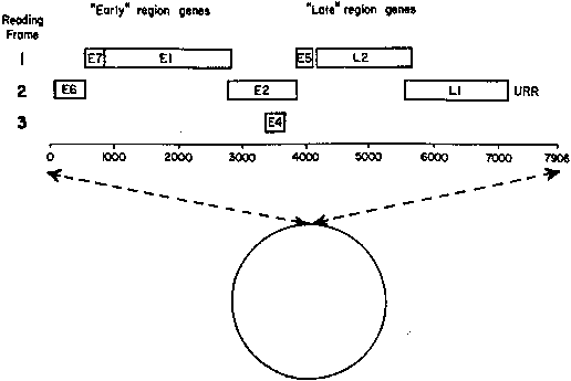
the viral subtype, involved organ, immune system, and local tissue factors.

Viral Subtype

According to their ability to induce cancers, HPV types often are grouped as low-risk types (HPV-6 and -11), medium- or intermediate-risk types (HPV33, -35, -39, -40, -43, -45, -51, -46, and -58), and high-risk types (HPV-16, -18, and -31). Intermediate- and high-risk viruses also are referred to as potentially oncogenic types. High-risk viruses commonly are isolated from high-grade dysplasias or condylomatous lesions with atypical mitoses. Up to 35% of bland-appearing condylomas, however, may contain potentially oncogenic viruses.69

Involved Organ

The HPV-6 and HPV-11 types cause exophytic condylomas (genital warts, condylomata acuminata) mostly on the vulva, vestibule, or perineum, and rarely on the cervix. The same viral types also may be associated with clinically inapparent or subclinical disease, which requires special methods for diagnosis, such as colposcopy. The cervical transformation zone appears particularly vulnerable to oncogenic viruses. Therefore, the most severe effects of high-risk infections generally occur on the cervix.

Immune System

An intact immune system prevents most HPV infections from becoming clinically evident. In immunosuppressed patients, by contrast, HPV virtually always produces recognizable epithelial proliferations, cellular atypia, or neoplasia. Pregnancy is a state of relative immunosuppression, permitting a higher replication rate of viral DNA with enhanced lesion formation and growth.

Local Tissue Factors

Epithelial abnormalities induced by HPV tend to be localized, although surrounding epithelium also is infected and may exhibit inconspicuous epithelial changes. Localized disease, therefore, may represent a focal breakdown of host control within a field of diffuse HPV infection.57

CERVIX.

On the cervix, the classic condylomata acuminata associated with HPV-6 and HPV-11 are uncommon and, when present, often are small and best assessed using the colposcope after application of 5% acetic acid. Most HPV-associated lesions are subclinical and present a large variety of colposcopic findings. In some patients, islands of acetowhite epithelium (satellite lesions) are found in the periphery of the cervix outside the transformation zone (Fig. 8).

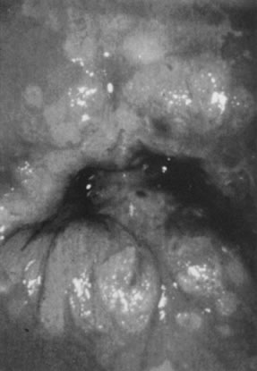
Fig. 8. Colpophotograph of flat condyloma of the cervix. Acetowhite epithelium covers much of the ectocervix. Satellite lesions are seen in the periphery of the lesion and outside the transformation zone.

The surface may appear micropapillary (spiked), flat, or sometimes microconvoluted to a brainlike epithelial arrangement (Fig. 9). Many of the flat lesions have a pure, shiny-white color reminiscent of pearls, in contrast to the full, oyster-white color of high-grade cervical intraepithelial neoplasia (Figs. 10 and 11). Some HPV infections produce coarse capillary loops with a horizontal or vertical orientation, giving the appearance of a mosaic or punctation. The regular spacing of the vessels helps to differentiate these arrangements 
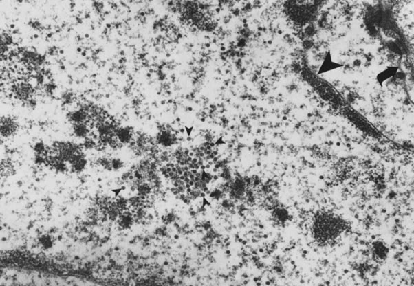
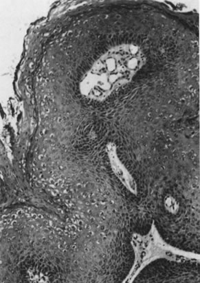
from invasive carcinomas.73,74 Histologically, the lesions contain koilocytotic and parakeratotic changes in the upper layers of the infected epithelium (Fig. 12).

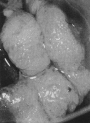
Fig. 9. Colpophotograph of a spiked condyloma on the ectocervix. Myriad tiny excrescences cause the surface of the lesion to be irregular.

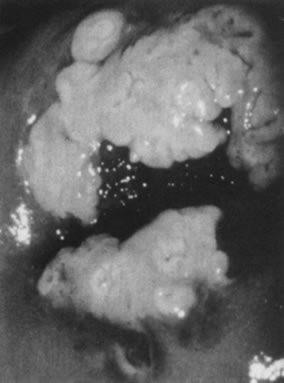
Fig. 10. Colpophotograph of a flat condyloma on the ectocervix. The surface is smooth and has a shiny white color reminiscent of pears.

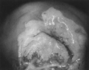
Fig. 11. Colpophotograph of high-grade cervical intraepithelial neoplasia on the ectocervix. The acetowhite epithelium has a dull, oyster-white color.

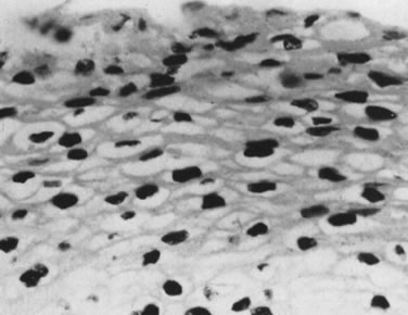
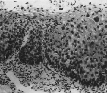
Fig. 12. Cervical intraepithelial neoplasia grade 1. Nuclear atypia, increased mitoses, nuclear crowding, and occasional binucleated forms are seen on the parabasal cell layers. Koilocytotic changes are prominent in the upper cell layers. (Hematoxylin-eosin, ×500.)

VAGINA.

Vaginal condylomata acuminata can be detected in one third of women with vulvar condylomata by careful examination. The distribution usually is patchy, with the proximal and distal thirds of the vagina being affected most commonly. Subclinical vaginal HPV-associated lesions include minute papillary epithelial projections (asperities), each containing a central capillary (Fig. 13). The projections may be isolated, in clusters, or diffuse, covering large areas of the vagina (micropapillomatosis vaginalis). These lesions most commonly are associated with HPV-6 and HPV-11. Acetowhite epithelium, by contrast, is more commonly found in infections due to potentially oncogenic HPV types, most notably HPV-16. Sometimes, minute capillaries are evident, giving rise to patterns reminiscent of punctation and a mosaic pattern on the cervix. Histologically, vaginal intraepithelial neoplasia often is present.75 A common subclinical finding of HPV infection is a pattern called reverse punctation. It consists of a myriad of slightly raised, tiny acetowhite dots, which are pronounced during pregnancy and may involve large areas of the vagina (Fig. 14). Acetic acid may accentuate the changes, but there usually is no sharp demarcation to normal epithelium. It is questionable whether this pattern is due to HPV infection or simply represents a nonspecific response to infectious, hormonal, or other stimuli. If HPV positive, HPV-6 and HPV-11 are isolated from these lesions. Histologic findings are characterized by mild and often focal koilocytosis, variable dyskeratosis, and prominent intraepithelial capillaries.

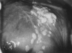
Fig. 13. Colpophotograph of vaginal condylomata. Multiple acetowhite papular lesions crowd the vaginal fornices. Minute papillary epithelial projections (asperities) are evident.

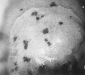
Fig. 14. Colpophotograph of reverse punctation on cervix and vagina in a pregnant woman. Note the acetowhitening of the cervical portion of the epithelium.

VULVA.

Condylomata acuminata in the vulvar area most commonly involve the perineum, the posterior portion of the vestibule, and the labia minora; less commonly involved are the labia majora, the clitoris, and the mons pubis. Lesions on moist, mucosal surfaces tend to be pink, vascular tumors with finger-like projections (Fig. 15). On keratinized skin, the condylomas are often white or dark because of keratin and pigment formation (Fig. 16). The lesions have a typical histologic appearance of a papillary growth with marked acanthosis, koilocytosis and hyperkeratosis, or parakeratosis. Condylomata acuminata most commonly are associated with HPV-6 and HPV-11.

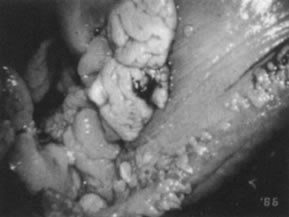
Fig. 15. Colpophotograph of condylomata acuminata of the vulva. The condylomata near the hymenal ring ( top left )are microconvoluted to a brainlike epithelial arrangement. Condylomata near the outside of the vestibule have an irregular papillary appearance.

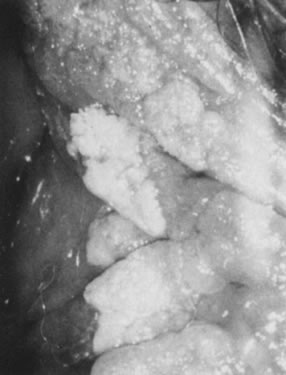
Fig. 16. Colpophotograph of condylomata on the inner aspect of the right labium majus. Some lesions are white because of keratin formation.

Potentially oncogenic HPV types, especially HPV-16, also may give rise to grossly visible lesions presenting as multiple, darkly pigmented, sometimes white, pale, or fleshy papules, formerly often referred to as bowenoid papulosis (Fig. 17). Histologic examination of the papules usually reveals vulvar intraepithelial neoplasia (Fig. 18).

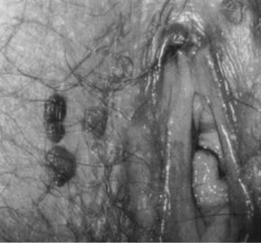
Fig. 17. Colpophotograph of several darkly pigmented papular vulvar lesions. Biopsy showed vulvar intraepithelial neoplasia, grade 3 (see Fig. 18).

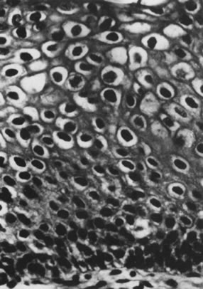
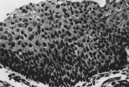
Fig. 18. intraepithelial neoplasia, grade 3. The entire thick- ness of the epithelium is replaced by atypical cells. The nuclei are enlarged but maintain some polarity. Increased cellularity and multiple mitoses are evident. (Hematoxylin-eosin, ×500.)

Other HPV-associated lesions of the vulva consist of papillary changes commonly visible with the naked eye but best appreciated colposcopically. The papillae are multiple, small villous projections from mucous membranes that may involve the entire vestibule and inner surface of the labia minora. When extensive, the papillary changes are referred to as micropapillomatosis vestibularis or labialis (Fig. 19). Some women with vestibular papillae have intense vulvar pain (vulvodynia), burning, irritation, and pruritus. Although the papillary formations have been associated with various HPV types, especially HPV-6, they frequently represent anatomic variants of vestibular mucosa.76 It is unlikely that HPV infections are responsible for vulvodynia, but overly aggressive therapy directed against vulvar or vaginal HPV-associated lesions may result in damage to the vulvar epithelium and chronic vulvar pain.

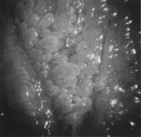
Fig. 19. Colpomicrograph of micropapillomatosis vestibularis. The entire vestibule is covered with myriad small villous projections. The epithelium tested negative for HPV.

Histologically, isolated papillary fronds are observed, with prominent fibrovascular cores associated with chronic inflammation and dilated capillaries. Koilocytotic transformation of superficial epithelial cells is variable.

Acetowhitening of the vulvar epithelium occurs most commonly within mucosal surfaces but is not confined to it (Fig. 20). Although it often represents a nonspecific epithelial reaction to chemical, mechanical, and infectious irritants, acetowhite epithelium frequently is associated with HPV-16 and HPV-18, particularly when it is multifocal and when significant epithelial atypia (vulvar intraepithelial neoplasia) is present.

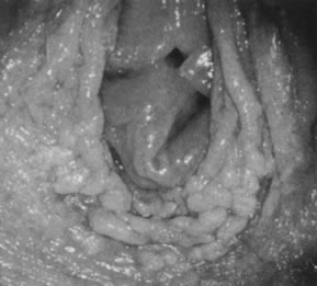
Fig. 20. Colpophotograph of acetowhitening of vulvar epithelium. Dots of acetowhite epithelium surround the vaginal introitus in a horseshoe-shaped configuration. The lesion tested positive for HPV-16. Histologically, vulvar intraepithelial neoplasia, grade 1, was present.

Back to Top
NATURAL HISTORY

Progression and Spontaneous Regression

Women with cytologic evidence of HPV in their Pap smears have a relative risk of developing cervical carcinoma that is 15.6 times higher than the general population.76 Condylomata acuminata can be expected to regress spontaneously in at least 20% of cases. Flat condylomata of the cervix resolve without therapy in 40% to 50% of cases; 20% to 40% progress to dysplasia or (rarely) carcinoma, and about 20% persist over an observation period of 1 year.77,78

Lesions with HPV-6 and HPV-11 are unlikely to develop significant epithelial atypia and are virtually assured a benign course. However, about 30% of flat condylomas and low-grade cervical dysplasias are associated with potentially oncogenic HPV types. Regression of these lesions is uncommon, and progression to invasive carcinoma may occur in up to 5% of cases. The findings emphasize that flat condylomas and low-grade dysplasia have a definite potential for malignant transformation and should not be ignored.79

More than 80% of invasive cervical cancers are preceded by high-grade cervical dysplasia for as long as 20 years. Many women with high-grade dysplasias do not develop cancer, but precise estimates of progression are confounded by length of follow-up and by biopsy altering the natural history of the disease. Estimates of progression of high-grade dysplasia to invasive cancer vary from 20% to more than 70%. The regression rate has been estimated to be as high as 32%.80 Some invasive carcinomas occur in the absence of a previously documented precursor lesion (that is, in the interval between a normal smear and the diagnosis of invasive carcinoma). These malignancies are sometimes referred to as interval cancer. They appear to be more common in younger women (25 to 35 years of age); are typically small cell carcinomas, adenocarcinomas, and adenosquamous carcinomas; are aggressive in their clinical course; and are strongly related to HPV-18.81 Others have argued that rapidly progressive carcinomas may be a manifestation of endocervical carcinomas that have been inadequately screened.82

Assessment of Malignant Potential

Histologic examinations of biopsy specimens from infected epithelium provide only a rough guide to the malignant potential of individual lesions. Although atypical mitoses are the hallmark of oncogenic HPV types, atypical mitoses also can be found in some middle-grade lesions caused by nononcogenic HPV types.73

High-risk HPV types commonly cause derangement in chromosomal content, resulting in aneuploidy. Aneuploid lesions rarely regress spontaneously. Polypoid lesions, by contrast, disappear in most cases. Thus, quantitation of cellular DNA content can identify a subset of lesions that are at much greater risk of malignant transformation. A proportion of HPV-16 or HPV-18 infections initially may present as polyploid lesions but subsequently evolve into aneuploid premalignancies.83 In addition, ploidy analyses are cumbersome and not suitable for routine clinical use.

Back to Top
PREVENTION

The high prevalence of latent HPV infections among sexually active men and women will continue to provide an abundant reservoir of HPV-related problems. Given a prevalence of HPV infections of 30% and a transmission rate of 65%, the risk of contracting the virus from sexual contacts with one person may be as high as 20%. Although prospective clinical studies are not available, condoms are likely to provide some protection but are incomplete barriers because they do not shield the scrotum, pubic area, and anus.

In the future, HPV-associated neoplasia may be prevented by polyvalent vaccines directed against various HPV types. Although the classic live, attenuated, and killed virus vaccines cannot be developed until an in vitro culture system for the propagation of HPV is found, the already existing recombinant DNA technology makes it possible to produce proteins derived from any of the viral genes in prokariotic and eukariotic vectors. HPV proteins are immunogenic in animals and could be used to induce humoral and cell-mediated immunity in humans. There are, however, no convenient methods to measure the effectiveness of the resultant immune response on infectivity.

Recently, viral capsids (also referred to as viral-like particles [VLPs]) encoded by the L1 ORF made it possible to induce high titers of strongly neutralizing type-specific antibodies to papilloma viruses. Antibodies to species-specific VLPs have been used successfully for immunization of rabbits, cows, and dogs. It is anticipated that a vaccine derived from VLPs of high-risk HPV types may be available in the near future for vaccination of a target population: young adults before initiation of sexual activity. Immunization therapy for active HPV infection is also under investigation.63 If it were possible to define an immunologic defect that explains ineffective recognition of certain HPV antigens, such as encoded by HPV-16 and HPV-18, it would be possible to screen uninfected populations to identify people at risk for development of HPV-associated neoplasia.84 Until women who lack immune responsiveness to HPV infections are detectable, the burden of screening for HPV-associated preneoplastic changes rests with the Pap smear.

Back to Top
PRINCIPLES OF MANAGEMENT
  1. Establish the diagnosis. Biopsy is required in all cases, except for the most obvious classic condylomata acuminata.
  2. Assess the extent of disease. Colposcopy and acetic acid application should be part of the initial assessment of HPV-associated epithelial changes, particularly when tissue destruction and ablation techniques, such as cryotherapy, local excision, electrocautery, or laser, are used. Colposcopy and other forms of examination under magnification are also useful during follow-up.
  3. Treat or eliminate associated problems. Immunosuppression related to wasting disease, diabetes mellitus, diseases of the immune system, stress, drugs, and hormones may strongly affect the course of HPV infections. Although avoidance of stress, better control of diabetes mellitus, and improvement of the nutritional status of patients with wasting disease often are feasible and effective, immunosuppressive drugs rarely can be discontinued. Most gynecologists also are reluctant to discontinue oral contraceptives because the danger associated with pregnancy usually outweighs the potential benefits of reduced HPV DNA replication. Treating coexistent trichomoniasis may also be helpful.
  4. Examine and treat the partner. Successful treatment of the male sexual partner of infected women may prevent future sex partners from becoming infected. There is no good evidence to suggest that male partner examination and treatment alters the course of the disease in the infected female partner.85
  5. Do no harm. Not all patients with HPV infections require treatment. The physician must consider the possibility of spontaneous regression and weigh the discomfort and risks of therapy against the existing disability.
  6. Select proper treatment. In choosing treatment for a particular patient, the physician should consider the clinical presentation and manifestations of the HPV infection and the effectiveness, availability, ease of application, side effects, long-term risks, and cost of the treatment method. The patient's tolerance and preferences, suitability for use in pregnancy, vaginal application, and control of latent infection also are of concern.
Back to Top
INDICATIONS FOR TREATMENT

Major Indications

ALLEVIATE IRRITATION, DISCOMFORT, OR PAIN.

Patients with symptomatic condylomata acuminata fall into this category.

PREVENT NEOPLASIA.

The known association of HPV with squamous neoplasia of the lower genital tract requires that precursor lesions be eradicated. Although most genital dysplasias do not transform into invasive carcinoma, there is general agreement to treat patients with high-grade (moderate and severe) dysplasia and carcinoma in situ. The role of therapy for low-grade lesions (flat condyloma, mild dysplasia) is less well defined. In view of the high rate of spontaneous regression of minor HPV-associated epithelial abnormalities, a watch-and-wait approach appears appropriate. For cervical lesions, the inconvenience, anxiety, time, and cost involved in repeated cytologic testing, colposcopy, and biopsies should be weighed against the relatively innocuous, effective ablation of the transformation zone by cryotherapy or other methods in the office.

Minor Indications

CONTROL SPREAD OF DISEASE.

Although most genital HPV-associated lesions are not genuine precursors of carcinoma, effective treatment may help to bring the troublesome HPV epidemic under control. Patients with asymptomatic HPV infections may be treated for this reason. Ablation of the products of HPV infection, such as exophytic or flat condylomata, however, by no means eliminates the virus. No method has been developed to eradicate latent or subclinical HPV infections, which, as previously emphasized, tend to involve the entire lower genital tract.86

ALLEVIATE ANXIETY.

Ablation of an abnormal transformation zone by cryotherapy reduces the risk of cervical carcinoma and may alleviate anxiety. The patient should be advised, however, that the virus is likely to persist in the periphery of the cervix and elsewhere in the lower genital tract, that she will remain potentially infectious, and that the virus probably cannot be completely eradicated. The understanding that HPV infections are extremely common and that the cancer risk in other areas of the genital tract and in male partners is very small should further alleviate mental anguish. The need for continued screening even after ablation of the transformation zone should be emphasized.

Back to Top
TREATMENT

The ideal therapy for HPV-associated lesions should be effective, readily available, easily applied, well tolerated, free of long-term risks, safe in pregnancy, suitable for vaginal use, and inexpensive. It should also take into consideration that genital HPV infection is a disease of both normal- and abnormal-appearing epithelium that often runs its course over several weeks to 8 months and spontaneously disappears. Therefore, the surgical approach to therapy is less than optimal. Destruction of the products of HPV infections, such as condylomas or dysplasias, leads to remission in about 85% of patients despite of the fact that HPV-associated lesions are often surrounded by areas of subclinical infection. Consequently, treatment methods with the potential to control subclinical disease, such as laser, 5-FU, and interferon, should be reserved for patients whose lesions are too extensive for control by caustic agents or outpatient destruction and for those who do not respond to simpler therapies.71

The carbon dioxide laser is considered the most effective form of treatment of extensive vulvar HPV-associated lesions. A trial with periodic 5-FU may still be warranted, however, because about 70% of patients with extensive HPV-associated disease respond to this regimen and may be spared the expense of laser therapy, which usually requires general anesthesia.87 The control rate of subclinical disease and the overall cure rate appear to be higher when laser is used in association with adjuvant (prophylactic) 5- FU.88 5-FU is probably superior to laser for the treatment of vaginal condylomata acuminata and is at least as effective as laser for multifocal vaginal dysplasia.89–91

In the future, antiviral drugs or immune modulators may be more desirable, and interferons were the first such agents to show promise. The efficacy of systematic interferon demonstrated so far may not warrant the toxic reactions observed, however, and the efficacy of topical interferon remains in doubt. Intralesional injection is no more convenient than destructive modalities and, unless subclinical virus is proved to be eliminated, shows no obvious therapeutic advantage.91

Imiquimod, an immune-response modifier supplied in a cream base, is applied by patients with condylomata acuminata. It is the first successful treatment that promotes a host immune response, resulting in complete clearance of the lesions in more than 50% of women treated.

Highly specific treatments for HPV-associated lesions would be based on the known biologic properties of HPVs and may consist of targeting oncoproteins expressed in malignant HPV-associated lesions. Experiments with antisense oligonucleotides targeting the E6 and E7 oncoproteins showed growth inhibition of cancer cell lines.92,93 At present, problems with cellular transfer and targeting using recombinant DNA technologies severely limit their clinical applications.

Back to Top
ACKNOWLEDGMENT

I thank Sonia M. Kheir for contributing to this chapter.

Back to Top
REFERENCES

1. World Heath Organization: Genital human papillomavirus infections and cancer: Memorandum from a WHO meeting. Bull WHO 65:817, 1987

2. zur Hausen H: Papillomaviruses as carcinomaviruses. Adv Viral Oncol 8: 1, 1989

3. Shope RE: Infectious papillomatosis of rabbits. J Exp Med 58: 607, 1933

4. Rous P. Beard JW: The progression of carcinoma of virus-induced rabbit papillomas (Shope). J Exp Med 62: 523, 1935

5. Meisels A, Fortin R: Condylomatous lesions of cervix and vagina. I. Cytologic patterns. Acta Cytol 20: 505, 1976

6. Purola E, Savia E: Cytology of gynecologic condyloma acuminatum. Acta Cytol 21: 26, 1977

7. Koss LG, Durfee GR: Unusual patterns of squamous epithelium of the uterine cervix: Cytologic and pathologic study of koilocytotic atypia. Ann NY Acad Sci 63: 1245, 1956

8. Della Torre G, Pilotti S, DePalo G et al: Viral particles in cervical condylomatous lesions. Tumori 64: 459, 1978

9. Laverty CR, Booth N, Hills E et al: Noncondylomatous wart virus infection of the postmenopausal cervix. Pathology 10: 373, 1978

10. Jenson AB, Rosenthal JD, Olson C et al: Immunological relatedness of papillomavirus from different species. JNCI 64: 495, 1980

11. Shah KV, Lewis MG, Jenson AB et al: Papillomavirus and cervical dysplasia. Lancet 2: 1190, 1980

12. Wright TC, Richart R: Role of human papillomavirus in the pathogenesis of genital-tract warts and cancer. Gynecol Oncol 37: 151, 1990

13. Schneider A, Holz M, Gissmarm L: Prevalence of genital HPV infections in pregnant women. Int J Cancer 40: 198, 1987

14. Kjaer SK, deVilliers EM, Haugaard BJ et al: Human papillomavirus, herpes simplex virus, and cervical cancer incidence in Greenland and Denmark: A population-based cross-sectional study. Int J Cancer 41: 518, 1988

15. Bauer H, Ting Y, Greer CE et al: Genital human papillomavirus infection in female university students as determined by a PCR based method. JAMA 265: 472, 1991

16. Melkert PWJ, Hopman E, van Den Brule AJC et al: Prevalence of HPV in cytomorphologically normal cervical smears as determined by polymerase chain reaction is age-dependent. Int J Cancer 53: 919, 1993

17. Fairley CK, Chen S, Ugoni A et al: Human papillomavirus infection and its relationship to recent and distant sexual partners. Obstet Gynecol 84: 755, 1994

18. Kjaer SK, Engholm G, Teisen C et al: Risks factor for cervical human papillomavirus and herpes simplex virus infection in Greenland and Denmark: A population-based study. Am J Epidemiol 131: 669, 1990

19. Wickenden C, Hanna N, Taylor Robinson D et al: Sexual transmission of human papillomaviruses in heterosexual and male homosexual couples, studied by DNA hybridization. Genitourin Med 64: 34, 1988

20. Krebs HB, Schneider V: Human papillomavirus associated lesions of the penis: Colposcopy, cytology and histology. Obstet Gynecol 70: 299, 1987

21. Pakarian F, Kaye J, Cason J et al: Cancer-associated papillomaviruses: Perinatal transmission and persistence. Br J Obstet Gynaecol 101: 514, 1994

22. Nuovo GJ, Pedemonte BA: Human papillomavirus types and recurrent genital warts. JAMA 263: 1223, 1990

23. Koutsky LA, Holmes KK, Critchlow CW et al: A cohort study of the risk of cervical intraepithelial neoplasia. grade 2 or 3 in relation to papillomavirus infection. N Engl J Med 327: 1272, 1992

24. Nuovo GJ, Blanco J S, Leipzig S, Smith D: Human papillomavirus detection in cervical lesions nondiagnostic for cervical intraepithelial neoplasia: Correlation with Papanicolaou smear, colposcopy, and occurrence of cervical intraepithelial neoplasia. Obstet Gynecol 75: 1006, 1990

25. Moscicki AB, Palefsky J, Smith G et al: Variability of human papillomavirus DNA testing in a longitudinal cohort of young women. Obstet Gynecol 82: 578, 1993

26. Coggin JR, zur Hausen H: Workshop on the papillomaviruses and cancer. Cancer Res 39: 545, 1979

27. Wright TC: Pathogenesis and diagnosis of preinvasive lesions of the lower genital tract. In Hoskins WJ, Perez C, Young RC (eds): Principles and Practice of Gynecologic Oncology, pp 744–752. 3d ed. Philadelphia, Lippincott William & Wilkins, 2000

28. zur Hausen, Schneider A: The role of papillomaviruses in human anogenital cancer. In Salzman HP, Howley PM (eds): The Papovaviridiae, p 245. New York, Plenum Press, 1987

29. Gissmann L, zur Hausen H: Partial characterization of viral DNA from human genital warts (condylomata acuminata). Int J Cancer 25: 605, 1980

30. Bonnez W: Murine models of human papillomavirus-infected human xenografts. Papillomavirus Rep 9: 27, 1998

31. Meyers C, Frattini MG, Hudson JB et al: Biosynthesis of human papillomavirus from a continuous cell line upon epithelial differentiation. Science 257: 971, 1992

32. Galloway DA: Papillomavirus capsids: a new approach to identify serologic markers of HPV infection. J Natl Cancer Inst 86: 474, 1994

33. Rose RA, Bonnez W, Reichman RC et al: Expression of human papillomavirus type 11 L1 protein in insect cells: in vivo and in vitro assembly of virus like particles. J Virol 67: 1936, 1993

34. Dürst M, Gissman L, Ikenberg H et al: A papillomavirus DNA from a cervical carcinoma and its prevalence in cancer biopsy samples from different geographic regions. Proc Natl Acad Sci USA 80: 3812, 1983

35. Boshart M, Gissman L, Ikenberg H et al: A new type of papillomavirus DNA, its presence in genital cancer and in cell lines derived from genital cancer. EMBO J 3: 1151, 1984

36. Southern EM: Detection of specific sequences among DNA fragments separated by gel electrophoresis. J Mol Biol 98: 503, 1975

37. Lörincz AT: Detection of human papillomavirus infection by nucleic acid hybridization. Obstet Gynecol Clin North Am 14: 451, 1987

38. Schneider A, Grubert T: Diagnosis of HPV infection by recombinant DNA technology. Clin Obstet Gynecol 32: 127, 1989

39. Reid RI, Lörincz AT: New generation of human papillomavirus tests. In Rubins SC, Hoskins WY (eds): Cervical Cancer and Preinvasive Neoplasia, p 27. Vol 3. New York, Lippincott-Raven, 1996

40. Hatch KD, Schneider A, Abdel Nour MW: An evaluation of human papillomavirus testing for intermediate- and high-risk types as triage before colposcopy. Am J Obstet Gynecol 172: 1150, 1995

41. Cox JT, Lörincz AT, Schiffman MH et al: Human papillomavirus testing by hybrid capture appears to be useful in triaging women with a cytologic diagnosis of atypical squamous cells of undetermined significance. Am J Obstet Gynecol 72: 946, 1995

42. Ritter D, Kadish AS, Vermund SH et al: Detection of human papillomavirus deoxyribonucleic acid in exfoliated cervicovaginal cells as a predictor of cervical neoplasia in a high-risk population. Am J Obstet Gynecol 159: 1517, 1988

43. Lörincz AT, Lancaster WD, Kurman RJ et al: Characterization of human papillomaviruses in cervical neoplasias and their detection in routine clinical screening. In Peto R, zur Hausen H (eds): Viral Etiology of Cervical Cancer, Banbury Report 21, p 225. Cold Spring Harbor, NY, Cold Spring Harbor Laboratory Press, 1986

44. Reid R: How HPV testing helps the clinician. Contemp Obstet Gynecol 2: 31, 1990

45. Kaufman RH, Adam E, Icenogle J et al: Relevance of human papillomavirus screening in management of cervical intraepithelial neoplasia. Am J Obstet Gynecol 176: 87, 1997

46. Wheeler CM, Greer CE, Becker TM et al: Short-term fluctuations in the detection of cervical human papillomavirus DNA. Obstet Gynecol 88: 261, 1996

47. Schiffman M: Arguments against routine clinical use of HPV assays. Contemp Obstet Gynecol 2: 31, 1990

48. Wright TC, Lörincz AT, Ferris DG et al: Reflex HPV deoxyribonucleic acid testing in women with abnormal Papanicolaou smears. Am J Obstet Gynecol 178: 926, 1998

49. Howley PM: Role of human papillomaviruses in human cancer. Cancer Res 51: 5019, 1991

50. Park DJ, Wilczynski SP, Paquette RL et al: Mutations in HPV-negative cervical carcinoma. Oncogene 9: 205, 1994

51. Gissman L, Dürst M, Oltersdorf T et al: Human papillomaviruses and cervical cancer. In Steinberg BM, Brandsma TL, Tacchman LB (eds): Cancer Cells, vol V, The Papilloma Viruses, p 275. New York, Cold Spring Harbor Laboratory, 1987

52. Pecoraro G, Morgan D, Defendi D: Differential effects of human papillomaviruses 6, 16, and 18 DNA's on immortalization and transformation of human cervical cells. Proc Nat Acad Sci USA 86: 563, 1989

53. Levine AJ: p53, cellular gatekeeper for growth and division. Cell 88: 323, 1997

54. Vogelstein B, Kinzler KE: p53 function and dysfunction. Cell 70: 523, 1992

55. zur Hausen H: Papillomavirus infections: A major cause of human cancers. Biochem Biophys Acta 1288: F55, 1996

56. Howley PM: Papillomavirinae: The viruses and their replication. In Fields BN, Knipe DM, Howley PM (eds): Fundamental Virology, p 947. Philadelphia, PA, Lippincott-Raven, 1966

57. Münger K, Werness BA, Dyson N et al: Complex formation of human papillomavirus E7 proteins with the retinoblastoma tumor suppressor gene product. EMBO J 8: 4099, 1989

58. Horowitz JM, Yandall DW, Park SH et al: Point mutational inactivation of the retinoblastoma antioncogene. Science 243: 937, 1989

59. Park TW, Fujawara H, Wright TC: Molecular biology of cervical cancer and its precursors. Cancer 76: 1902, 1995

60. Dürst M, Kleinheinz A, Holz M et al: The physical state of human papillomavirus type 16 DNA in benign and malignant genital tumors. J Gen Virol 66: 1515, 1985

61. Smotkin D: Virology of human papillomavirus. Clin Obstet Gynecol 32: 117, 1989

62. Clavel C, Huu VP, Durlach AP et al: Mucosal oncogenic human papillomaviruses and extragenital Bowen's disease. Cancer 86: 282, 1999

63. Alani RM, Munger K: Human papillomaviruses and associated malignancies. J Clin Oncol 16: 330, 1998

64. Evander M, Frazer IH, Payne E et al: Identification of the alpha-6 integrin as a candidate receptor for papillomaviruses. J Virol 71: 2449, 1997

65. Stoler MH, Mills SE, Gersell DJ, Walker AN: Small cell neuroendocrine carcinoma of the cervix: A human papillomavirus type 18 associated cancer. Am J Pathol 15: 28, 1991

66. Smotkin D, Berek JS, Fu YS et al: Human papillomavirus deoxyribonucleic acid in adenocarcinoma and adenosquamous carcinoma of the uterine cervix. Obstet Gynecol 68: 241, 1986

67. Tate JE, Resnick M, Sheets E, Crum CP: Absence of papillomavirus DNA on normal tissue adjacent to most cervical intraepithelial neoplasms. Obstet Gynecol 88: 257, 1996

68. Nuovo GH, Nuovo MA, Cottral S et al: Histological correlates of clinically occult human papillomavirus infection of the cervix uteri. Am J Surg Pathol 12: 198, 1988

69. Campion MJ, Franklin EW, Stacy LD et al: Human papillomavirus and anogenital neoplasia: A fresh look at the association. South Med J 82: 35, 1989

70. Fischer RG, Silverton J: The virus-induced papilloma-to-carcinoma sequence. IV. Carcinoma in domestic rabbits infected while in uterus. Cancer Res 11: 737, 1951

71. Jarrett WFH: The natural history of bovine papillomavirus infections. Adv Virol Oncol 5: 83, 1985

72. Orth G, Jablonska S, Favre M et al: Epidermodysplasia verruciformis: A model for viral oncogenesis in man. Cold Spring Harbor Conf Cell Prolif 7: 259, 1980

73. Reid R, Greenberg M, Jenson AB et al: Sexually transmitted papillomaviral infections. I. The anatomic distribution and pathologic grade and neoplastic lesions associated with different viral types. Am J Obstet Gynecol 156: 212, 1987

74. Reid R, Campion MJ: HPV-associated lesions of the cervix: Biology and colposcopic features. Clin Obstet Gynecol 32: 157, 1989

75. Campion MJ: Clinical manifestations and natural history of genital human papillomavirus infections. Obstet Gynecol Clin North Am 14: 363, 1987

76. Bergeron C, Ferenczy A, Richart RM et al: Micropapillomatosis labialis appears unrelated to human papillomavirus. Obstet Gynecol 70: 281, 1990

77. Mitchell H, Drake M, Medley G: Prospective evaluation of risk of cervical cancer after cytological evidence of human papillomavirus infection. Lancet 2: 573, 1986

78. Nash JD, Burke TW, Hoskins WJ: Biologic course of cervical human papillomavirus infection. Obstet Gynecol 69: 160, 1987

79. Evans AS, Monaghan JM: Spontaneous resolution of warty atypia: The relevance of clinical and nuclear DNA features. A prospective study. Br J Obstet Gynaecol 92: 165, 1985

80. Campion MJ, McCance DJ, Cuzick J et al: Progressive potential of mild cervical atypia: Prospective cytological, colposcopic, and virologic study. Lancet 2: 168, 1986

81. östör AG: Natural history of cervical intraepithelial neoplasia: A critical review. Int J Gynecol Pathol 12: 186, 1993

82. Crum CP, Cibas ES, Lee KR: Pathology of Early Cervical Neoplasia, Chap 2, p 7. New York, Churchill Livingstone, 1997

83. Schwartz PE, Hadjimichael O, Lowell DM et al: Rapidly progressive cervical cancer: The Connecticut experience. Am J Obstet Gynecol 175: 1105, 1996

84. Reid R, Fu YS, Hershman BR et al: Genital warts and cervical cancer. VI. The relationship between aneuploid and polyploid cervical lesions. Am J Obstet Gynecol 150: 189, 1984

85. Lancaster WD, Jenson AB: Human papillomavirus infection and anogenital neoplasia: Speculations for the future. Obstet Gynecol Clin North Am 14: 601, 1987

86. Krebs HB, Helmkamp BF: Does the treatment of genital condylomata acuminata in men decrease the treatment failure rate of cervical dysplasia in the female sexual partner? Obstet Gynecol 76: 660, 1990

87. Riva JM, Sedlacek TV, Cunnane MF, Mangan CE: Extended carbon dioxide laser vaporization in the treatment of subclinical papillomavirus infection of the lower genital tract. Obstet Gynecol 73: 25, 1989

88. Krebs HB: Treatment of extensive vulvar condylomata acuminata with topical 5-fluorouracil. South Med J 83: 762, 1990

89. Krebs HB: Combination of laser plus 5-fluorouracil for the treatment of extensive genital condylomata acuminata by weekly topical application of 5-fluorouracil. Obstet Gynecol 70: 68, 1987

90. Krebs HB: Treatment of vaginal condylomata acuminata by weekly topical application of 5-fluorouracil. Obstet Gynecol 70: 68, 1987

91. Krebs HB: Treatment of vaginal intraepithelial neoplasia with laser and topical 5-fluorouracil. Obstet Gynecol 73: 657, 1989

92. Ferenczy A: Comparison of 5-fluorouracil and CO2 laser for treatment of vaginal condylomata. Obstet Gynecol 64: 773, 1984

93. Kirby P. Editorial. JAMA 259:570, 1988

Back to Top