Chapter 12
Endometrial Hyperplasia and Neoplasia: Definition, Diagnosis, and Management Principles
Alex Ferenczy and George Mutter
Main Menu   Table Of Contents

Search

Alex Ferenczy, MD
Professor of Pathology, Obstetrics and Gynecology, McGill University and The Sir Mortimer B. Davis Jewish General Hospital, Montreal, Quebec, Canada (Vol 4, Chap 12; Vol 5, Chap 18)

George Mutter, MD
Associate Professor of Pathology, Harvard Medical School, Brigham and Women's Hospital, Boston, Massachusetts (Vol 4, Chap 58)

 
DEFINITION, NOMENCLATURE, AND CLINICAL IMPLICATIONS OF ENDOMETRIAL NEOPLASIA AND ITS PRECURSORS
EMERGING CONCEPTS
POPULATION AT RISK
SCREENING AND DIAGNOSIS
TREATMENT OF ENDOMETRIAL HYPERPLASIA
CONTROVERSIAL ISSUES
PROGESTERONE CHALLENGE TEST
CONCLUSIONS
REFERENCES

DEFINITION, NOMENCLATURE, AND CLINICAL IMPLICATIONS OF ENDOMETRIAL NEOPLASIA AND ITS PRECURSORS

By definition, adenocarcinoma of the endometrium is an invasive disease, invading either the endometrial stroma or the underlying myometrium of extrauterine tissues. Most endometrial carcinomas maintain endometrioid differentiation; these also can contain areas of mucinous or squamous differentiation. Other nonendometrioid subtypes seen in routine practice include clear cell carcinoma, papillary serous carcinoma, and other rare variants. According to the U.S. Gynecologic Oncology Group histologic grading system,1 grade 1, well-differentiated carcinoma, consists of a neoplasm with less than 5% of solid cancer; grade 2, moderately differentiated carcinoma, contains 6% to 50% solid cancer; and grade 3, poorly differentiated carcinoma, contains more than 50% of solid tumor.

Tumor grading is of greater independent prognostic value for endometrioid endometrial adenocarcinoma and its related types (i.e., endometrial, secretory, mucinous, squamous) than for papillary serous and clear cell adenocarcinomas. Papillary serous and clear cell cancers do not show the grade-dependent changes in aggressiveness seen with the endometrioid tumors; instead, as a group they are consistently aggressive. Division of endometrial adenocarcinomas into the clinicopathologic classes of endometrioid and nonendometrioid types is paralleled further by differences in epidemiologic risk factors and precursor lesions.2,3 Type I, or endometrioid, endometrial adenocarcinomas are more frequent in women taking exogenous estrogen and often are preceded by precursor lesions, which is the subject of this discussion.

It has traditionally been suggested that endometrioid endometrial adenocarcinoma is preceded by endometrial hyperplasia (EH).4 EH previously was considered a continuum of morphologic changes often beginning with simple glandular/stromal overgrowth (simple hyperplasia) and ending with complex, highly atypical histologic and cytologic proliferations, variously referred to as atypical adenomatous hyperplasia, dysplasia, or carcinoma in situ. The figure most often cited in the literature for progression of atypical adenomatous hyperplasia to carcinomas was 30% at 10 years.4

When the relevant laboratory and clinical evidence was critically reviewed, it became apparent that the only important morphologic feature distinguishing endometrial lesions from those without significant invasive potential is cytologic atypia of gland cells.5,6,7,8,9 Histologically, lesions without invasive potential lack cytologic atypia (Fig. 1). Their nuclear characteristics are similar to those of normal, cyclic, proliferative gland cell nuclei. Development of useful architectural criteria to be used in conjunction with cytologic atypia for precancer diagnosis is an area of active investigation (see Emerging Concepts later).

Fig. 1. Histology of endometrial hyperplasia (without atypia). A. Voluminous proliferative-type glands and abundant stroma. B. Higher power magnification of tall, columnar gland cells with regular nuclear pseudostratification and abundant intranuclear receptors for estrogen. There is an increased number of crowded and irregularly shaped glands relative to stroma.

Traditionally the most frequently used terms for hyperplastic endometrium without atypia include cystic glandular hyperplasia and adenomatous hyperplasia. The International Society of Gynecologic Pathologists adopted the terms simple and complex hyperplasia instead of cystic glandular and adenomatous hyperplasia.10 Clinically, most cases of EH regress either spontaneously or after conservative progestogen-suppressive therapy.7

In contrast to EH, glandular proliferations with significant nuclear atypia are endometrial carcinoma precursors.5,6,7,8,9,11 In a prospective study with a mean follow-up of 7 years, none of the 65 women with EH without atypia developed carcinoma, whereas carcinoma was diagnosed in 5 of 20 women with endometrial intraepithelial neoplasia (EIN).7 Cytologically and by morphometric analysis, these cells resemble9,11,12,13 and many times are indistinguishable from cells in well-differentiated invasive carcinoma (Fig. 2). These lesions have been variously referred to as atypical adenomatous hyperplasia and severe or atypical complex hyperplasia. The International Society of Gynecologic Pathologists proposed the term atypical hyperplasia (adenomatous with atypia).10 The morphologic criteria for distinguishing among the various subsets of hyperplastic endometrial lesions are presented in Table 1. Excluded from hyperplasias are anovulatory persistent proliferative endometrium, focal glandular crowding without atypia, postmenstrual regenerative endometrium, and focal cystic dilation of glands. These conditions should not be considered part of or be confused with EH.14 Although the term atypical hyperplasia (AH) is the generally accepted nomenclature, we prefer EIN.12 Lesions devoid of atypia are not cancer precursors, whereas lesions with cytologic atypia may progress to carcinoma if untreated; it is logical to use two different terms for lesions that have different biologic behavior. EIN is not synonymous with invasive carcinoma but indicates a lesion that may regress, persist, or progress to invasion. Progression rates of surgically untreated EIN range from 25%7 to 58%.11 Morphologically, stromal invasion distinguishes invasive carcinoma from EIN. Stromal invasion (intramucosal carcinoma) usually manifests with inflammation or necrosis of the area being invaded or still desmoplasia of the stroma, or the glands have a ragged contour or replace large portions of preexisting endometrial stroma.

Fig. 2. Histology of endometrial intraepithelial neoplasia (atypical hyperplasia). A. Voluminous gland with multiple intraluminal microlumens (cribriform pattern). The lining epithelium has pleomorphic, round nuclei devoid of regular pseudostratification. Macronucleoli are present. B. High magnification of hyperplastic (left) and neoplastic (right) epithelium. Note regular, nuclear pseudostratification of tall columnar gland lining epithelium in hyperplasia without atypia. Endometrial intraepithelial neoplasia (atypical hyperplasoa) has pleomorphic, round nuclei with macronucleoli, and nuclear pseudostratification is lost.


TABLE 1. WHO Classification and Diagnostic Criteria of Endometrial Hyperplasia

  Simple Hyperplasia Without Cytologic Atypia*
  Increased number of glands relative to stroma
  Dilated glands with irregular outlines
  Crowded, clustered glands
  Tall, columnar epithelium with nuclear pseudostratification
  Complex Hyperplasia Without Cytologic Atypia
  Increased number of glands relative to stroma
  Back-to-back glands (crowded glands with little or no intervening stroma)
  Hyperplasia With Cytologic Atypia
  Variation of size and shape of nuclei
  Nuclear enlargement
  Loss of polarity
  Coarse chromatin clumping
  Prominent nucleoli
  Hyperchromatism


*Two of the four criteria must be present.
Both criteria must be present.
Three of the first four criteria must be present.


Back to Top
EMERGING CONCEPTS

Endometrial precancers first were identified as premalignant lesions by virtue of their temporal and spatial association with cancer in large patient series. 25% Of all women with atypical EH, 25% have an adenocarcinoma at hysterectomy.14 Although these strategies generally have been successful in defining broad classes of morphologic lesions most likely to be associated with cancer, clinical outcomes are highly insensitive in detection of precancers. A low precancer-to-cancer progression efficiency predicts that most premalignant lesions will never display a malignant end point. Further difficulty in standardizing diagnosis of endometrial precancers comes from poor reproducibility by pathologists of histopathologic criteria used for lesion classification.15 This situation has spurred development of novel diagnostic strategies applicable to lesional tissues of individual patients that are capable of accurately discriminating between biologic precancers and non-precancers. Even if such a laboratory approach were impractical for everyday use, it would constitute a powerful tool for critical evaluation and refinement of current histologic diagnostic practices.

Monoclonal growth and mutation of tumor-suppressor genes are measurable features of the premalignant phase of endometrial tumorigenesis that can be directly ascertained in paraffin-embedded tissues and correlated with histology on a case-by-case basis. The idea that endometrial precancers are monoclonal proliferative products of a single transformed cell is based on a multistep model of tumorigenesis16 in which progression is driven by sequentially acquired mutations manifest as altered morphology and increasing aggressiveness. Although initial stages may not show an invasive phenotype, it is anticipated that premalignant lesions have sufficient growth advantage relative to their source tissues that they expand monoclonally. This expansion has now been shown to be the case for putative endometrial precancers using a variety of polymerase chain reaction–based molecular genetic methodologies applied to DNA isolated from targeted regions of paraffin sections: nonrandom X chromosome inactivation,17 clonal propagation of altered microsatellites in microsatellite-unstable tissues,18 and clonal propagation of acquired mutations of tumor-suppressor genes such as K-ras18 and PTEN.19 Monoclonal growth seems to be one of the seminal qualities of premalignant tissues at a variety of sites, including the oral mucosa, cervix, skin, stomach, and vulva.

Early stages of carcinogenesis are characterized by incremental growth advantages, which are necessarily small in relation to normal tissues and exquisitely sensitive to environmental modification. Hormonally mediated selection20 of latent transformed clones is one mechanism that might link genetic and endocrine events in genesis of this disease. This selection may occur through changes in precancer clone proliferation rates or remodeling of adjacent normal tissues. In the case of precancers confined to the functionalis, persistence is enhanced by absence of regular shedding (anovulation). Shedding is also a key part of progestin therapy for precancers because patients who have biopsies before a withdrawal bleed often have persistent lesions, albeit with an altered cytology. For this reason, repeat biopsy for confirmation of postprogestin precancer ablation is best accomplished after a withdrawal bleed to realize the full benefit of shedding and to avoid the confounding effects of progestins on histopathology interpretation.

Multiple marker systems (X inactivation, novel microsatellites) used together are approximately 80% sensitive in detection of monoclonal precancers from paraffin sections, a significant improvement over the 25% sensitivity of precancer detection realized when using a clinical standard of progression to carcinoma. In cases that do have an associated carcinoma, conservation of acquired genetic changes between matched premalignant and malignant tissues has provided a highly specific basis to conclude evolution from the former to the latter. Detailed lineage reconstruction, including hierarchical ordering of steps from precancer to cancer, has been accomplished in cases in which the repertoire of informative genetic markers is sufficiently rich.18 High cost and technical complexity place a molecular genetic laboratory standard of precancer diagnosis beyond the reach of a routine diagnostic setting.

Careful histopathologic study of genotypically ascertained endometrial precancers explains prior problems in diagnosis and provides specific directions for improvement.21 Close correlations between histopathology and genotype are possible by isolating DNA from delineated regions of a paraffin section, which is also available as a serially sectioned stained slide. Epithelial differentiation of monoclonal precancers is usually endometrioid, but foci of squamous, mucinous, and tubal differentiation may be present. Changes in the hormonal environment, such as progesterone administration, may reduce the degree of cytologic atypia. Although most genetic precancers are diagnosed as atypical EH, poor reproducibility15 of this diagnosis compromises consistent management and raises the possibility that existing diagnostic criteria are inadequate. A particular void in precancer diagnosis has been absence of informative architectural criteria. Computerized morphometric analysis of monoclonal endometrial precancers,21 using algorithms that previously were shown to predict relevant clinical outcomes of concurrent22 or future23 endometrial adenocarcinoma, has broken this stalemate. When the endometrial glands become so crowded that they comprise more than half of the sectioned surface, they predict monoclonality with a sensitivity and specificity at least equal to the clinical judgment of experienced subspeciality gynecologic pathologists.21

The term endometrial intraepithelial neoplasia accurately describes endometrial precancers because monoclonal origin from a single transformed cell is the pathognomonic feature of all neoplasms. The superb performance of computerized morphometric analysis in classifying all genetically21 and clinically23 defined precancers into one group reaffirms the feasibility of using routine hematoxylin and eosin–stained tissue sections to define a singular category of precancers. It is not anticipated that computerized morphometric analysis will become the predominant method of diagnosis, but rather that practicing pathologists will be able to emulate its performance by extracting new criteria for subjective application in a routine setting (Table 2). Implementation of revised diagnostic and nomenclature standards is likely in the near future, a process that will be facilitated through electronic dissemination of reference images and educational materials.24


TABLE 2. Functional Classification of Premalignant and Malignant Endometrial Disease


Nomenclature

Criteria

Functional Category

Management

Endometrial hyperplasia (EH)

1. Irregular glands with cysts

Estrogen effect

Hormonal therapy


and


2. VPS > 55%


and


3. No atypia


Endometrial intraepithelial neoplasia (EIN)

1. Atypia

Precancer

Hormonal or surgical therapy


and


2. VPS < 55%


Adenocarcinoma

1. Invasion

Cancer

Surgical and/or radiotherapy


or


2. Solid epithelium


or


3. Mazelike glands


Classification of endometrial disease based on lesion biology aspires to place all precancers into a single group (EIN) and contrast with mutually exclusive entities corresponding to different management options. Introduction of refined precancer diagnostic criteria, such as volume percentage stroma
22,25 (VPS) (that function of the sectioned tissue occupied by stroma), may improve histopathologic resolution between benign anovulatory (EH) and premalignant (EIN) disease.


Classification of endometrial disease based on lesion biology aspires to place all precancers into a single group (EIN) and contrast with mutually exclusive entities corresponding to different management options. Introduction of refined precancer diagnostic criteria, such as volume percentage stroma22,25 (that function of the sectioned tissue occupied by stroma), may improve histopathologic resolution between benign anovulatory (EH) and premalignant (EIN) disease.

Back to Top
POPULATION AT RISK

Most patients (75%) with endometrial cancer present with postmenopausal bleeding; however, only 10% of women with postmenopausal bleeding have endometrial carcinoma.25 The remaining women with postmenopausal bleeding have atrophic or inactive endometrium or benign endometrial conditions. The clinical predictive model proposed by Feldman and colleagues26 (i.e., 70 years of age or older, diabetes, nulliparity, and postmenopausal status) is not predictive enough to distinguish between women with perimenopausal or postmenopausal bleeding at low versus high endometrial carcinoma risk.27 The traditional risk indicators associated with EH/carcinoma are shown in Table 3. Most of the indicators are estrogen related, either from endogenous or exogenous sources. In women with these risk factors, the relative risk of developing carcinoma is 1.2 to 35.28 Experience also suggests that a significant proportion of patients fail to have these risk indicators but develop endometrial carcinoma. In these cases, either the disease may not be hormone related, or hyperestrogenism is metabolically inapparent. In a literature review, 74% of patients with adenocarcinoma of the endometrium were not obese, 58% were not nulliparous, 22% experienced menopause before age 49 years, and 43% to 89% were not exposed to hormone replacement therapy (HRT).29 It seems that the only constant endometrial carcinoma risk indicator is age. In women aged 65 years or older, endometrial cancer is generally aggressive and has a high mortality rate (75%) compared with that (15%) in the younger age group with hormone-related cancer.30


TABLE 3. Risk Indicators for Endometrial Cancer and Precursors

  Age≥ 60 years
  Obesity (with upper body fat pattern)*
  Estrogen-only replacement therapy
  Previous breast cancer
  Tamoxifen therapy for breast cancer
  Chronic liver disease
  Infertility

  Low parity
  Chronic anovulation (polycystic ovarian disease, estrogensecreting ovarian stroma or tumors)


*With or without diabetes and hypertension.


Back to Top
SCREENING AND DIAGNOSIS

In the case of endometrial carcinoma, the current consensus among experts in the field of periodic health examinations is not to recommend screening for endometrial cancer and its precursors because there is no scientific evidence to support such examinations in menopausal and postmenopausal women.31 The arguments against screening for endometrial carcinoma are the following:

  Although endometrial carcinoma is common, morbidity rates are low, comparatively less important in number than breast carcinoma, colon carcinoma, lung carcinoma, leukemia, lymphoma, brain carcinoma, pancreas carcinoma, and ovary carcinoma.
  Based on the incidence of endometrial carcinoma in asymptomatic women, it would take about 1000 procedures to detect a single case of either a carcinoma or its precursor, AH.32,33
  The techniques available for diagnosing endometrial disease in asymptomatic women suffer from pitfalls in interpretation or instrumentation. One is the difficulty in interpreting relatively inexpensive cytologic material34; the other is that office biopsy aspiration techniques are relatively expensive and uncomfortable to painful, and tissue insufficient for diagnosis rates may be 25%.
  No controlled randomized trials have been done to evaluate the effectiveness of screening in endometrial carcinoma. Even in high-risk menopausal women, screening would detect only 50% of all cases of endometrial carcinoma.35
  Most patients with disease eventually become symptomatic (i.e., presenting with abnormal uterine bleeding, yet have early clinical stage disease at the time of surgical diagnosis and treatment). This contention is supported by the excellent 5-year survival rates of patients with stage I endometrial carcinoma (i.e., 80% to 91%).36 The clinicopathologic and epidemiologic data suggest that about 80% of endometrial carcinomas are slow growing with a favorable course,30 and earlier treatment of asymptomatic carcinomas would be no more effective than treatment given when symptoms appear.
  Elderly people are difficult to enroll into screening programs, and the dropout rate is relatively high. This is particularly true if painful techniques are used for endometrial evaluation.
  The incidence of endometrial carcinoma and its precursors is low in women aged younger than 50 years and in women receiving combination-type HRT (estrogen/progestins).28

Screening for endometrial carcinoma or its precursor, AH, in asymptomatic, postmenopausal women is presently not recommended because of the low incidence of endometrial carcinoma in this group of women, estimated to be 1.7 cases per 1000 women per year, and the low prevalence, in the order of 1 per 1000 women.32 In the Postmenopausal Estrogen/Progestin Intervention (PEPI) trial, no patients developed endometrial carcinoma while on daily estrogen-only replacement therapy, 0.625 mg, during a follow-up of 36 months versus 1% of women who developed endometrial carcinoma on placebo.37 The mean transit time of AH to endometrial carcinoma has been estimated to be 4.56 to 5.5 years.7

Who Should Be Screened

Women receiving unopposed estrogens need endometrial sampling once every 2 years (relative risk increases only after 2 years of estrogen use), particularly if endometrial hyperstimulation has been documented previously and has not been treated by short-term administration of progestins. Also, if the informed, high-risk individual requests an endometrial evaluation before or during HRT or at any time during her periodic health examinations, she should not be deprived of an office-based investigative procedure to rule out endometrial pathology. An endometrial evaluation also should be performed in women at high risk for endometrial carcinoma, such as women with history of Lynch II syndrome.38

The term diagnosis, as opposed to screening, refers to the application of a test to women presenting with symptoms (most commonly abnormal uterine spotting or bleeding) that presumably are related to endometrial carcinoma or its precursors. A study addressed the optimal evaluation strategy for patients with a first period of postmenopausal bleeding at various risks for endometrial carcinoma and AH.39 Among four options—office endometrial biopsy, dilation and curettage (D&C), hysterectomy, and observation alone (unless bleeding recurred)—office biopsy with the Vabra technique was the most cost-effective initial means, costing less than $41,000 U.S. per year of life saved for patients with a 10% risk of having endometrial carcinoma or AH. For patients at 5% risk, the cost of endometrial biopsy increased, however, to $66,000 U.S. per year of additional life saved for 60-year-old patients. Neither D&C nor hysterectomy was as cost-effective as office biopsy as an initial diagnostic evaluation procedure in patients with any risk for carcinoma/AH and abnormal uterine bleeding. Based on this decision-analytic model, the patient’s age and the risk for endometrial carcinoma/AH seem to be important determinants for the use of a given endometrial evaluation technique.

Screening and Diagnostic Techniques

At present, seven methods exist for assessing the endometrium: cervical/vaginal cytology, endometrial cytology, endometrial biopsy, transvaginal ultrasonography (TVS), magnetic resonance imaging, hysteroscopy, and D&C.

CERVICAL/VAGINAL CYTOLOGY.

The main drawbacks of this method are that it detects mainly advanced endometrial carcinoma and has a high false-negative rate (80%) in postmenopausal, asymptomatic endometrial carcinoma patients. In one study, the odds ratio of endometrial carcinoma in symptomatic postmenopausal women was three times greater in the presence of histiocytes with phagocytosis of acute inflammatory and red blood cells compared with controls.40 Histiocytes alone failed to predict either endometrial carcinoma or hyperplasia. Endometrial cells on cervical smears carried a fourfold odds ratio for EH. Vaginal cytology may detect recurrent cancer in women treated for endometrial carcinoma. Because the risk of recurrence of endometrial carcinoma (11% to 17%) and adjunct radiotherapy complications (70%) are greatest during the first 3 postoperative years and because most patients with recurrence are symptomatic and only few survive their recurrent disease, annual follow-up examination that includes vaginal cytology is sufficient.36

It is generally accepted that the best yield is obtained with tests that directly sample the endometrial lining.34 Numerous endometrial cell samplers are available commercially. Most of them obtain cellular samples either by brushing or by aspirating the superficial endometrial mucosa (Table 4). All endometrial cell samplers have been used under experimental conditions; the results in detection rates do not represent detection rates 
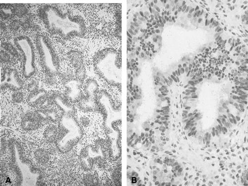
at large. Nevertheless, if cytologic atypia is the only feature to look for, endometrial cytology may be highly accurate in distinguishing carcinoma from normal or hyperplasia without cytologic atypia (Fig. 3). In one study, endometrial cytology using plastic brushes yielded 79% sensitivity, 95.4% specificity, and 80.5% negative predictive value.41 If the smear contains normal endometrial cells, the patient may have either a normal or a hyperplastic uterine lining. Often, hyperplasia without cytologic atypia is indistinguishable from normal proliferative endometrium. Because this form of hyperplasia is not a carcinoma precursor, however, the patients with symptoms such as uterine bleeding can be treated conservatively. Most cytologic laboratories lack expertise for distinguishing cytologic atypia related to neoplasia from atypia associated with degeneration or repair. As a result, false-positive rates may be too high to justify the routine use of cytology for endometrial disease. Also, the screening of an endometrial smear is time-consuming, and interpretation is difficult because of the complexity of endometrial gland cell morphology. Many carcinoma mimics lead to false-positive results.33,34


TABLE 4. Accuracy of Endometrial Cytologic Methods for Detecting Neoplasia*


Method

Diagnostic Accuracy(%)

Unsatisfactory Specimen(%)

Brushing (Endo-Pap, Gynecyte, Endocyte, Endoscan)

91–100

4–10

Aspiration (Isaacs cell sampler, Gravlee-jet washer)

96–100

10–12


*Histologically verified.
Including carcinoma and atypical hyperplasia.
In postmenopausal women.


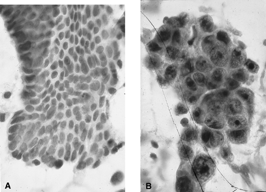
Fig. 3. Endometrial cytology obtained by endometrial aspiration. A. Hyperplastic endometrial cells with regular cytologic pattern are indistinguishable from normal endometrial cells of the proliferative phase of the cycle. B. Atypical endometrial cells with pleomorphic, hyperchromatic nuclei and macronucleoli. This obese patient was asymptomatic and had a FIGO stage 1A well-differentiated invasive adenocarcinoma.

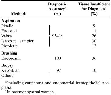
HISTOLOGIC METHODS.

At present, histologic sampling is the best means to diagnose either asymptomatic or symptomatic (abnormal uterine bleeding) endometrial neoplasia. Plastic disposable or metal reusable devices using brushing, aspiration biopsy, suction curettage, or stroke biopsy have been used with similarly high diagnostic accuracy (Table 5).34 The pitfalls of histologic methods lie in their relatively high cost and degree of discomfort. The latter leads to low compliance rates for repeat testing. Conventional curettage is much too costly yet not 100% foolproof as far as diagnostic accuracy is concerned.42 According to current experience including our own, the endometrial devices that seem to be the most cost-effective and are associated with the least discomfort for patients are the endometrial aspirators.34 In cases in which tissue is not obtained with one of the low-vacuum, suction-type aspirators, particularly in an elderly postmenopausal woman whose endometrium is more often than not atrophic, aspirators with a powerful vacuum suction force (e.g., Vabra aspirator; Tis-u-Trap; or sharp-bladed, four-stroke biopsy curette) provide diagnostic tissues.


TABLE 5. Accuracy of Endometrial Histologic Methods for Diagnosing Neoplasia


We have had success in using the endometrial brush Gynecyte (Looper Surgical, Inc; European version of Endocyte) for cytologic sampling of the endometrium and the endometrial Pipelle (Unimar, Wilton, CT) or Endocell (Wallach Surgical, Inc, Milford, CT) (Figs. 4 and 5) and, when appropriate, the Kevorkian curette (EuroMed, Redmond, WA) (see Fig. 5) for histologic sampling in asymptomatic and symptomatic women at risk for endometrial carcinoma and its precursors.34 In about 10% of postmenopausal women, the endometrial cavity is difficult or impossible to penetrate because of severe stenosis of the external/internal os or because of internal os spasm. In these cases, placing the patient on sequential cyclic therapy with conjugated estrogens (Premarin) (0.625 mg for 25 days) and medroxyprogesterone (Provera) (5 mg for 11 to 12 days) for 3 consecutive months often results in adequate dilation of the external/internal os to allow penetration of the endometrial cavity. Another alternative is to perform TVS and assess the thickness of the endometrium (see Transvaginal Ultrasonography later). Finally, traction of the uterus with the endocervical Emmett’s tenaculum or a skin (Iris) hook (see Fig. 5) is of considerable help for entering the endometrial cavity in the office. If an endometrial aspirator of the Pipelle type is used, it is important to move and rotate the cannula under negative action suction force within the endometrial cavity at least six times to sample the greatest surface area of the endometrium. In a comparison of the Pipelle versus the Vabra aspirator, the percentage of endometrial surface mucosa sampled with the Pipelle was 4.2% versus 42% with the Vabra aspirator and 60% with D&C under general anesthesia.43 The difference in percentages of area sampled is likely due to the comparatively greater suction force of the Vabra than the Pipelle device.

Fig. 4. Endometrial Endocell. Flexible polypropylene suction cannula with a 24-cm-long, 3-mm-wide outer sheath and a fine-caliber piston (top). Detailed view of the 2.5-mm distal side port through which the specimen is aspirated (bottom).

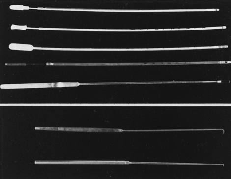
Fig. 5. Endometrial aspirators and curette. Pipelle, Z-xampler (BEI/Zinnanti, Chatsworth, CA), Uterobrush (Medscand USA, Hollywood, FL), Endocell, and Kevorkian curette without baskets (top). Emmet tenaculum and iris hook (Cooper Surgical, Shelton, CT) for uterine traction (bottom).

Sampling for Histology: A Step-by-Step Guide

  1. Bimanually examine the uterus to determine its position.
  2. Clean the cervix and vagina with acetic acid or other aseptic solution.
  3. Insert an Emmett’s tenaculum or Iris hook (see Fig. 5) into the outer one third of the endocervical canal and pull gently to obtain traction of the uterus.
  4. Insert an endometrial aspirator into the endocervical canal (see Figs. 4 and 5). When the aspirator is at the lower uterine segment level, push and rotate it to facilitate entering the endometrial cavity.
  5. When it is at the fundus, pull the plunger back rapidly and completely in the cannula to create a high negative pressure gradient.
  6. Move the cannula back and forth 6 to 12 times in the endometrial cavity and rotate it at the same time.
  7. Remove the cannula with the plunger pulled back (retain suction) from the endometrial cavity. Empty the material on a lens paper by pushing the plunger forward and place it in 10% buffered formalin tissue fixative.

If little or no tissue is obtained, the procedure can be repeated once. If still no tissue has been obtained, endometrial sampling can be performed using either the Vabra or other powerful aspirators or a metal curette (see Fig. 5). If still no tissue is obtained and the uterus is small, one can assume endometrial atrophy or fibrous pedunculated polyps are present. TVS or hysteroscopy, if the patient is symptomatic, may be performed.

In current practice, staging of endometrial carcinoma is surgical (hysterectomy, bilateral salpingo-oophorectomy, and pelvic node biopsy)44 and includes the histologic assessment of invasion of the endocervical mucosa (International Federation of Gynecology and Obstetrics [FIGO] stage IIA) versus the stroma (FIGO stage IIB) in the hysterectomy specimen. As a result, the preoperative evaluation of the endocervical canal is no longer necessary. The exception to the rule is a younger, premenopausal woman in whom a fractional sampling of the uterus can determine whether the patient has an endocervical or an endometrial primary tumor.

TRANSVAGINAL ULTRASONOGRAPHY.

TVS can visualize the endometrium on a monitor when a 5-MHz probe is placed against the vaginal fornix. The thickness of the endometrium can be measured with precision because the endometriomyometrial junction has a distinct halo-like appearance (Fig. 6). TVS is highly sensitive but also has high false-positive rates (low specificity) for identifying endometrial carcinoma. Studies suggested that specificity may be improved without jeopardizing sensitivity rates if the cutoff values were based on length of time since menopause.41 When the endometrial thickness is 4 mm for women less than 5 years since menopause and 3 mm for women more than 5 years since menopause, TVS had a 97.4% sensitivity, 75.7% specificity, and 99.7% negative predictive value.

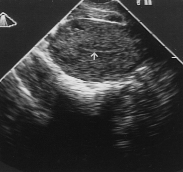
Fig. 6. Transvaginal ultrasonography of the uterus. Note the atrophic endometrial lining (arrow).

MAGNETIC RESONANCE IMAGING.

At present, magnetic resonance imaging and computed tomography have been proved to be useful for obtaining preoperative data on the extent and depth of myometrial invasion by endometrial carcinoma rather than in the primary diagnosis of endometrial carcinoma and its precursors.45 Its role in the primary diagnosis of endometrial cancer and its precursors remains to be determined.

HYSTEROSCOPY.

The value of hysteroscopy in the diagnosis and directed biopsy of a variety of intracavitary or endometrial lesions in women with postmenopausal bleeding has been extensively documented (see Chap. 36). If insufficient tissue is obtained on suction curettage, or if a patient continues to have abnormal bleeding, a formal D&C is often recommended, despite the fact that its superiority over office procedures in the diagnosis of cancer has not been established.42

Absolute indications for hysteroscopy have not been established (Fig. 7). When available, however, hysteroscopy is indicated in any woman with abnormal uterine bleeding in whom an intrauterine abnormality is suspected. Other indications include recurrent miscarriages, infertility caused by endometrial pathology, removal of an impacted intrauterine device, and suspected submucous leiomyomas before abdominal myomectomy. Hysteroscopy is contraindicated in the presence of active infection and intrauterine pregnancy. Active bleeding is a relative contraindication to office hysteroscopy only because blood interferes with vision if carbon dioxide is used as a distending medium. In patients who have severe medical problems, it is prudent to perform hysteroscopy in an outpatient setting where full monitoring and resuscitation facilities are available.

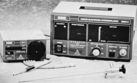
Fig. 7. Hysteroscope with light source (left) and insufflator (right).

DILATION AND CURETTAGE.

D&C essentially has been replaced by office-based endometrial biopsy using flexible aspiration devices. The latter is more cost-effective than D&C, and the diagnostic yield in symptomatic and asymptomatic women is similar to D&C with sensitivity and specificity rates of 90% and 95%.34 Cervical stenosis prevents successful endometrial sampling in about 10% of cases.

FALSE-NEGATIVE HISTOLOGY.

Even direct sampling of the endometrium for histology may fail to detect adenocarcinoma. In several studies, D&C under general anesthesia missed 10% of endometrial carcinomas.34,42 This is not surprising for, as was stated earlier, only 60% of mean surface area is sampled with D&C versus 40% for Vabra curettage and 4% for endometrial biopsy with the Pipelle endometrial aspirator.43 Others found 4 of 86 (4.6%) women with postmenopausal bleeding with endometrial carcinoma who had either a negative endometrial biopsy result or D&C within 2 years before cancer diagnosis.46 In another study from Australia,47 the false-negative rate of endometrial biopsy of focal adenocarcinomas of the endometrium was 47%.

Back to Top
TREATMENT OF ENDOMETRIAL HYPERPLASIA

Although there are no bona fide treatment regimens for EH, most current guidelines recommend either medical or surgical treatment of women with EH (Fig. 8). The choice depends on the histopathologic form of hyperplasia, the reproductive status of the woman, whether the patient is on estrogen-only replacement therapy, and her general health. In women who desire to conceive, the initial treatment should include progestational suppression, regardless of whether the lesion is classified as hyperplasia with or without atypia or even well-differentiated adenocarcinoma. Although this approach may undertreat an adenocarcinoma, previous experience suggests that the risk of these women having myometrial invasion is exceedingly small (0.6%).48 Although some cases of AH/early intramucosal adenocarcinoma respond to exogenous progestogens, ovulation inducers, or both, in most cases the lesions tend to recur within a few months to a few years after delivery of the newborn. Medical hormone therapy is also given to women whose general health is unsuitable to withstand surgery.

Fig. 8. Management scheme for endometrial hyperplasia. *If on, estrogen-only replacement therapy (ERT), discontinue, and switch the patient to progestin (P) therapy. **If complete response (CR), prescribe continuous combined hormone replacement therapy (CC-HRT), if required. NR, no response.

Surgery (i.e., transabdominal hysterectomy) with or without bilateral salpingo-oophorectomy is recommended for women who have persistent EH without atypia but are symptomatic (abnormal uterine bleeding) and women in the postreproductive age group with AH. Surgery is justified in this group in the face of 25% to 35% progression rates to invasion and an 80% failure rate to respond to progestational therapy.7 There is no compelling evidence to indicate that women with complex hyperplasia without atypia (formerly known as adenomatous hyperplasia) respond less favorably to progestational therapy than women with simple hyperplasia.7 Women who develop EH with or without atypia during estrogen-alone replacement therapy may benefit from the addition of progestins into their replacement regimen. The rare patient (1%) who develops EH while on combined cyclic or continuous HRT37 may benefit from either higher doses of combined HRT or simply switching to a progestin-only replacement therapy for 3 months to attempt reverting the hyperplastic endometrium to normal.

It has been shown that duration of progestin administration is crucial for inhibiting endometrial mitotic activity; this is important because control of endometrial growth is primarily related to control of epithelial mitotic activity. Inhibition of endometrial mitotic activity is noted after 11 days of progestin treatment.49 The most frequent hormone preparations used for medical treatment of evaluated hyperplasia with or without atypia are presented in Table 6.


TABLE 6. Treatment Regimens for Endometrial Hyperplasia by Type of Hormone, Dosage, and Duration*


Endometrial Hyperplasia

Hormone Preparations

Without Atypia

With Atypia

Medroxyprogesterone acetate

10 mg PO × 14 days/month

100 mg PO


1000 mg/week IM

Micronized progesterone

300 mg PO × 14 days/month

300 mg/day PO

Megestrol acetate

80 mg PO × 14 days/month

160 mg/day PO


Abbreviations: IM, intramuscularly; PO, orally.
* All regimens are given for 3 months.


EH without atypia responds well to medroxyprogesterone acetate (MPA), 10 mg orally, or micronized progesterone, 300 mg orally, once a day for 14 days per month for 3 months. Such cyclic regimens lead to withdrawal bleeding; a biopsy specimen is obtained at the end of the progestin therapy at 3 to 4 months. Complete responders should be maintained on cyclic progesterone therapy or, if appropriate, combined cyclic or continuous HRT. If a partial response is obtained, another 3-month trial with MPA, 10 mg orally four times per day, or megestrol acetate, 80 mg, for 3 months may be carried out. Nonresponders and patients with intractable breakthrough bleeding may have transabdominal hysterectomy. Progestin therapy for premenopausal women with AH calls for larger doses of MPA, 100 mg orally daily; megestrol acetate, 160 mg; or 1 g/week of MPA intramuscularly for 12 weeks. The biopsy specimen should show progestational-type endometrium with marked stromal decidualization. Induction of ovulation should follow the progestational therapy.

Gonadotropin-releasing hormone (GnRH) agonists (leuprolide acetate or triptorelin) have been tried (mainly in Europe) to treat EH with or without atypia at a dose of 1 ampule/3.75 mg intramuscularly every 28 days for 6 months.50 The rationale for this form of experimental therapy is that the endometrium in health and disease, similar to the breast, ovary, and prostate, contains GnRH receptors and that GnRH agonist may down-regulate GnRH receptors. This down-regulation results in a direct antiproliferative effect of endometrial growth. Of patients, 25% recurred with hyperplasia within 16 months after the end of the 6-month GnRH agonist therapy.

Back to Top
CONTROVERSIAL ISSUES

Should women who are to be placed on the combined variant of HRT receive endometrial evaluation, and if so, should endometrial biopsy or TVS be used? There may be two factors in favor of assessing the endometrium in these women, preferably by obtaining histologic material. There is growing evidence of discovering preexisting endometrial carcinoma in women on either combined cyclic or combined continuous HRT.51,52,53,54 It is important to avoid legal responsibilities in subjecting a patient with preexisting endometrial carcinoma to HRT. Although in most of these cases the endometrial carcinoma is early-stage FIGO IA/B, well-differentiated FIGO grade I with excellent prognosis, some are advanced carcinomas with poor prognostic factors. Also, some patients are asymptomatic, and it is possible at least theoretically that earlier detection could lead to better survival than later diagnosis of disease.

Screening for endometrial pathology with TVS has been suggested in women receiving long-term tamoxifen therapy.55 Others caution that endometrial TVS may have high false-positive rates because tamoxifen exerts an echogenic and sonolucent effect in the endometrial stroma and myometrium, masquerading as hyperplasia or carcinoma.56 Ultrasonographic studies found no evidence of increased prevalence of endometrial pathology in a series of 108 women on tamoxifen therapy who had biopsies.57 The various endometrial changes in postmenopausal breast carcinoma patients on tamoxifen treatment include endometrial polyps of the adenomatous type, hyperplasia, carcinoma, and sarcoma.58

Back to Top
PROGESTERONE CHALLENGE TEST

Historically, Padwick and colleagues59 introduced the progesterone challenge test into clinical practice in Europe, particularly in the United Kingdom, to the extent that bleeding on or after the 11th day of progestin administration was considered assurance of effective endometrial protection. This study was based on only 96 women, however. In a more recent study of 413 postmenopausal women on combined HRT using progestins for 10 days, there was no correlation between endometrial histology including hyperplasia and timing of onset of bleeding.60 In our experience of 25 years in practice and clinical research, although many women with an estrogenized endometrium including hyperplasia may experience withdrawal bleeding, others do not.61

Back to Top
CONCLUSIONS

Invasive carcinoma of the endometrium is preceded by AH (intraepithelial neoplasia), which by genetic markers is monoclonal and morphologically is identified by significant cytologic atypia and 50% or greater reduction of stroma versus endometrial glands. EH without atypia is not a carcinoma precursor lesion.

Although endometrial carcinoma and its precursors are significant because of their morbidity, mortality resulting from carcinoma is low. As a result, mass screening for asymptomatic endometrial carcinoma and its precursors is not cost-effective and is not recommended. Nevertheless, if screening for endometrial carcinoma is desired in a private practice, it should focus on women aged 55 years old and older and women with high carcinoma risk indicators.

Cytologic sampling of the endometrium directly is limited to communities in which cytologic expertise is available. The most often used method to evaluate the endometrium is histology; to reduce cost, it should be carried out in the office, and the device used should employ vacuum suction force, be disposable, and be of low cost. TVS seems to be a potentially useful alternative to histology for screening and diagnosing endometrial carcinoma and hyperplasia. Hysteroscopy is the diagnostic method of choice for patients in whom office biopsy and TVS failed to provide a definite diagnosis. Patients with EH without cytologic atypia and patients with atypia who desire to conceive should have progestational therapy. Patients with atypia or intractable uterine bleeding without atypia benefit from hysteroscopy.

Back to Top
REFERENCES

1. Mittal KR, Schwartz PE, Barwick KW: Architectural (FIGO) grading, and other prognostic indicators in stage 1 endometrial adenocarcinoma with identification of high-risk and low-risk groups. Cancer 61:538, 1988

2. Bokhman J: Two pathogenic types of endometrial carcinoma. Gynecol Oncol 15:10, 1983

3. Deligdisch L, Holinka C: Endometrial carcinoma: Two diseases? Cancer Detect Prev 10:237, 1987

4. Gusberg SB: Precursors of corpus cancer. In Gusberg SB, Shingleton H, Deppe G (eds): Female Genital Cancer. pp 337, 378 New York, Churchill Livingstone, 1988

5. Fox H, Buckley CH: The endometrial hyperplasias and their relationship to endometrial neoplasia. Histopathology 6:493, 1982

6. Kurman RJ, Kaminski PF, Norris HJ: The behavior of endometrial hyperplasia: A long-term study of “untreated” hyperplasia in 170 patients. Cancer 56:403, 1985

7. Ferenczy A, Gelfand MM: The biologic significance of cytologic atypia in progestogen-treated endometrial hyperplasia. Am J Obstet Gynecol 160:126, 1989

8. Colgan TJ, Norris HJ, Foster W, et al: Predicting the outcome of endometrial hyperplasia by quantitative analysis of nuclear features using a linear discriminant function. Int J Gynecol Pathol 1:347, 1983

9. Baak JPA: The use and disuse of morphometry in the diagnosis of endometrial hyperplasia and carcinoma. Pathol Res Pract 17:287, 1981

10. Silverberg SG: Hyperplasia and carcinoma of the endometrium. Semin Diagn Pathol 5:135, 1988

11. Sherman AI, Brown S: The precursors of endometrial carcinoma. Am J Obstet Gynecol 135:947, 1979

12. Ferenczy A: Endometrial hyperplasia and neoplasia: A two disease concept. In Cohen CJ, Kase NG (eds): Contemporary Issues in Obstetrics and Gynecology, Gynecologic Oncology. pp 197, 213 New York, Churchill Livingstone, 1988

13. Kurman RJ, Norris HJ, Hendrickson MR, et al: Endometrial stromal invasion in the diagnosis of well-differentiated carcinoma (letter). Am J Surg Pathol 8:719, 1984

14. Winkler B, Alvarez S, Richart RM, et al: Pitfalls in the diagnosis of endometrial neoplasia. Obstet Gynecol 64:185, 1984

15. Kendall BS, Ronnett BM, Isaacson C, et al: Reproducibility of the diagnosis of endometrial hyperplasia, atypical hyperplasia, and well-differentiated carcinoma. Am J Surg Pathol 22:1012, 1998

16. Vogelstein B, Kinzler KW: The multistep nature of cancer. Trends Genet 9:138, 1993

17. Mutter GL, Chaponot M, Fletcher J: A PCR assay for non-random X chromosome inactivation identifies monoclonal endometrial cancers and precancers. Am J Pathol 146:501, 1995

18. Mutter GL, Boynton KA, Faquin WC, et al: Allelotype mapping of unstable microsatellites establishes direct lineage continuity between endometrial precancers and cancer. Cancer Res 56:4483, 1996

19. Maxwell G, Risinger J, Gumbs C, et al: Mutation of the PTEN tumor suppressor gene in endometrial hyperplasia. Cancer Res 58:2500, 1998

20. Tate JE, Mutter GL, Boynton KA, Crum CP: Monoclonal origin of vulvar intraepithelial neoplasia and some vulvar hyperplasias. Am J Pathol 150:315, 1997

21. Mutter GL, Baak JPA, Crum CP, et al: A multidisciplinary approach to diagnosis of endometrial precancers: Clonal analysis, histopathology, and computerized morphometry. Am J Pathol 190:462, 2000

22. Dunton C, Baak J, Palazzo J, et al: Use of computerized morphometric analyses of endometrial hyperplasias in the prediction of coexistent cancer. Am J Obstet Gynecol 174:1518, 1996

23. Baak JPA, Nauta J, Wisse-Brekelmans E, Bezemer P: Architectural and nuclear morphometrical features together are more important prognosticators in endometrial hyperplasia than nuclear morphometrical features alone. J Pathol 154:335, 1988

24. Mutter GL: Endometrial precancer type collection. Accessed 1999 Available at http://www.endometrium.org

25. Karlsson B, Granberg S, Wikland M, et al: Transvaginal ultrasonography of the endometrium in women with postmenopausal bleeding: A Nordic multicenter study. Am J Obstet Gynecol 172:1488, 1995

26. Feldman S, Cook EF, Harlow BL, Berkowitz RS: Predicting endometrial cancer among older women who present with abnormal vaginal bleeding. Gynecol Oncol 56:376, 1995

27. Weber AM, Belinson JL, Piedmonte MR: Risk factors for endometrial hyperplasia and cancer among women with abnormal bleeding. Obstet Gynecol 93:594, 1999

28. Ferenczy A: Endometrial carcinoma and its precursors in relation to hormone replacement therapy. In Lorrain J, Plouffe L Jr, Ravnikar V, et al (eds): Comprehensive Management of Menopause. pp 254, 268 New York, Springer-Verlag, 1994

29. Richardson GS, MacLaughlin DT (eds): Hormonal Biology of Endometrial Cancer. A Series of Workshops on the Biology of Human Cancer. Report No. 8. Geneva, International Union Against Cancer, 1978

30. Homesley HD, Zaino R: Endometrial cancer: Prognostic factors. Semin Oncol 21:71, 1994

31. Pritchart KI: Screening for endometrial cancer: Is it effective? Ann Intern Med 110:177, 1989

32. Archer DF, McIntyre-Seltman K, Wilborn WW Jr, et al: Endometrial morphology in asymptomatic postmenopausal women. Am J Obstet Gynecol 165:317, 1991

33. Koss LG, Schriber K, Oberlander SG, et al: Detection of endometrial carcinoma and hyperplasia in asymptomatic women. Obstet Gynecol 64:1, 1984

34. Ferenczy A: Methods for detecting endometrial carcinoma and its precursors. In Buchsbaum HJ, Sciarra JJ (eds): Gynecology and Obstetrics. pp 1, 8 Vol 4:New York, Harper & Row, 1982

35. MacMahon B: Risk factors for endometrial cancer. Gynecol Oncol 2:122, 1974

36. Burke TW: How should we monitor women treated for endometrial carcinoma? Gynecol Oncol 65:377, 1997

37. The Writing Group for the PEPI Trial: Effects of hormone replacement therapy on endometrial histology in postmenopausal women: The Postmenopausal Estrogen/Progestin Interventions (PEPI) trial. JAMA 275:370, 1996

38. Hakala T, Mecklin J-P, Forss M, et al: Endometrial carcinoma in the cancer family syndrome. Cancer 68:1656, 1991

39. Feldman S, Berkowitz RS, Tosteson ANA: Cost-effectiveness of strategies to evaluate postmenopausal bleeding. Obstet Gynecol 81:968, 1993

40. Nguyen TN, Bourdeau J-L, Ferenczy A, Franco EL: Clinical significance of histiocytes in the detection of endometrial adenocarcinoma and hyperplasia. Diagn Cytopathol 19:89, 1998

41. Tsuda H, Kawabata M, Yamamoto K, et al: Prospective study to compare endometrial cytology and transvaginal ultrasonography for identification of endometrial malignancies. Gynecol Oncol 65:383, 1997

42. Grimes DA: Diagnostic dilation and curettage: A reappraisal. Obstet Gynecol 142:1, 1982

43. Rodriguez GC, Yaqub N, King ME: A comparison of the Pipelle device and the Vabra aspirator as measured by endometrial denudation in hysterectomy specimens: The Pipelle device samples significantly less of the endometrial surface than the Vabra aspirator. Am J Obstet Gynecol 168:55, 1993

44. Homesley HD: Revised 1988 International Federation of Gynecology and Obstetrics staging systems for endometrial and vulvar cancer: An assessment. Clin Obstet Gynecol 35:89, 1992

45. Takahashi S, Murakami T, Narumi Y, et al: Preoperative staging of endometrial carcinoma: Diagnostic effect of T2-weighted fast spin-echo MR imaging. Radiology 206:539, 1998

46. Feldman S, Shapter A, Welch WR, Berkowitz RS: Two-year follow-up of 263 patients with post/perimenopausal vaginal bleeding and negative initial biopsy. Gynecol Oncol 55:56, 1994

47. Quinn MA, Kneale BJ, Fortune DW: Endometrial carcinoma in premenopausal women: A clinicopathological study. Gynecol Oncol 20:298, 1985

48. Longacre TA, Chung MH, Jensen DN, et al: Proposed criteria for the diagnosis of well-differentiated endometrial carcinoma: A diagnostic test for myoinvasion. Am J Surg Pathol 19:371, 1995

49. Moyer DL, de Lignieres B, Driguez P, Pez J-P: Prevention of endometrial hyperplasia by progesterone during long-term estradiol replacement: Influence of bleeding pattern and secretory changes. Fertil Steril 59:992, 1993

50. Agorastos T, Bontis J, Vakiani A, et al: Treatment of endometrial hyperplasia with gonadotropin-releasing hormone agonists: Pathological, clinical, morphometric, and DNA-cytometric data. Gynecol Oncol 65:102, 1997

51. McGonigle KF, Karlan BY, Barbuto DA, et al: Development of endometrial cancer in women on estrogen and progestin hormone replacement therapy. Gynecol Oncol 55:126, 1994

52. Shipley CF III, Smith ST, Dennis EJ III, et al: Evaluation of pretreatment transvaginal ultrasonography in the management of patients with endometrial carcinoma. Am J Obstet Gynecol 167:406, 1992

53. Comerci JT, Fields AL, Runowicz CD, Goldberg GL: Continuous low-dose combined hormone replacement therapy and the risk of endometrial cancer. Gynecol Oncol 64:425, 1997

54. Dallenback-Hellweg G, Hahn U: Mucinous and clear cell adenocarcinomas of the endometrium in patients receiving antiestrogens (Tamoxifen) and gestagens. Int J Gynecol Pathol 14:7, 1995

55. Jordan VC, Morrow M: Should clinicians be concerned about the carcinogenic potential of tamoxifen? Eur J Cancer 30:1714, 1994

56. Goldstein SR: Unusual ultrasonographic appearance of the uterus in patients receiving tamoxifen. Am J Obstet Gynecol 170:447, 1994

57. Cecchini S, Ciatto S, Bonardi R, et al: Screening by ultrasonography for endometrial carcinoma in postmenopausal breast cancer patients under adjuvant tamoxifen. Gynecol Oncol 60:409, 1996

58. Kennedy MM, Baigrie CF, Manek S: Tamoxifen and the endometrium: Review of 102 cases and comparison with HRT-related and non-HRT-related endometrial pathology. Int J Gynecol Pathol 18:130, 1999

59. Padwick ML, Pryse-Davies J, Whitehead MI: A simple method for determining the optimal dosage of progestin in postmenopausal women receiving estrogens. N Engl J Med 315:930, 1986

60. Sturdee DW, Barlow DH, Ulrich LG: Is the time of withdrawal bleeding a guide to endometrial safety during sequential oestrogen-progestagen replacement therapy? Lancet 344:979, 1994

61. Gelfand MM, Ferenczy A: A prospective 1-year study of estrogen and progestin in postmenopausal women: Effects on the endometrium. Obstet Gynecol 74:398, 1989

Back to Top