Chapter 14
Endocrine Aspects of Adenocarcinoma of the Endometrium
Charles B. Hammond and Michael R. Soules
Main Menu   Table Of Contents

Search

Charles B. Hammond, MD
Professor, Department of Obstetrics and Gynecology, Duke University Medical Center, Durham, North Carolina (Vol 4, Chaps 14, 48)

Michael R. Soules, MD
Professor and Director, Reproductive Endocrinology and Department of Obstetrics and Gynecology, University of Washington, Seattle, Washington (Vol 4, Chap 14; Vol 5, Chap 56)

INTRODUCTION
EFFECTS OF ESTROGEN AND PROGESTERONE ON THE ENDOMETRIUM
ESTROGEN SYNTHESIS AND METABOLISM
ESTROGEN LEVELS AND LIFE STAGES
ESTROGENIC POTENCIES
ESTROGEN CONVERSIONS
ENDOCRINOLOGY OF PATIENTS WITH ENDOMETRIAL CANCER
REFERENCES

INTRODUCTION

Adenocarcinoma of the endometrium is the most common genital malignancy in the United States and accounts for over 95% of endometrial malignancies. Although patients often respond well to various therapies, it is estimated that 3,000 to 4,000 women will die annually from these malignancies1. Debate continues as to whether the incidence of adenocarcinoma of the endometrium is rising or whether the increased number of patients with this disease merely reflects the greater number of people living to older ages and a more aggressive diagnostic surveillance by their physicians. Endometrial cancer is often preceded by or associated with various types of hyperplasia of the endometrium, and although seen in younger women, it is usually a disease of the postmenopausal years. The discussion in this chapter regarding hormonal influences on cancer of the uterine corpus will be confined to endometrial adenocarcinoma.

A number of findings suggest that some type of endocrinopathy, yet undetermined, may be a factor in the genesis of endometrial cancer. Such data include the results of endocrinologic manipulations in animals, the association of endometrial cancer in women with certain physical and biochemical characteristics, and the more recent controversial findings regarding estrogen replacement therapy. All of these factors suggest an important role for the sex steroids estrogen and progesterone in patients with these diseases.

This chapter will attempt to explore the endocrinology of patients with adenocarcinoma of the endometrium and to discuss data regarding the possible etiologic and therapeutic roles of estrogen and progesterone.

Back to Top
EFFECTS OF ESTROGEN AND PROGESTERONE ON THE ENDOMETRIUM

The endometrium, or uterine mucosa, responds promptly to changes in the hormonal milieu, notably estrogen and/or progesterone. The hormonal concept of the endometrial cycle dates from the mid 19th century and has been reviewed by Novak and Novak2. Pfleuger, in 1863, first ascribed menstruation to the presence of the ovaries and it was believed to be caused by a reactive pelvic hyperemia secondary to a spinal reflex originating from the ripening follicles in the ovary. The transplant work of Knauer negated the necessity for a neural linkage in this system, which reaffirmed a hormonal role for the ovary in the endometrial cycle. In 1903, Frankel demonstrated the endocrine importance of the corpus luteum, which previously had been thought to be functionless. Hitschman and Adler in 1908 described the histochemical cycle of the endometrium, and it then became possible to integrate the ovarian and endometrial cycles.

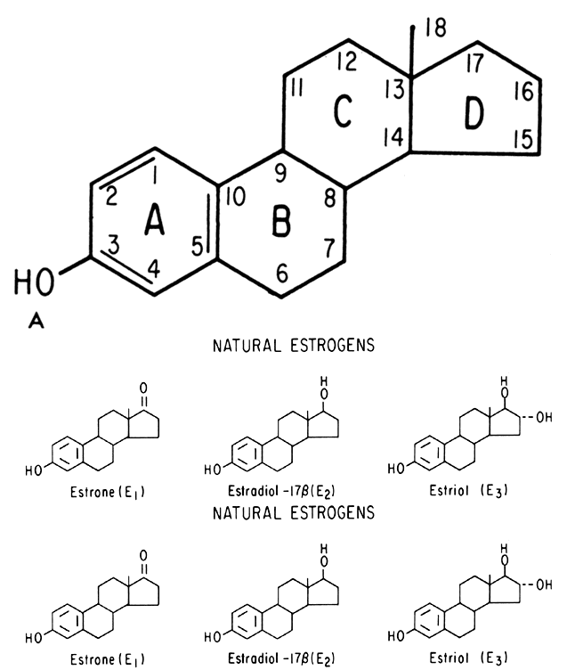
In 1917, Frank called attention to the estrus-producing effects of a hormone contained in ovarian follicular fluid. Allen and Doisy in 1923 made more elaborate demonstrations of the müllerian-stimulating effects of this hormone. After Zondek demonstrated large amounts of this substance in the urine of pregnant women, two groups simultaneously in 1929 (Doisy, Veter, and Thayer from the United States and Butenandt from Germany) isolated and characterized the purified urinary extract. This urinary extract was later to be known as estrogen. The term estrogen has now been broadened to include all of the natural and synthetic derivatives of this female sex hormone.

Long before either the follicular or corpus luteal hormones were isolated, it seemed certain that two separate and distinct ovarian hormones must exist, simply on the basis of the histologic changes of the endometrium. Frankel's demonstration of the importance of the corpus luteum in 1903 was followed by the isolation and purification of progesterone by Butenandt, Allen, and others in 1929. Entwined in these discoveries was the identification of the pituitary gonadotropic hormones (follicle-stimulating hormone [FSH] and luteinizing hormone [LH] and their role in the ovarian cycle and the ultimate control of endometrial growth, maturation, and menstruation.

Research in the next 4 decades was involved with further purification and identification of the various natural estrogens and progesterone, their synthesis for experimental and therapeutic use, the development of highly sensitive and specific assays applicable to small samples of biologic fluid, and the development of molecular analogues which not only could be used orally but also possessed varying degrees of differing biologic effects. Simultaneous with these advances in steroid endocrinology were further explorations into the natural history of endometrial adenocarcinoma and hyperplasia. Endometrial hyperplasia, which can arise after prolonged estrogen stimulation, was found to be associated with and perhaps a precursor of endometrial cancer.

Effects of Estrogen

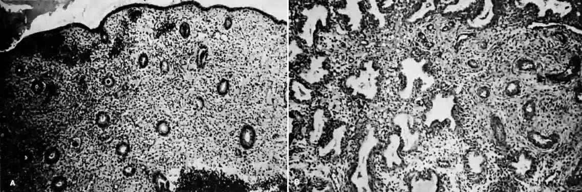
Estrogens are growth hormones for tissues derived from the müllerian ducts, the endometrium being one of the most sensitive of these tissues. During the follicular phase of the endometrial cycle (proliferative) the endometrium regenerates from perivascular “collars” about residual vessels remaining after menstruation. This epithelium gradually thickens, becomes taller and more dense, and is characterized in this phase by increased mitotic activity in glands and stroma. This estrogenic stimulation results in straight tubular glands and a compact stroma when follicular phase tissue samples are microscopically examined (Fig. 1A).

Fig. 1. A. Early proliferative endometrium (days 3–6). Surface epithelium is intact. Glands are straight and tubular without mitotic figures or pseudostratification. This normal endometrium was exposed only to estrogen stimulation at the time of biopsy. B. Late secretory endometrium (days 25–26) in a normal menstrual cycle. Tissue has been predominantly stimulated by progesterone for 11 to 12 days. Glands are convoluted and have expended most of their secretory products. The stroma has undergone an extensive decidual reaction.

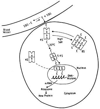
It is now known that estrogens exert their end-organ effect by activating a complex intracellular mechanism. Tissues which respond to estrogen possess intracytoplasmic proteins (receptors) that preferentially bind specific steroids (Fig. 2). For instance, a cell from the uterus will possess 5,000 to 15,000 estrogen receptors whereas a cell from the spleen will have none. These receptors recognize estrogens by their three dimensional and chemical characteristics and bind it with high affinity (KD =10-10), specificity, and saturability. The estrogen molecules present in the circulation are relatively loosely bound to intravascular carrier proteins (sex-steroid-binding globulin [SBG]) (KD = 10-8) or to albumin. In excess of 95% of the estrogen in the circulation is found in the bound form. The estrogen readily diffuses across the cell membrane in its active free form due to a concentration and a binding gradient. The estrogen molecule is relatively small (molecular weight is 300) and lipophilic and probably passes through the cell membrane by simple diffusion. Once in the cell, the estrogen is promptly bound to the intracellular (intracytoplasmic) receptor protein, which then undergoes a series of complex spatial changes prior to intranuclear transport. This nuclear transport occurs within 30 to 45 minutes after the target tissue is exposed to estrogen. The following system of nuclear interactions between receptor and DNA is a model that has been proposed by McCarty3. The activated receptor-estrogen complex then nonspecifically binds to the DNA and protein of dispersed chromosomes (euchromatin) and stimulates acetylation of the histone protein. This acetylation of the histones in nucleosomes causes the nucleosome to “open up” and expose specific DNA segments for transcription. The “estrogen message” is transcribed into new messenger RNA which then migrates back into the cytoplasm and activates various cellular processes including new protein synthesis. The now “freed” receptor protein is probably recycled back into the cytoplasm for further use3,4. The estrogen receptor recognizes a molecule as being “estrogen” if its size, three-dimensional configuration, and charge are similar to the parent molecule. Therefore, the nonsteroidal synthetic estrogens may not resemble the “prototype” estrogen (estradiol-17β) on paper diagrams but are very similar in shape and other properties as seen by the cellular receptor. Estrogen receptors are perhaps the determinants of potency for estrogenic substances. The estrogen receptors preferentially bind estradiol over estriol (2x) and estrone (3x)5. This receptor also discriminates among the estrogens by binding estradiol within the cellular nucleus longer than the weaker estrogens estriol and estrone4,6. Therefore, estradiol is the most potent of the natural estrogens probably because of the greater affinity and duration of its receptor-binding compared with the other available estrogens. Receptors for estrogen and other steroid hormones can now be accurately quantified and studied. Estrogen in physiologic concentrations stimulates the synthesis of estrogen receptors and of progesterone and testosterone receptors7. Progesterone and testosterone, however, inhibit estrogen receptors. Progesterone inhibits its own receptor population in the secretory phase of human endometrium8. Thus, it is apparent that for estrogen to bind and influence a tissue, the specific estrogen receptors must be present. The potency of a particular estrogen in a tissue roughly parallels and is probably dependent on the quantity of the estrogen receptor in the cells of that tissue. Studies with estrogen receptors in breast cancers are being successfully utilized to predict the responsiveness of these tumors to hormonal manipulation9,10.

Fig. 2. Estrogen receptor kinetics. Estrogen (E) must exist in the free form separate from its carrier protein (sex-steroid-binding globulin [SBG]) prior to diffusion into the cell. The estrogen receptor has two (a and b) components (dimer) which can become an 8S tetramer under certain conditions. The 4S receptor undergoes a temperature-dependent conformational change to the 5.4S form prior to nuclear transport. After interacting with the euchromatin, the receptor complex is probably recirculated.

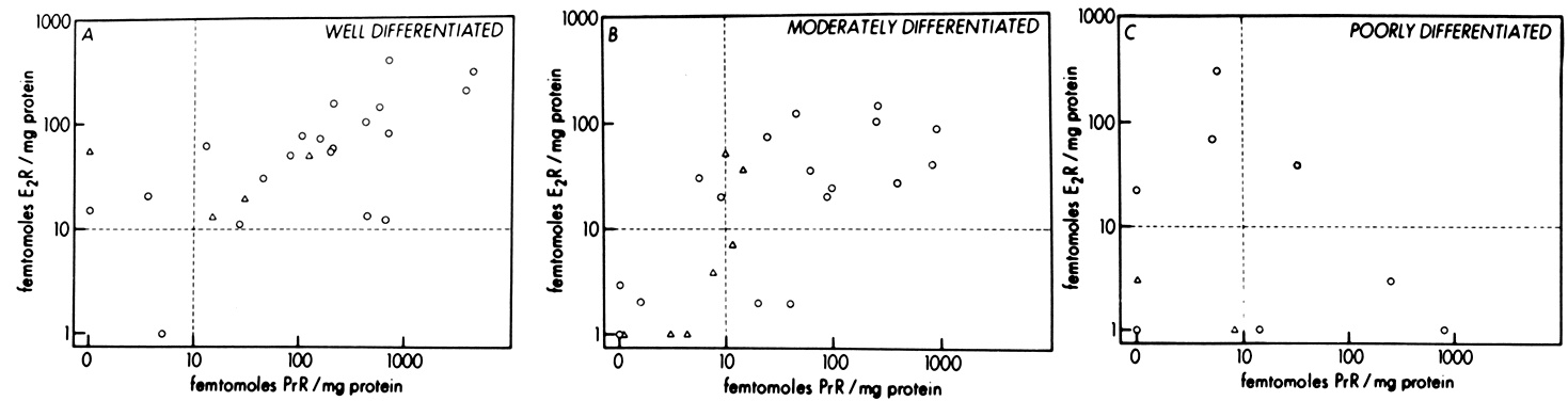
Endometrial carcinoma is another malignancy that can be hormonally dependent and manipulated by endocrine therapy. Both normal endometrium and endometrial carcinoma possess active estrogen receptors. There have been few reports showing qualitative or quantitative changes in estrogen receptors in hyperplastic endometrium or endometrial cancer, but the potential exists and detailed investigation is beginning.

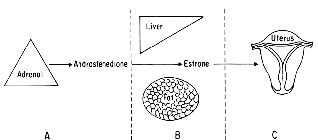
An intriguing theory has been proposed linking endometrial carcinoma to the relative levels of estrone and estriol in the postmenopausal female11. This theory ascribes a protective effect to estriol over estrone. Clearly, estrone is the dominant estrogen in postmenopausal women and also in younger women with polycystic ovarian disease; therefore, it has been implicated in the etiology of endometrial carcinoma. A protective effect from cancer for estriol was proposed when epidemiologic studies demonstrated higher estriol-estrone ratios in control groups as compared with groups with endometrial or breast cancer12,13, but other studies have failed to demonstrate these findings14,15. As noted, the receptor data show estrone and estriol competing for binding on about an equimolar basis, with similar decreases in duration of nuclear binding as compared with estradiol. There is no evidence that estriol has a different or less carcinogenic induced cellular response than estrone. Therefore, in postmenopausal patients, the unopposed levels of estrone and estriol are probably inducing the same cellular response. The concept that estriol binds to the receptor and produces a “benign” product or that it occupies the receptor for a long period of time and blocks estrone binding is not substantiated by current data. Therefore, the theory of a protective effect of estriol must rest on epidemiologic data which is conflicting and weak.

Effects of Progesterone

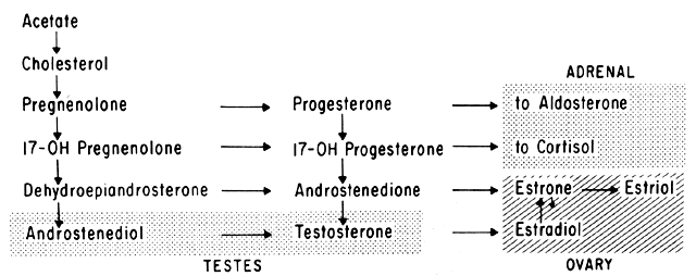
Progesterone is produced and secreted by the adrenal cortex, ovary, and placenta. From the ovary, the progesterone secretory rate is negligible during the follicular phase and increases dramatically after ovulation, yielding 30 mg per day between the 18th and 24th day of the cycle. Greater than 90% of the progesterone is removed by the liver within 25 minutes16. Progesterone acting alone has little effect on unstimulated endometrium. However, if the endometrium has been “primed” by estrogen (proliferated), then progesterone causes a secretory change in the glands, stromal edema, regression and stromal pseudodecidual formation (Fig. 1B). Under the influence of progesterone, the total thickness of the endometrium decreases due to fluid loss and three layers become defined: the basalis next to myometrium, the compact layer immediately beneath the endometrial surface, and the spongy layer between the other two layers. The basal layer undergoes little if any histologic alteration during the menstrual cycle, but mitoses are found in the glands of this layer. The spongy layer comprises a lacy labyrinth with little stroma between the glands, which are tortuous and serrated. Little change occurs in the compact layer except that gland lumens are filled with secretion. By late in the secretory phase, the endometrium has become extremely vascular and succulent, ripe for implantation. If no implantation occurs, then 2 to 3 days before menstruation the reticular framework of the endometrium begins to disintegrate and vascular compression of the coiled spiral arteries occurs with vasoconstriction and decreased blood flow. Subsequently, vascular relaxation occurs at which time bleeding and endometrial sloughing begin. Thus, after endometrial proliferation induced by estrogen, progesterone ultimately causes secretion, regression, and sloughing. Progesterone acts through specific intracellular receptors, as described for estrogen.

Abnormal Endometrial Changes

The histopathologic picture of endometrial hyperplasia differs from that of normal proliferative endometrium as a result of sustained and unopposed estrogen stimulation. It is characterized by an increased endometrial thickness, on occasion threefold or greater, and both qualitative and quantitative changes in glandular patterns which exceed similar changes in the stroma. A number of types of endometrial hyperplasia have been reported and include cystic and adenomatous hyperplasia. A further subdivision of adenomatous hyperplasia is the atypical variety. It is thought that endometrial hyperplasia represents uninterrupted growth of a tissue which normally should undergo cyclic menstrual degeneration and sloughing1.

Endometrial hyperplasia is usually found in women who experience 3 to 6 months or more of uninterrupted (by progesterone) estrogen stimulation. Continuous estrogen stimulation most often occurs in anovulatory women who maintain significant estrogen production. Women with chronic anovulation include the adolescent (in whom Frasier17 has reported that up to 50% of early cycles are anovulatory), the perimenopausal female, patients with the polycystic ovary syndrome, obese women, and a large group of females who idiopathically fail to ovulate. Another group of patients exposed to unopposed estrogen is women receiving estrogen replacement therapy without added progesterone. The clinical impact of endometrial hyperplasia falls into two basic areas: dysfunctional uterine bleeding and, perhaps, the subsequent development of adenocarcinoma of the endometrium. It has been estimated that 1% to 2% of patients with cystic endometrial hyperplasia will ultimately develop endometrial cancer1, while 5% to 15% of patients with adenomatous hyperplasia18 and 20% to 25% of women with atypical adenomatous endometrial hyperplasia19 will also ultimately develop this malignancy. While the precise incidence and mechanism of progression of carcinoma of the endometrium following in situ carcinoma is unknown, it is certainly significant.

Thus, it appears that tonic,. prolonged, and/or higher concentrations of unopposed estrogen can at least initiate hyperplasia of the endometrium. Whether the next step to endometrial malignancy can be linked to estrogen is unclear. Certain animal data support the contention that estrogen can be carcinogenic in terms of the endometrium, but the human data are less certain. Perhaps the best representation of these changes was based on data by Sommers20 and presented by Kistner21 (Fig. 3), which illustrates endometrial polyps progressing to cystic hyperplasia and then to adenomatous hyperplasia under the influence of exogenous estrogen. All three of these steps are clearly capable of being stimulated by estrogen. The linkage of estrogen to the remaining step from hyperplasia to neoplasia is less certain, but it appears to be related in some manner.

Fig. 3. There is general agreement that relatively high or sustained levels of estrogen result in endometrial polyps and endometrial hyperplasia (cystic and adenomatous). Although there is strong evidence to suggest that estrogen induces anaplasia in susceptible individuals, this step is not proved.

Back to Top
ESTROGEN SYNTHESIS AND METABOLISM

There are three naturally occurring estrogens: estrone (El), estradiol-17β (E2), and estriol (E3). Every natural estrogen molecule has 18 carbon atoms. As depicted in Figure 4A, the aromatic A ring is common to them all along with a phenolic group at C-3. They differ only in the number of hydroxyl groups: estrone, OH- at C-3; estradiol, OH- at C-3 and C-17; and estriol, OH- at C-3, C17, and C-16. The synthetic estrogens are chemically manufactured and have various additions to the basic C-18 molecule or do not chemically resemble natural estrogens at all. Natural estrogens may be artificially synthesized but the C-18 molecule contains no additions other than the natural hydroxyl groups (Figure 4B). All natural estrogen molecules originate from C-19 androgen precursors (androstenedione or testosterone). This C-19 to C-18 conversion step depends on a series of aromatizing enzymes which are present in the adrenal gland, the ovary, the placenta, and certain body tissues which do not primarily produce steroid hormones (peripheral conversion sites). Peripheral conversion of androstenedione or testosterone to estrogen occurs most efficiently in adipose tissue and in the liver. Only the adrenal gland and the ovary are capable of synthesizing estrogens from acetate and cholesterol. This is not to say that they synthesize equal amounts of estrogen: each steroid-producing gland has major and minor synthetic pathways leading to vastly different amounts of end products. These pathways are controlled by enzyme concentrations and kinetics. An example is the adrenal gland, which is enzymatically “programmed” to primarily produce glucocorticoids and only secondarily to make sex steroids. The basic pathway of sex steroid synthesis is illustrated (Fig. 5).

Fig. 4. A. Basic estrogen molecule. All natural estrogen molecules have 18 carbon atoms and a phenolic A ring with a hydroxyl group on C-3. B. The three natural and three common synthetic estrogen molecules.(From Hammond CR, Soules M: Clinical significance of estrogen metabolism and physiology. Contemp OB/GYN 11:41, 1978.)

Fig. 5. Steroid synthetic pathway. Those portions of the overall steroid pathway that are dominant in a given organ are indicated by the shaded areas. Although the adrenal gland and the testes produce estrogen, the primary organ for direct estrogen production is the ovary.

When the ovary secretes estrogen as it does during the reproductive years, the ovarian follicle is the site of steroid synthesis (Fig. 6). The follicular theca cell has long been a known source for estrogen production, but only recently have in vitro ovarian tissue culture experiments implicated the follicular granulosa cell as a significant estrogen producer. The granulosa cells lack a C-21 to C-19 step because of a relative desmolase deficiency. But when provided with a C-19 androgen precursor, they quickly convert it to estrogen. The relative contributions of theca and granulosa cells to ovarian estrogen secretion are unknown at this time. The ovary containing the dominant follicle, that follicle which will go on to eventual rupture and ovulation, produces the most estrogen, as determined by simultaneous sampling of both ovarian veins during the proliferative phase of the: menstrual cycle. The unruptured preovulatory follicle contains estrogen in 1,000-fold concentrations over peripheral blood, but the physiologic function is uncertain.

Fig. 6. The follicles and corpus luteum are the ovarian sources of estrogen in the proliferative and secretory phases of the cycle, respectively. An antral follicle in the proliferative phase is shown ( top ). The theca layer has long been a known source of estrogen in the preovulatory phase, but investigations have demonstrated a striking propensity for preovulatory granulosa cells to convert adrogens to estrogen. The ability of granulosa cells to make estrogen is demonstrated by the thousandfold increase in estrogen found in preovulatory follicular fluid.

Estrogens can be measured in body fluids (serum and urine) by hormone assays. The bioassays use a quantitative change in an estrogen responsive animal organ (ie, rat uterine weight change) and are now of only limited usefulness in clinical medicine. The modern assay technique is by radioimmunoassay. Since estrogens are lipid soluble, an ether extraction step is used to separate steroids from other serum components. Careful technique requires quantitating the efficiency of ether extraction which is usually about 80%. A specific estrogen antibody is then used along with tritium-tagged estrogen tracer to measure the antibody-tracer displacement potential of the unknown sample. The amount of tracer displaced varies directly with the amount of estrogen in the unknown sample, as shown in the following equation:

A specific antibody (Ab) generated against the estrogen molecule will bind a given amount of radio-labeled estrogen (E*). The estrogen in a serum sample will displace a certain amount of the radio-labeled estrogen from the antibody. The quantity of labeled estrogen displaced by an unknown serum sample is compared with the displacement obtained by a known amount of estrogen. The results are commonly expressed in picograms per milliliter. Depending on the specificity of the antibody used to bind estrogen in a radioimmunoassay, individual estrogens (E1, E2, or E3) or total estrogens (all three molecules) will be measured.

It is necessary to define several terms relating to estrogen synthesis. Production rate, expressed in mass of hormone per 24 hours, is the product of direct estrogen secretion by the ovary and adrenal plus any estrogen synthesized by peripheral conversion. The production rate is derived by multiplying the metabolic clearance rate by the average serum concentration of that hormone. The hormonal metabolic clearance rate is analogous to renal clearance, such as that for creatinine, and is expressed in liters of blood per unit time. The production rate of estrogen is the sum of hormone produced by glandular secretion and by peripheral conversion (Fig. 7). In experimental situations, the production rate is calculated after the metabolic clearance rate and concentration have been determined by doing an infusion of tritiated estrogen until an equilibrium is attained. Estrone has a higher metabolic clearance rate than estradiol secondary to less binding with the carrier protein SBG. In general, the more a hormone is bound to a carrier protein, the slower its metabolism; thus, it will have a lower clearance rate. The weaker estrogens, E1 and E3, exist more in the free state with less SBG binding. This decreased binding besides increasing the metabolic clearance rate also results in an initially larger volume of distribution within the body for estrone and estriol as compared with estradiol.

Fig. 7. Three sources that can contribute to the production of a given estrogen in women at any point in time are the adrenal gland, the ovary, and the peripheral conversion of precursor compounds. Peripheral conversion primarily occurs in the liver and adipose tissue. The formula for calculating the production rate is given. PR, production rate; MCR, metabolic clearance rate: C, serum concentration.

An estrogen can only bind to a receptor and exert a biologic effect when it is in the free (unbound) state. Serum estrogens are bound nonspecifically to albumin or to a specific glycoprotein (SBG). Estrogens share this binding globulin with testosterone. Serum binding proteins are not necessary to enable steroid hormones to circulate in the plasma but primarily serve as a buffering mechanism by binding more or less steroid as the plasma steroid concentrations vary. Estrogen stimulates the synthesis of SBG as it does other plasma proteins. The usual assays for estrogens measure the total concentration of both bound and free. Estrogens that have been metabolized from a lipophilic to a hydrophilic (water-soluble) form by the addition of a sulfate or glucuronide group cannot spontaneously dissociate into the free active form.

The metabolism of estrogens involves molecular interconversions, conjugation, and excretion. The 16- and 17-hydroxyl groups can undergo oxidation and reduction reactions with the production of numerous inactive epimeric forms22. The C-3 phenolic group is not altered by these reactions. Whether or not an oxidation-reduction reaction occurs first, all of the estrogen molecules must undergo conjugation in order to be excreted. Whereas E3 is present in insignificant quantities as an active estrogen, it is the major excretory form of estrogen. El and E2 are also excreted in significant quantities. Estrogens undergo conjugation to a glucosiduronate or a sulfate in the liver. There is a significant bile-enterohepatic recirculation system with estrogens, but the gut is an insignificant pathway for final estrogen excretion. The conjugated estrogens are primarily cleared by the kidney23 (Fig. 8).

Fig. 8. Estrogen metabolism. Prior to conjugation, the three basic estrogen molecules may undergo interconversions to more active or less active forms. During the interconversion phase, any of the estrogen molecules may also undergo a number of oxidation-reduction reactions leading to numerous epimeric forms. These natural or epimeric forms of estrogen are then conjugated in the liver into water-soluble forms prior to renal excretion. Conjugation with glucosidurinate is more prevalent than sulfate conjugation. The quantities of conjugated natural estrogens in the urine of a woman in the early follicular phase of her menstrual cycle is indicated (the epimeric forms of estrogens are excluded)

A significant proportion of total estrogen circulates in the plasma compartment as estrone sulfate. This is not measured by radioimmunoassay because it is excluded by the ether extraction step. Estrone sulfate is not secreted from any organ; it is a product of the peripheral conversion of estradiol and estrone. This conversion to the sulfate form probably takes place in the liver, as muscle and adipose tissue do not have sulfate enzymatic capabilities. This inactive sulfate form of estrogen is quite water soluble and circulates easily in the plasma and perhaps acts as a buffer to the active forms. Although sulfated estrogens are inactive, they are not primarily an excretory form. The kidney does not clear sulfated estrogens to any appreciable degree. When the sulfate group is cleared from estrone (probably in the splanchnic bed), it either assumes the role of an active estrogen or it is excreted after being conjugated in the liver.

Back to Top
ESTROGEN LEVELS AND LIFE STAGES

The three natural estrogens vary in their relative concentrations throughout the lifespan of the human female. Each stage of life has its dominant estrogen: prepubertal, estrone; reproductive, estradiol; and postmenopausal, estrone. The two extremes of life with the absence of menstrual function are associated with the weak estrogen estrone.

Puberty

The normal girl under 8 years of age has no clinical signs of estrogen function. Average peripheral levels for E1 are 25 pg/ml and for E2 are 10 pg/ml. In early puberty, the increased E1 levels occur secondary to adrenal function. Estrone is elevated prior to the onset of pituitary gonadotropic stimulation of the ovaries and the emergence of E2 as the dominant estrogen in the reproductive female. Therefore, thelarche (breast budding) as the classical first sign of puberty is primarily a function of the adrenal gland24. This estrone is probably secondary to peripheral conversion of adrenal androgenic precursors.

Reproductive Age

Normal levels of estrogen in menstruating women are presented in Table 1. After puberty is well established, the ovaries become the dominant organs for estrogen production. Their principal product is the most potent form of the natural estrogens, ie, estradiol. There is a small contribution to total E2 from conversion of El, perhaps from direct E2 secretion by the adrenals, and from peripheral conversion from testosterone, but the bulk (90%) of E2 is from direct ovarian secretion (Fig. 9A). In comparing the changes in serum estrogen concentrations after oophorectomy in normal cycling females, it is apparent that the adrenal contributes about 10% to the estradiol pool25.

TABLE 1. Average Estrogen Concentrations During the Menstrual Cycle


Plasma


Metabolic Clearance


Concentrations

Production Rate

rate


(pg/ml)

(μg/day)

(liters/24 hr)


Follicular

Luteal

Follicular

Luteal

Follicular

Luteal

Estrone

50–200

70–100

109

151

1,750

1,750

Estradiol

50–400

100–200

116

204

1,005

1,055

Estriol

7

10.9


From Hammond CB, Soules M: Clinical significance of estrogen metabolism and physiology. Con-temp OB/GYN 11:41,1978.

Fig. 9. Estrogen sources in premenopausal women. A. Estradiol (E2) is primarily secreted from the ovary; another known source is peripheral conversion of testosterone (T), estrone (E1 ), and androstenedione (A). The adrenal gland may directly secrete small amounts of E2. B. E1 primarily comes from peripheral conversion of A and E2.(Modified from Hammond CB, Soules M: Clinical significance of estrogen metabolism and physiology. Contemp OB/GYN 11:41, 1978.)

Estrone is produced in quantities comparable to estradiol during the reproductive years. Of course, estrone is a less potent estrogen than estradiol, The estrone production rate varies throughout the menstrual cycle with a maximum output of about 160μg/day. The adrenals contribute 10% to 25% to the total estrone pool, depending on which phase of the menstrual cycle is being considered. Therefore, the ovaries are the principal source of estrone as well as estradiol in this age-group. Peripheral conversion of ovarian and adrenal androstenedione is a major source of estrone in women of reproductive age (10% to 50% of the total). Women of this age have a constant rate of peripheral conversion of androstenedione to estrone of 1.2%, whether or not they have intact adrenals or ovaries26. A second major source for estrone in the premenopausal woman is peripheral conversion from ovarian estradiol; a minor source is direct ovarian secretion of estrone (Fig. 9B).

Estriol is produced in insignificant amounts by the menstruating woman. It is primarily an excretory form of estrogen in all but the pregnant woman,

After Menopause

Estrogen dynamics begin to change in the older woman even before the menopause. The short cycles that are often present before menopause may be ovulatory. These short cycles have normal early follicular but low late follicular and luteal total estrogen levels27. Anovulatory cycles are also common prior to the menopause and are associated with irregular estrogen (estradiol) fluctuations. When functional ovarian follicles are depleted at the menopause, estradiol ceases to be a significant component in the estrogen picture.

Estradiol production falls to negligible levels after the menopause (Table 2). The source for the remaining E2 is conversion from estrone, conversion from testosterone, and some minimal direct ovarian excretion (Fig. 10A). The postmenopausal ovary still accounts for approximately half of the circulating peripheral testosterone, but the senescent ovary only makes negligible amounts of androstenedione and estradiol, as determined by both comparative oophorectomy and ovarian vein gradient studies25. The small amount of estradiol produced after the menopause comes from a number of sources, of which the adrenal gland is most significant (Fig. 10).

TABLE 2. Average Estrogen Concentrations After Menopause


Metabolic


Production

Clearance


Concentration

Rate

Rate


(pg/ml)

(μg/day)

(Liters/24 hr)

Estrone

30–60

45

1,610

Estradiol

7–15

12

910


From Hammond CB, Soules M: Clinical significance of estrogen metabolism and physiology. Contemp OB/GYN 11:41, 1978.

Fig. 10. Estrogen sources in postmenopausal women. A. The relatively small amounts of estradiol (E2) produced after the menopause primarily come from peripheral conversion of adrenal precursors androstenedione (A) and testosterone (T). B. The dominant postmenopausal estrogen is estrone (E1) which primarily comes from peripheral conversion of adrenal A. The ovary only makes a minor contribution to postmenopausal E1 from the sources identified.(Modified from Hammond CB, Soules M: Clinical significance of estrogen metabolism and physiology. Contemp OB/GYN 11:41, 1978.)

The adrenal gland is the primary source of estrogen in postmenopausal women, and estrone is the dominant estrogen, the E2/E1 ratio being reversed after menopause. In comparison to those of cycling women, estrone levels are reduced to low follicular phase levels. There is an insignificant contribution to the estrone pool from estradiol conversion, ovarian estrone secretion, and conversion of ovarian androstenedione (Fig. 10B). However, virtually all the total estrone production can be accounted for by peripheral conversion of androstenedione in adipose tissue and liver26. There is a strong correlation with age and obesity in the conversion efficiency of androstenedione to El. The nonobese postmenopausal woman has an average androstenedione to E1 conversion rate of 2.7%, compared with 5.1% for the obese postmenopausal patient with uterine bleeding secondary to increased endogenous estrogen26.

Comparison of androstenedione levels before and after oophorectomy has shown that the adrenal gland contributes about 85% of the total androstenedione after menopause. Production of androstenedione (and thus the serum level) decreases about 50% due to the minimal ovarian secretion. However, the estrone pool remains significant after menopause because there is a compensatory mechanism of doubling the peripheral conversion rate of milligram quantities of androstenedione to estrone (1.2% to 2.7%) (Table 2 and Fig. 10). The reason for the preservation of estrone production out of proportion to estradiol production (for which there is no compensatory mechanism) is unknown.

Back to Top
ESTROGENIC POTENCIES

Much confusion has arisen about the biologic potencies of the various endogenous and exogenous estrogens. It is necessary to understand the testing mechanisms of estrogenic potency to comprehend the variabilities. The only satisfactory way to compare the relative strengths of interrelated compounds is to test their ability to produce a quantifiable effect unique to those compounds within an animal or an in vivo system. This is called a bioassay. Various bioassays of relatively pure estrogenic effects have been developed. The more precise bioassays, of necessity, use animal models. The extrapolation of animal studies to the human situation is always difficult. There are interpretive and inherent methodologic errors in each bioassay method used. For the sake of discussion, let us suppose that a universally acceptable bioassay for estrogen becomes available, ie, one with an easily defined end point that allows for minimal observer variation. There still would be many difficulties: First, the estrogenic compounds available for testing would need to be pure; this is obviously a problem when natural (nonsynthetic) estrogens, such as the conjugated estrogens, are tested. Second, the biologic effect produced can vary with the route of administration (oral, subcutaneous, intravenous) and the dosage schedule within the same assay system and with the same compound. For instance, Pedersen-Bjergaard28 found a twofold to 620-fold increase in the end organ effect of equal doses of 17β-estradiol when given parenterally rather than orally to various animals28. Third, in an intact animal, there is considerable interconversion of the various estrogens which varies with the route of administration. Therefore, one might not really be testing the compound in question but one of its products. Fourth, even though the bioassay system may appear flawless, it will show variation to the extent of the genetic variation and hormonal balance of the population under study. This variation in genetic potential, receptor populations, and hormonal milieu is prominent in human bioassays.

Animal bioassays have been used to measure the biologic effects of various estrogens by 1) vaginal cornification changes, 2) gonadotropin suppression in castrated rats as measured by an inhibited change in ovarian or uterine weight, 3) the inhibition of zygote implantation in various mammals treated with estrogens after fertilization, and 4) the ability to inhibit ovulation by counting ova in the fallopian tubes of the female rats exposed to exogenous estrogens during the estrus cycle (Table 3). It is apparent that the effect of estrogens is being measured at the level of the vagina, the hypothalamus-pituitary, the fallopian tube, and the ovary in these four examples. The genetic purity, age, and hormonal balance of these animals can be controlled during the test situation. The relative potencies of the three natural and two synthetic estrogens (ethinyl estradiol and mestranol) are compared in Table 3 29,30. These potencies are based on the milligram per kilogram quantities of drug necessary to achieve a certain level of effect within the assay system, using estradiol as the index compound. For instance, it would require 10 times the milligram quantity of mestranol to equal the vaginal cornification effect of estradiol in the female rat. Some general conclusions from Table 3 are possible: 1) The relative general potencies of the natural estrogens is estradiol > estrone > estriol. 2) The synthetic estrogens have a unique propensity to suppress gonadotropins at the hypothalamic level out of proportion to their general estrogenic potencies. Table 3 is based on in vivo tests. In the same species, a series of in vitro tests which quantitated the induction of a protein in an immature female rat uterine cell culture system found different results5. These investigators found E3 to be more potent than E1 in protein stimulation and receptor binding, with E2 still the dominant estrogen. These tests appeared to establish estrone as an active estrogen in its own fight and not dependent on intracellular conversion to estradiol, as had been postulated.

TABLE 3. Results of Animal Bioassays Comparing Biologic Effects of Estrogens on Estrogen-Sensitive Sites


Vaginal

Anti-

Anti-

Anti-


Cornification

gonadotropin

implantation

ovulation

Estradiol

100

100

100

100

Estrone

30

30

70

150

Estriol

3

10

12

15

Ethinyl estradiol

100

300

70

170

Mestranol

10

100

20

85


* Last four estrogens are compared with
estradiol on a weight per weight basis, eg, mestranol is one tenth as potent as estradiol in effecting vaginal cornification.
From Hammond CB, Soules M: Clinical significance of estrogen metabolism and physiology. Contemp OB/GYN 11:41, 1978.

In the human bioassay, the situation with regard to relative potency is more complex (Table 4). The species variation and experimental end points cannot be as rigorously controlled in the human. An example of comparing estrogen effects between natural and synthetic estrogens is presented in Table 4 31. The investigators used the lowest dose of oral estrogen that reportedly gave maximal uterine-vaginal stimulation to test comparative ovulation suppression effects. The subjects were normal cycling females under 36 years of age. The uterine-vaginal stimulatory response was assessed by endometrial biopsy, the ferning quality of cervical mucus, and maturation index. Ovulation suppression, demonstrated by the absence of an endometrial progesterone effect, was determined by biopsy or pregnanediol measurement in the luteal phase. The doses of estradiol, conjugated estrogens (Premarin), and stilbestrol listed that reportedly achieved maximum stimulation to the pelvic organs seem relatively high, with maximum uterovaginal responses achieved at lower doses in our clinical experience. Nevertheless, it is apparent that the synthetic estrogens ethinyl estradiol and mestranol, in microgram quantities, are efficient inhibitors of ovulation. This agrees with the animal studies already cited. In the same study, estriol was also studied in five volunteers, who were given daily 5-mg oral doses. It had only a minor uterovaginal estrogen effect and caused no inhibition of ovulation. From this human study, it appears that ethinyl estradiol and mestranol have about 100 times the potency of natural estradiol in a comparison by weight, both in uterine stimulation and hypothalamic suppression, but it is mandatory to keep in mind that these synthetic drugs were designed for maximum gastrointestinal absorption and oral effectiveness. Plain estradiol is poorly absorbed and much of it is converted to estrone (a weaker estrogen) by the gut. It is apparent that animal bioassays with human extrapolations are superior to most human estrogen bioassay systems because there seem to be more consistent results compatible with clinical observations.

TABLE 4. Results of Human Bioassays Comparing the Effectiveness of Estrogens in Inhibiting Ovulations*


Estrogen

Daily Dose

Ovulations

Estradiol

5 mg

3/24

Conjugated estrogens

3.75 mg

1/17

Stilbestrol

5 mg

1/12

Ethinyl estradiol

0.05 mg

1/44

Mestranol

0.08 mg

1/60


From Hammond CB, Soules M: Clinical significance of estrogen metabolism and physiology. Contemp OB/GYN 11:41, 1978.

When mestranol and ethinyl estradiol were recently compared in women of reproductive age in well-conducted studies, they were shown to be of equal potency by weight in stimulating endometrial proliferative changes, in quantitatively suppressing gonadotropins, and in rates of ovulation suppression32. These studies were in young women taking 20 to 100μg mestranol or ethinyl estradiol over several cycles. The study on endometrial proliferation used the estrogen alone whereas the gonadotropin suppression and antiovulatory studies used the particular estrogen under study combined with a constant amount of nortestosterone progestational agent.

Several points seem to be consistent enough regarding estrogen potencies in animals and humans to allow for some generalizations:

  1. The relative potencies of natural estrogens are E2 > E1 > E3.
  2. The natural estrogens preferentially stimulate the müllerian end organs.
  3. The synthetic estrogens ethinyl estradiol and mestranol preferentially suppress the hypothalamus.
  4. Mestranol and ethinyl estradiol are equally potent in human bioassay systems.
Back to Top
ESTROGEN CONVERSIONS

Before the advent of sophisticated metabolic studies, little was known about the extent of interconversion among the natural estrogens. To measure these changes, pure estrogen compounds with a radioactive label (tritium) must be injected into normal individuals, with subsequent determinations of production rates, metabolic clearance rates, and serum concentrations of E1, E2, E3 and E1-sulfate. For example, after allowing sufficient time for equilibrium, the extent of conversion of radioactively labeled estradiol to estrone can be determined by measuring the amount of tritium incorporated into estrone. A summary of these interconversions is presented in Figure 11 33,34. The principal source of estrone sulfate is from estrone and estradiol33. A significant amount of estradiol (27%) is converted to estrone after intravenous injection in normal females between 21 and 35 years old34. The majority of estrogens are converted to estriol prior to excretion.

Fig. 11. Estrogen interconversions. These figures are based on calculations of production rates after intravenous injection of tritium-labeled precursors into normal individuals of reproductive age and allowing sufficient time for equilibrium. Estrone sulfate is exclusively a product of the peripheral conversion of estrone and estradiol.

These interconversions to forms of estrogen with more or less potency are pertinent to the clinical pharmacology of estrogen. One study examined serum levels of estrone-estradiol and estriol as a function of the oral dose of conjugated estrogens in normal postmenopausal females35. At the lowest dose examined (0.625 mg), levels of estriol became detectable in contrast to untreated controls. The plasma levels of E1-E2 (measured together) also increased with this dosage. As the oral dose was increased to 1.25 mg per day and 2.5 mg per day, the levels of E1-E2 responded positively but the E3 levels remained in the same range. The E1-E2 determinations were in a therapeutic range (premenopausal levels) at the 0.625-mg dosage. The higher doses only increased the ratio of E1-E2 to E3. If the protective effect of estriol is a valid concept, as suggested by some epidemiologic studies, then the lower dosage would appear to be superior. Conjugated estrogen compounds on the market contain 50% to 60% estrone sulfate and 20% to 30% equilin sulfate. This drug obviously exerts a biologic effect and serum levels of E1, E2, and E3 are increased after oral ingestion of this drug. Obviously, the sulfate groups are being cleared from these molecules to give the active forms of estrogens.

The route of estrogen administration is crucial to the interconversion equation. In 1975, it was demonstrated that micronized 17β-estradiol primarily appeared in plasma as estrone after oral ingestion36. Micronized means that 80% of the particles present are 20 × 10-6 or less in size. Nine normal postmenopausal females were given 2 mg micronized estradiol orally, with plasma samples for E1 and E2 measured over a 6-hour period after the drug was ingested. The peak estrone level rose to 11 times baseline, but the estradiol peak was only four times its baseline. Major elevations of E1 and E2 were present at both 1 hour and 24 hours after ingestion and the E1/E2 ratio remained markedly elevated at all times measured. Previous investigators demonstrated a pronounced conversion of E2 to E1 in tissue slices of cultured ileum as opposed to other body tissues37. It is apparent that the gut has a propensity to convert orally ingested estradiol to estrone.

Apparently, the vagina does not have this propensity for estrogen conversion, although estrogens are efficiently absorbed from this mucosal surface. In a recent study, normal postmenopausal women were treated with conjugated estrogen vaginal cream (3.4 to 3.75 mg/day) for 7 days, after which urine was collected for 24 hours and analyzed for total estrogen. The mean response was about 80μg/day, which is equivalent to premenopausal late follicular levels38. Estrone and estriol composed about 90% of the excreted estrogens when the urine was frationated. A similar study measured plasma E1 and E2 in normal postmenopausal females for 6 hours after the deposition of solubilized micronized 17β-estradiol (0.5 mg) in the vagina39. In contradistinction to the oral route, with the vaginal route, peak values of plasma E2 were 29 times baseline at 1 hour. The simultaneous E1 plasma levels gradually rose over 4 hours to four times baseline; this is consistent with normal conversion of E2 to E1 found in the plasma compartment. Therefore, it appears that the vaginal mucosa rapidly absorbs estrogens with minimal tissue conversions and results in physiologic plasma levels of active estrogens.

Back to Top
ENDOCRINOLOGY OF PATIENTS WITH ENDOMETRIAL CANCER

The background of adenocarcinoma of the endometrium remains a subject of lively debate. One group presents an argument for the pathogenesis of the disease as arising in women with certain inherited phenotypes accompanied by certain endocrinologic disturbances. The cancer itself, according to this hypothesis, is the end product of an abnormal histologic process which begins as simple hyperplasia and progresses stepwise to adenocarcinoma of the endometrium (Fig. 3). The role of ovulatory failure in this neoplastic process is stressed. Ovulatory failure with the resultant lack of progestational modification of constant estrogen stimulation is thought to be contributory to or directly cause endometrial cancer. The other side of this debate presents endometrial cancer as arising in a certain area of the endometrium as a result of causes yet unidentified but not specifically related to hormonal functions. The cancer is presumed to arise by some mutation of the endometrial epithelium which gives it a malignant character. While perhaps endometrial carcinoma may be found coexistent with hyperplasia, it is not necessarily derived from it. There are increasing data to support the former argument.

Interest in a common somatotype and endocrine profile in patients with endometrial carcinoma has been present for years.

Clinical Profile

AGE.

Patients are usually postmenopausal.

HEREDITY.

Gusberg and Frick40 have drawn attention to a positive family background for cancer in patients with endometrial cancer. This, plus the finding of corpus cancer in twins, suggests a hereditary linkage (predisposition) for this malignancy.

ECONOMIC STATUS.

A number of studies have pointed out that patients with endometrial cancer tend to be from the higher socioeconomic groups. Whether this reflects nutritional or activity differences, different medication exposure or other factors is unknown.

SOMATOTYPE.

Sheldon, Hartl, and McDermott41 in 1949 drew attention to the fact that women with breast cancer were of the “burgeoning” type, ie, big, tall, and obese. Similar somatotypic descriptions are quite common among patients with endometrial cancer. Such patients are often massively obese, and there are numerous reports documenting obesity as a common finding among patients with endometrial malignancy. Chronic anovulation with unopposed estrogen is more common in obese women.

PARITY.

Speert42 in 1948 demonstrated the relatively low parity found in women with this malignancy. Many others have made similar reports and suggested that this lower parity is often due to infertility rather than the elective avoidance of childbearing.

MENSTRUAL HISTORY.

Reports by Hertig et al43, Dockerty and co-workers44, and others have illustrated that patients in whom endometrial cancer was found often had a history of significant menstrual dysfunction. Analysis of these problems suggests a common theme: anovulation and excessive bleeding due to hyperestrogenism and possibly hyperplasia of the endometrium. Thus, it would appear that abnormalities of menstruation, particularly those of excessive flow, have an important place in the background of the woman with endometrial adenocarcinoma.

PRIOR AND ACCOMPANYING DISEASES.

There seems to be a significantly higher incidence of several diseases among patients with endometrial cancer when compared with controls without neoplasia. These include diabetes mellitus (16 times increased, Palmer45), hypertension (although close comparison after age and obesity correction reduces this association), fibromyoma uteri (3 times increased in earlier studies but now thought only to be coexistent and not associated), and a variety of anovulatory and hyperestrogenic states including the polycystic ovarian syndrome and functioning ovarian neoplasms (predominantly estrogen- and androgen-producing).

All of these findings have suggested certain common symptoms and endocrine features in women with endometrial cancer. These data plus data derived from animal models and from human responses all suggest prolonged estrogenicity may be a cause of endometrial cancer.

Endocrinologic Profiles

Detailed endocrinologic studies of patients with adenocarcinoma of the endometrium were rare before 1965. Rapid advances in laboratory methodology are now providing such studies, but in many areas the answers remain unclear.

GONADOTROPINS.

The observation by Sherman and Woolf46 of increased gonadotropin excretion by patients with endometrial cancer has not been confirmed by subsequent studies. Dillman et al47, however, have reported that there may be a significant increase in immunologically reactive luteinizing hormone as compared with biologically active luteinizing hormone when patients with endometrial cancer are compared with normals. He also suggested a qualitative difference in. the carbohydrate moiety of the gonadotropins in these two groups of patients. The role of gonadotropins in these diseases remains to be determined.

GROWTH HORMONE.

Because of the association between diabetes, obesity, and endometrial carcinoma, a possible role for growth hormone in this disease was postulated. Benjamin and Romney48 reported that mean growth hormone levels during 6-hour glucose tolerance tests were significantly higher in patients with adenocarcinoma of the endometrium than in controls. Further investigation is required to assess the significance of this finding.

ESTROGEN AND PROGESTERONE.

O'Malley49 and Lucas50 have reviewed the roles of estrogen and progesterone at the cellular level in patients with and without endometrial cancer. Estradiol-17β is rapidly bound to DNA chromatin in endometrial cells. Therefore, the template capacity for RNA synthesis is increased as it is for RNA polymerase activity. This information suggests a direct carcinogenic effect of estrogen at the level of DNA transcription.

In general, no major differences in serum concentrations, production rates, and metabolic clearance rates for androgens and estrogens in normal women and women with endometrial carcinoma have been defined. The studies by Terenius et al51 of the binding of estradiol-17β to endometrial cancer are of interest. Hyperplastic endometrium and well-differentiated endometrial cancers were found to have very high estrogen-receptor content, while poorly differentiated tumors had very low receptor content. Other studies investigating preferential nuclear binding of estradiol with respect to estrone have only led to further confusion. The confusion appears to be due to laboratory methodology. More recent investigations establish multiple concentration saturation analysis and sucrose density gradient studies as the only reliable methods to quantitate estrogen and progesterone receptors. Recent work by McCarty et al52 employing these techniques quantitated the estrogen and progesterone receptors in 55 women with endometrial adenocarcinoma (Fig. 12). Their data support the findings of Terenius that receptor content varies in direct proportion to tumor differentiation. These receptor studies have implications for the treatment of persistent, recurrent, or metastatic endometrial carcinoma. If endometrial carcinoma proves to be analogous to breast carcinoma, the more differentiated lesions should respond better to progestational agents and cytotoxic chemotherapeutic agents could be reserved for the less differentiated tumors. The clinical studies regarding therapeutic response to chemotherapy as a function of receptor content are ongoing at this time.

Fig. 12. Endometrial receptor content was determined in 55 patients with endometrial carcinoma. The neoplastic tissue was categorized by three separate pathologists as well differentiated, moderately differentiated, and poorly differentiated. The receptor quantification was by dextran-coated charcoal analysis analyzed by a Scatchard plot. The estrogen receptor is plotted on the y-axis and progesterone receptor on the x-axis. Triangles indicate the receptor level in patients with metastatic disease, while the circles indicate patients with carcinoma confined to the uterus. Dotted lines indicate the receptor level content in breast carcinoma below which no predictable response to hormonal therapy occurred.(From McCarty JS, Jr, Barton TK, Fetter BF, Creasman WT, McCarty JS, Sr: Correlation of estrogen and progesterone receptors with histologic differentiation in endometrial adenocarcinoma. Am J Pathol 96:171, 1979.)

MacDonald53 and Siiteri54 have explored the role of altered peripheral conversion of prehormones to estrogen, proposing this as a possible linkage to endometrial cancer. They found that young women with polycystic ovarian disease and obese women with endometrial cancer tend to produce excessive amounts of estrone via increased aromatization of androstenedione. This conversion rate was found to be independent of the ovarian, adrenal, or uterine function in the subjects studied. Although they found that the production rate of androstenedione decreases in the postmenopausal woman to about half that of the premenopausal patient, the conversion of androstenedione to estrone almost doubles in the normal patient. The adrenal thereby provides essentially all of the estrone produced (Fig. 13). A striking observation was the marked elevation in the amount of androstenedione converted to estrone in patients with endometrial hyperplasia or cancer. These findings led to the “estrone hypothesis,” which proposes that estrone is the etiologic agent in endometrial carcinoma. This hypothesis is based on the observed fact that estrone is the dominant estrogen in the clinical syndromes at risk for these diseases. In polycystic ovarian disease (PCOD) there is a definite increased incidence of endometrial neoplasia (80% of the cases in women under 40 years old). Women with PCOD have increased androstenedione production rates. With increased levels of androstenedione, the estrone/estradiol ratio is increased in these women, An increased conversion rate has been demonstrated to closely correlate with age and obesity after the menopause has been reached. The very obese older woman may have a 15- to 20-fold increase in the androstenedione conversion rate. Therefore, a woman with a genetic and/or environmental predilection to develop endometrial adenocarcinoma may develop this disease if she is exposed to relatively high sustained levels of estrone such as those found in patients with PCOD or in older obese postmenopausal females. There is no evidence that estrone stimulates the receptor and alters protein metabolism in a different manner than estradiol. As noted previously, estrone appears to bind to the estrogen receptor less actively and for a shorter period of time than estradiol. But in the normal cycling female with a dominance of estradiol there are inherent natural interruptions with an opposing steroid, ie, progesterone. If the more generally potent estrogen, ,estradiol, had the natural opportunity for long unopposed stimulation of susceptible endometrium as does estrone, then it too may have been implicated in the genesis of endometrial adenocarcinoma.

Fig. 13. Estrone hypothesis. Because estrone is the dominant estrogen in postmenopausal women, it has been implicated in the genesis of endometrial cancer. A. The adrenal gland is the source of essentially all androstenedione in this age-group. B. The efficiency of conversion of androstenedione to estrone increases with age and obesity after the menopause. C. Endometrial pathology occurs when a given critical level of estrone stimulation is exceeded.

Animal Data

Greene55 described in 1941 a strain of rabbits which developed a toxemia of pregnancy, suffered liver damage, became infertile, and later developed anovulation, endometrial hyperplasia, and carcinoma of the endometrium. In 1952, Blurrows and Horning56 reported the induction of endometrial cancer with prolonged administration of exogenous estrogen to rabbits. Griffiths and coworkers57 showed that such neoplasia could be prevented by progesterone therapy. Similar phenomena have been observed in mice and rats. Cancer of the endometrium has not yet been induced by experimental hormonal therapy involving subhuman primates, but work continues in this important area.

Human Therapeutic Data

Less data are available to evaluate the possible role of chronically higher estrogen levels on the likelihood of development of endometrial carcinoma in humans. Certainly, the increased incidence of this neoplasm in patients with polycystic ovarian disease or functioning ovarian neoplasms (both of which involve high estrogen levels, anovulation, and no progesterone) suggests a possible linkage. A number of studies have focused attention upon a possible causal relationship between chronic estrogen replacement therapy and endometrial cancer58,59,60,61,62,63,64. In six of these studies, it was estimated that the incidence of endometrial carcinoma was increased from 7.5 to 12.5 times among patients who received estrogen therapy58,59,60,61,62,63. In general, these studies purport that the causal relationship only became manifest after long-term estrogen administration and that it was dosage related. One study questions the validity of this latter observation63, while others have not demonstrated any linkage at all between estrogen replacement therapy and endometrial adenocarcinoma65,66. Finally, several studies exist which have reevaluated prior positive studies and seemingly negated their results67,68. Thus, while the question of a causative role of estrogen therapy in the development of endometrial cancer remains to be absolutely proved, there is at least significant concern at this time. There are no major studies proving an increased incidence of other neoplasms elsewhere in the body in patients with estrogen-producing ovarian neoplasms or patients receiving estrogen replacement therapy.

Several authors have drawn attention to the fact that the patients who receive estrogen therapy have, in general, a better prognosis and more well differentiated adenocarcinoma of the endometrium63,69. Whether this increased survival is solely due to the differentiation of the lesion or because of increased surveillance and earlier diagnosis of the estrogen-treated patient remains to be answered. Certainly, the patient who receives estrogen therapy warrants close supervision and both prompt and appropriate endometrial assessment for any abnormal bleeding. It may even become advisable to routinely sample the endometrium, even in asymptomatic patients on such therapy.

Of great importance to this controversy regarding the possible linkage between estrogen therapy and adenocarcinoma of the endometrium is the possible protective role of added progesterone. Several studies have suggested that the regular addition of a potent synthetic progestin to estrogen therapy might reduce the incidence of endometrial cancer70,71. A recent study from this institution showed a major protective effect from such treatment63. To seemingly counter this thesis, Silverberg and Makowski72 have reported the development of adenocarcinoma of the endometrium in young women (<40 years) who were treated with sequential oral contraceptives prior to diagnosis. These contraceptive agents had relatively high dosages of a potent synthetic estrogen (100μg/tablet) and a short duration (5 days) of a very weak progestin. To date, no studies have reported an increased incidence of this malignancy in patients treated with the combination oral contraceptives, which, as a rule, contain a lower estrogen dosage and have a more potent progestin in each tablet. There are no good data to compare the effects of the several types and varieties of estrogen available for therapeutic use in regard to their potential carcinogenicity.

In 1959, Kelly73 and Baker proposed the possible chemotherapeutic value of progesterone for the treatment of adenocarcinoma of the endometrium. Many reports since that time have demonstrated that potent synthetic progestins in larger doses induce objective remission in approximately 30% of patients with metastatic endometrial cancer. Other studies have shown that progestin therapy can produce regression or disappearance of early in situ lesions. All of these studies demonstrate that the response correlates well with the degree of differentiation of the tumor. Adenocarcinomas of the tube, ovary, and cervix as a rule do not respond to this therapy.

Anderson74 has postulated the beneficial effect of progestins on endometrial cancer as potentially due to three mechanisms: an immunosuppressive effect, a change in the endogenous steroid milieu, or a direct effect on the neoplastic cells. Nordquist75 has reported from in vivo and in vitro studies that progesterone induces a decrease in DNA and RNA formation in both normal and neoplastic endometrial cells. Studies investigating the changes in estrogen receptors and the roles of these receptors in endometrial carcinoma will likely add to the understanding of such hormonal effects49,76.

Back to Top
REFERENCES

1. Boronow RC: Endometrial cancer and endometrial hyperplasia. In Rutledge F, Boronow RC, and Wharton JT: Gynecologic Oncology. New York: Wiley, 1976, pp 97

2. Novak E, Novak ER: Textbook of Gynecology, ed 5. Baltimore: Williams & Wilkins, 1966, pp 55–60

3. McCarty KS: Steroid hormone receptors in the regulation of differentiation. Am J Pathol 86: 705, 1977

4. Gorski J, Raher B: Estrogen action in the uterus: The requisite for sustained estrogen binding in the nucleus. Gynecol Oncol 2: 249, 1974

5. Ruh TS, Katzenellenbogen BS, Gorski J: Estrone interaction with the rat uterus: In vitro response and nuclear uptake. Endocrinology 92: 125, 1973

6. Anderson JN, Peck E J, Clark JH: Estrogen-induced uterine responses and growth: Relationship to receptor estrogen binding by uterine nuclei. Endocrinology 96: 160, 1975

7. Boulieu EE: Steroid receptors and hormone receptivity: New approaches in pharmacology and therapeutics. Biochem Pharmacol 24: 1743, 1975

8. Kontula K: Progesterone-binding protein in human myometrium: Binding site concentration in relation to endogenous progesterone and estradiol levels. J Steroid Biochem 6: 1555, 1975

9. McGuire WL, Carbone PP, Vollmer EP: Estrogen Receptors in Human Breast Cancer. New York: Raven Press, 1975

10. Horowitz KB, McGuire WL: Specific progesterone receptors in human breast cancer. Steroids 25: 497, 1975

11. Siiteri PK, MacDonald PC, Schwartz BE: Estrogen receptors and the estrogen hypothesis in relation to endometrial and breast cancer. Gynecol Oncol 2: 228, 1974

12. MacMahon B, Cole P, Brown JB, et al: Estrogen profiles of Asian and North American women. Lancet 2: 700, 1971

13. Lemon HM: Endocrine influences on human mammary cancer formation. Cancer 23: 781, 1969

14. Haushnecht RV, Gusberg SB: Estrogen metabolism in patients at high risk for endometrial carcinoma. Am J Obstet Gynecol 105: 1161, 1969

15. Arguelles AE, Hoffman C, Poggi UL: Endocrine profiles and breast cancer. Lancet 1: 165, 1973

16. Goldman BD, Zarrow MX: The physiology of progestin. In Greep RD, Astwood EB (eds): Handbook of Physiology. Washington, DC: American Physiology Society, 1973, p 547

17. Fraser IS, Baird DT: Endometrial cystic glandular hyperplasia in adolescent girls. J Obstet Gynaecol Br Commonw 79: 1009, 1972

18. Gusberg SB, Kaplan AL: Precursors or corpus cancer. Am J Obstet Gynecol 87: 659, 1963

19. Chambin DL, Taylor HB: Endometrial hyperplasia in young women. Obstet Gynecol 36: 659, 1970

20. Sommers SC: The clinical significance of endometrial hyperplasia and its early diagnosis. In Barber HRK, Graber EA (eds): Gynecologic Oncology. Baltimore: Williams & Wilkins, 1970

21. Kistner RW: The effects of progestational agents on hyperplasia and carcinoma in situ of the endometrium. In Barber HRK, Graber EA (eds): Gynecologic Oncology. Baltimore: Williams & Wilkins, 1970

22. Ryan KJ: Biosynthesis and metabolism of ovarian steroids. In Berman SJ, Kistner RW (eds): Progress in Infertility, ed 2. Boston: Little Brown, 1975, p 281

23. Slaunwhite WR, Kirdani RY, Sandbert AA: Metabolic aspects of estrogens in man. In Greep RD, Astwood EB (eds): Handbook of Physiology: section 7. Endocrinology, vol 2, part 1. Baltimore: Williams & Wilkins, 1973, p 485

24. Collu R, Ducharme JR: Role of adrenal steroids in the regulation of gonadotropin secretion at puberty. J Steroid Biochem 6: 869, 1975

25. Judd HL: Hormonal dynamics associated with the menopause. Clin Obstet Gynecol 19: 775, 1976

26. Siiteri PK, MacDonald PC: Role of extraglandular estrogen in human endocrinology. In Greep RD, Astwood EB (eds): Handbook of Physiology: section 7. Endocrinology, vol 2, Baltimore: Williams & Wilkins, 1973, p 615

27. Sherman BW, Korenman SG: Hormonal characteristics of the human menstrual cycle throughout reproductive life. J Clin Invest 55: 699, 1975

28. Pedersen-Bjergaard K: Comparative Studies Concerning the Strengths of Estrogenic Substances. London: Oxford Univ Press, 1939

29. Desaulles PA, Krahenbuhl C: Comparison of antifertility and sex hormonal activities of sex hormones and their derivatives. Acta Endocrinol 47: 444, 1964

30. Fotherby K: Pharmacology of natural and synthetic estrogens. In Campbell S (ed): The Management of the Menopause and Post-Menopausal Years. Lancaster. England: MTP Press, 1976, p 87

31. Martinez-Manautou J, Rudel H W: Antiovulatory activity of several synthetic and natural estrogens. In Greenblatt RB (ed): Ovulation Stimulation, Suppression, Detection. Philadelphia: Lippincott, 1966, p 243

32. Goldzieher JW, et al: Comparative studies of the ethinyl estrogens used in oral contraceptives. Am J Obstet Gynecol 122: 615, 1976

33. Longcope C: The metabolism of estrone sulfate in normal males. J Clin Endocrinol Metab 34: 113, 1972

34. Longcope C, Williams KIH: The metabolism of estrogens in normal women after pulse injections of 3 H-Estradiol and 3 H-Estrone. J Clin Endocrinol Metab 38: 602, 1974

35. Rose DP, Fern M, Liskowski L, et al: Effect of treatment with estrogen conjugates on endogenous plasma steroids. Obstet Gynecol 49: 80, 1977

36. Yen SCC, Martin PL, Burnier AM, et al: Circulating estradiol, estrone and gonadotropin levels following the administration of orally active 17B-estradiol in postmenopausal women. J Clin Endocrinol Metab 40: 518, 1975

37. Ryan KJ, Engel LL: The interconversion of estrone and estradiol by human tissue slices. Endocrinology 52: 287, 1953

38. Widholm O, Vortiainen E: The absorption of conjugated oestrogen and sodium oestrone sulfate from the vagina. Ann Chir Gynaecol Fenn 63: 186, 1974

39. Schiff I, Tulchinsky D, Ryan KJ: Prompt vaginal absorption of estrone and estradiol 17-B. Fertil Steril 28: 1063, 1977

40. Gusberg SB, Frick HC: Cancer of the endometrium: Diagnosis and histogenesis. In Gusberg SB, Frick HC (eds): Corscaden's Gynecologic Cancer, ed 4. Baltimore: Williams & Wilkins, 1970, p 363

41. Sheldon WH, Harth EM, McDermott E: Varieties of Delinquent Youth. New York: Harper, 1949

42. Speert H: Corpus cancer: Clinical, pathologic and etiologic aspects. Cancer 1: 584, 1948

43. Hertig AT, Sommers SC, Benghoff H: Genesis of endometrial cancer. Cancer 2:957, 964, 1949

44. Dockerty MB, Lovelady SB, Foust GT: Carcinoma of the corpus uteri in young women. Am J Obstet Gynecol 61: 966, 1951

45. Palmer JP: A statistical study of cancer of the corpus uteri. Am J Obstet Gynecol 58: 457, 1959

46. Sherman AI, Woolf RB: An endocrine basis for endometrial cancer. Am J Obstet Gynecol 77: 233, 1959

47. Dilman VM, Goluber VN, Kryder NY: Association of hormonal activity of LH excreted by endometrial carcinoma patients. Am J Obstet Gynecol 126: 966, 1973

48. Benjamin F, Romney SL: Disturbed carbohydrate metabolism in endometrial carcinoma. Cancer 17: 386, 1974

49. O'Malley BW: Mechanism of action of steroid hormones. N Engl J Med 284: 370, 1971

50. Lucas WE: Causal relationship between endocrine-metabolic variables in patients with endometrial cancer. Obstet Gynecol Survey 29: 507, 1974

51. Terenius L, Lindell A, Persson BH: Binding of estradiol-17-beta to human cancer tissue of the female genital tract. Cancer Res 31: 1895, 1971

52. McCarty JS. Jr, Barton TK, Fetter BF, Creasman WT, McCarty JS. Sr: Correlation of estrogen and progesterone receptors with histologic differentiation in endometrial adenocarcinoma. Am J Pathol 96: 171, 1979

53. MacDonald PC, Rombaut RP, Siiteri PK: Plasma precursors to estrogen. J Clin Endocrinol 27: 1103, 1967

54. Siiteri PK, Ashby R, Schwartz B, MacDonald PC: The mechanism of estrogen action studies in the human. J Steroid Biochem 3: 459, 1972

55. Greene HSN: Uterine adenomata in the rabbit. J Exp Med 73: 273, 1941

56. Burrows H, Horning ES: Oestrogens and Neoplasia. Springfield, IL: Thomas, 1952

57. Griffiths CT, Tomic M, Craig JM, Kistner RW: Effects of progestin, estrogens and castration on induced endometrial cancer in the rabbit. Surg Forum 14: 399, 1963

58. Mack TM, Pike MC, Henderson BE, Pfeiffer RJ, Gerkins VR, Arthur M, Brown SE: Estrogen and endometrial cancer in a retirement community. N Engl J Med 294: 1262, 1976

59. Smith DC, Prentice R, Thompson DJ, Herrman WL: Association of exogenous estrogen and endometrial cancer. N Engl J Med 293:1164. 1975

60. Zeel HK, Finkle WD: Increased risk of endometrial cancer among users of conjugated estrogens. N Engl J Med 293: 1167, 1975

61. McDonald TW, Annergers JF, O'Fallon WM, Dockerty MB, Malkasian GD, Kirkland LT: Exogenous estrogen and endometrial carcinoma: Case-controlled and incidence study. Am J Obstet Gynecol 127: 572, 1977

62. Gray LA, Christopherson WM, Hoover RN: Estrogens and endometrial carcinoma. Obstet Gynecol 49: 385, 1977

63. Hammond CB, Jelovsek FR, Lee KL, Creasman WT, Parker RT: Effects of long term estrogen replacement therapy: II. Neoplasia. Am J Obstet Gynecol, in press

64. Weiss ND, Szekely DR, Austin DF: Increasing incidence of endometrial cancer in the United States. N Engl J Med 294: 1259, 1976

65. Donn LJ, Bradbury JT: Endocrine factors in endometrial cancer. Am J Obstet Gynecol 97: 465, 1967

66. Greenblatt RB, Bryner JR: Estradiol pellet implantation in the management of menopause. J Reprod Med 18: 307, 1977

67. Gordon GS, Greenberg BG: Exogenous estrogen and endometrial cancer: An invited review. Postgrad Med 59: 67, 1976

68. Feinstein AR, Horiwitz R: New Methods On Analytic Critique of Five Studies Investigating the Relationship of Estrogens and Endometrial Cancer, read before American Society for Clinical Investigations, Sheraton Park Hotel, Washington, DC, May, 1977

69. Gusberg SB, Hall RE: Precursors of corpus cancer: III. The appearance of cancer of the endometrium in estrogenically conditioned women. Obstet Gynecol 17: 397, 1961

70. Gambrell RD, Jr: Estrogen, progestagens and endometrial cancer. In Von Krep PA, Greenblatt RB, Albeaux-Fermet M (eds): Consensus on Menopause Research. Baltimore: University Park Press, 1976, pp 152–163

71. Kistner RW, Griffiths CT, Craig JM: Use of progestational agents in the management of endometrial cancer. Cancer 18: 1563, 1965

72. Silverberg SQ, Makowski EL: Endometrial carcinoma in young women taking oral contraceptives. Obstet Gynecol 46: 503, 1975

73. Kelly RM: Discussion. Proceedings of the second conference on steroids and cancer, Council on Pharmacy and Chemistry. JAMA, vol 116, 1959

74. Anderson DG: The possible mechanisms of actions of progestins on endometrial adenocarcinoma. Surg Forum 20: 41 l, 1969

75. Nordquist S: The synthesis of DNA and RNA in human carcinomatous endometrium in short term incubation in vitro and its response to oestradiol and progesterone. J Endocrinol 48: 29, 1970

76. Tseng L, Guspide E: Effect of estrone and progesterone on the nuclear uptake of estradiol by slices of human endometrium. Endocrinology 93: 245. 1973

Back to Top