Chapter 28
Natural History and Detection of Ovarian Carcinoma
Elizabeth I. O. Garner, Audrey P. Garrett and Ross S. Berkowitz
Main Menu   Table Of Contents

Search

Elizabeth I.O. Garner, MD, MPH
Department of Obstetrics and Gynecology, Brigham & Women' Hospital, Boston Massachusetts (Vol 4, Chap 28)

Audrey P. Garrett, MD, MPH
Clinical Instructor in Obstetrics and Gynecology and Reproductive Medicine, Harvard Medical School; Gynecologic Oncology Fellow, Brigham and Women' Hospital, Boston, Massachusetts (Vol 4, Chap 28)

Kathleen M. Berkowitz, MD
Clinical Assistant Professor, Department of Obstetrics and Gynecology, University of California–Irvine, Long Beach, California (Vol 2, Chap 54)

 
EMBRYOLOGY
PROGNOSTIC FACTORS
HISTOLOGY AND GRADE
SCREENING
NEW TECHNOLOGIES FOR MARKER DISCOVERY
CONCLUSION
ACKNOWLEDGMENTS
REFERENCES

Epithelial ovarian cancer remains the most lethal of all gynecologic malignancies, accounting for approximately 13,900 deaths among 23,300 cases annually.1 It is the fifth most common cancer among American women and accounts for more than one half of the mortality ascribable to gynecologic cancers. For a clinician to improve early diagnosis and provide optimal therapy, a thorough appreciation of the natural history of ovarian cancer is essential.

Back to Top
EMBRYOLOGY

Under direction of the XX karyotype, the ovary differentiates from the early genital ridge on the posterior wall of the celomic cavity during the sixth week of gestation. The celomic cavity is lined by a mesothelium, which specializes to form the serosal epithelium of the ovary. The mesothelial lining lateral to the genital ridge invaginates, forming the müllerian ducts from which the fallopian tubes, uterus, and upper vagina arise.

The serosal or germinal epithelium extends into the ovarian stroma to form inclusion glands and cysts. This, in addition to the stigmata of ovulation, imparts the characteristic wrinkled surface appearance of the adult ovary. It is generally accepted that the common epithelial tumors of the ovary arise from this single layer of flattened cells referred to as the serosa. Epithelial tumors have various cell types, which reflect the metaplastic potential of the differentiated mesothelial tissue. Cell types in decreasing order of frequency are as follows: (1) serous, resembling Fallopian tube; (2) mucinous, resembling endocervix; (3) endometrioid, resembling endometrium; (4) clear cell, resembling endometrial glands in pregnancy; and (5) Brenner tumors, exhibiting urothelial metaplasia.

Anatomic Considerations

The ovary is an almond-shaped organ that lies on the pelvic sidewall in the shallow peritoneal fossa of Waldeyer, which is marked by the angle formed between the external iliac vein and the ureter. The average dimensions of the ovary in women of reproductive age are 3.5 × 2 × 1.5 cm, decreasing to 2 × 1 × .5 cm in postmenopausal women. It is attached to the uterus by the ovarian ligament, to the posterior leaf of the broad ligament by the mesovarium, and to the pelvic sidewall by the infundibulopelvic ligament. The blood vessels, lymphatics, and nerves exit and enter the ovary at the hilum. The hilum projects into the medulla, which is surrounded by the ovarian cortex.

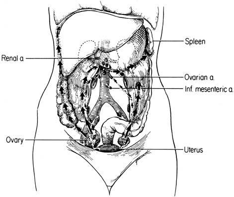
The ovaries have the most extensive lymphatic drainage of all the pelvic organs. Injection of dye into the ovarian stroma outlines a rich lymphatic network that drains the external theca of the follicles and corpora lutea converging on the hilus.2 As the lymphatic channels approach the hilus, they envelop the ovarian veins in a helical pattern. Six to eight collecting lymphatic trunks converge in the mesovarium to form the subovarian lymphatic plexus. The plexus also receives efferent channels from the Fallopian tube and uterine fundus. The lymphatics then follow the ovarian vein in the infundibulopelvic ligament. They traverse the ureter and external iliac artery with the ovarian vessels and continue cephalad, lateral to the ureter. At the lower pole of the kidney they turn medially, cross the ureter, and enter the periaortic nodes at the level of the renal hilus (Fig. 1). Although the primary drainage is the periaortic nodes, accessory channels that traverse the broad ligament are present, draining into the upper interiliac nodes in 25% of patients. Retrograde flow may also occur by the uterine fundal and tubal lymphatics to the inguinal nodes when the normal lymphatic flow is disrupted.

Fig. 1. Patterns of dissemination of ovarian cancer.

A review of the lymphatic drainage of the peritoneal cavity is necessary to explain common sites of involvement in advanced disease. When a tumor cell or particle is injected intraperitoneally into experimental animals or humans, the particle is removed almost exclusively by the lymphatic plexus lining the inferior diaphragmatic surface. In humans, this is densest in the muscular portion of the right diaphragm. The right paracolic gutter is the main pathway of flow from the pelvis to the abdomen. Flow in the left paracolic gutter is impeded by the phrenicocolic ligament that fixes the colonic splenic flexure to the left hemidiaphragm. This cephalad migration is related to negative subphrenic pressure generated with respiration, intestinal peristalsis, and position of the small bowel mesentery. From the diaphragmatic lymphatics, peritoneal lymph is carried by retrosternal lymphatics to the anterior mediastinal nodes. These drain to the right thoracic duct that empties into the right subclavian vein. The right thoracic duct receives 80% of the peritoneal lymph, with the remainder being carried by diaphragmatic lymphatics to the perihilar pulmonary node, thoracic duct, and upper lumbar retroperitoneal nodes.

Natural History

The natural history of early ovarian cancer is unknown. Early disease is a silent, asymptomatic process, and most cases are diagnosed incidentally. There are no known precursor lesions to ovarian cancer, and the time interval required for disease localized to the ovary to disseminate is unknown. Several authors have noted distinct histologic features in the ovaries considered to be at increased risk for ovarian carcinoma and suggest that this association may represent a precursor lesion similar to other epithelial dysplasia found in the female genital tract. Gusberg and Deligdisch3 identified three ovarian cancer patients with identical twin sisters; the unaffected sisters underwent prophylactic oophorectomy after menopause, and their ovarian surface epithelium was found to have histologic features such as stratification, loss of polarity, and nuclear pleomorphism, which are suggestive of a preneoplastic disorder. Fraumeni and colleagues4 described similar changes in ovaries from ovarian cancer prone families, as did Werness and colleagues5 and Dyck and associates.6 Salazar and coworkers7 and Lu and associates,8 in separate studies, examined the ovaries of women known to be at high risk by either positive linkage analysis to BRCA1 or BRCA2 or strong family history, and not only found these histologic changes but also identified unanticipated microscopic malignant neoplasms. Mittal and associates9 adopted a slightly different tactic, examining the contralateral ovary in patients with unilateral ovarian carcinoma, and found a statistically significantly increased number of inclusion cysts within the normal ovaries of cancer patients compared with age-matched controls. In a retrospective review, Plaxe and coworkers10 found nuclear atypia and cellular atypia in noncancerous tissue adjacent to the primary ovarian tumor in stage I cancers, whereas Zheng and partners11,12 identified molecular characteristics of high-grade carcinomas in histologically benign or low-grade malignant tumors adjacent to high-grade ovarian carcinomas. This association may represent a precursor lesion similar to other epithelial dysplasia found in the female genital tract.

The natural history of early ovarian cancer could alternately be explained by multifocal tumorigenesis, with multiple tumors developing simultaneously in the peritoneal epithelium or unifocal tumorigenesis when the tumor develops in the ovary and spreads to other sites. Woodruff and Julian13 studied a large number of ovarian malignancies thought to be metastatic, but after review of histology and analysis of survival, they reclassified them as synchronous primary lesions. Russell and colleagues14 reviewed 128 cases of primary ovarian carcinoma. They found 8 of 10 (80%) borderline carcinomas and 37 of 75 (49%) invasive serous carcinomas to have evidence of independent primary neoplasia at more than one anatomic site.

Several recent molecular biologic studies indicate that invasive ovarian cancer is indeed unifocal in origin, however. Polymerase chain reaction (PCR)-based methods have revolutionized the ability to copy fragments of nuclear DNA and have allowed much more precise evaluation of cancer tissues than was available to Woodruff and Julian in 196913 or even Russell and coworkers in 1985.14 These fragments of DNA can serve as unique genetic fingerprints that may be used to trace the origin of cells. DNA from cancer specimens, duplicated precisely to measurable quantities, can be analyzed for alterations when compared with specimens of normal controls. Early in the use of PCR techniques, the genome was searched for areas of frequent loss of heterozygosity (LOH); if cancer specimens consistently showed LOH in a specific region and the normal controls did not, this suggested the possible presence of a gene related to tumorigenesis. As the genome has become better understood, a library of such regions has developed. Researchers have shown identical patterns of allelic loss, identical patterns of X-chromosome inactivation, and identical codons involved in the mutation of the p53 gene in stage III serous ovarian cancer collected from the primary tumor and metastatic sites from the same patient.15,16,17 This suggests a unifocal origin of serous ovarian carcinoma in more than 90% of cases. Newer molecular biologic techniques have been employed in papillary serous carcinoma of the peritoneum to determine whether these tumors are unifocal or multifocal in origin. Unlike the published experience with invasive ovarian cancer, papillary serous carcinoma of the peritoneum appears to be multifocal in origin, based on differing patterns of allelic loss, X-chromosome inactivation, and p53 mutation at different tumor sites within the same patient.18,19,20,21

Recent advances in cell biology and molecular genetics have been applied to the study of ovarian cancer to elucidate genetic events involved in the malignant transformation of ovarian epithelium. There appear to be two classes of genes associated with tumorigenesis. Protooncogenes are normal cellular genes involved in regulation of cell growth and differentiation. Protooncogenes may be activated by point mutation, gene amplification, or translocation. More than 60 oncogenes have been described in various tumors. Although oncogenes tend to behave in a dominant manner, activation of a single oncogene is unlikely to result in malignant transformation of the cell. Thus far, no single mutation has been found common to all epithelial ovarian cancers, even when histologic subtypes have been examined separately. The heterogeneity of the genetic mutations involved in ovarian tumorigenesis has further complicated the understanding of the natural history of ovarian cancer. Tumor suppressor genes, which constitute the second class of genes, exert inhibitory effects on normal cell growth and differentiation. Inactivation of tumor suppressor genes leads to unregulated cell growth. These genes usually act in a recessive fashion, requiring inactivation of two alleles before an alteration of the gene product can be realized. This model of gene mutation led to the study of loss of heterozygosity at specific chromosomal sites as a guide to identify potential tumor suppressor genes.

Some oncogenes encode for growth factors or growth factor receptors that may act as self-stimulants (exhibiting autocrine control), or stimulants of nearby cells (paracrine control), or stimulants of distant cells (endocrine control). Other oncogenes perform critical roles in intracellular signal transduction. Protooncogenes studied in ovarian cancer include K-ras, Her-2/neu, epidermal growth factor receptor, c-fms, and c-myc. K-ras is the most commonly identified proto-oncogene in human carcinomas.22 Mok and coworkers showed that K-ras mutations are found in borderline and invasive ovarian tumors but noted a nonuniform distribution of mutations when compared by histologic classification.23,24 In both borderline and invasive ovarian tumors, K-ras mutations occurred more commonly in mucinous than in serous tumors. K-ras mutation was observed in approximately 50% of mucinous tumors, regardless of grade or stage, whereas most ras mutations identified in tumors of serous differentiation were found in stage II or III invasive disease.25,26,27 These data suggest that the K-ras mutation may be involved in mucinous differentiation of ovarian epithelial tumors. Given the relatively small prevalence of mucinous ovarian cancers, ras abnormalities are overall less frequent among invasive ovarian cancers (<20%) than among cancers that arise at other sites, such as the pancreas (>90%).

The amplification and overexpression of the Her-2/neu proto-oncogene in approximately 30% of ovarian cancers were first reported by Slamon and associates.28 This was shown to be associated with poor survival rates not only in association with ovarian cancer but also in cervical, vaginal, vulvar, and breast cancer.28,29,30,31,32 Expression of EGF-R occurs in normal ovarian epithelial cells. Continued expression of EGF-R in advanced ovarian carcinoma has been associated with a poor prognosis.33 Experiments with EGF-R ligands have attempted to define a possible autocrine loop that may regulate ovarian cancer growth.34 The c-fms protooncogene codes for the colony-stimulating factor-1 (CSF-1) receptor. An in situ hybridization study of 23 benign and malignant tumors showed that c-fms expression was associated with high-grade advanced stage tumors.35 Amplification and overexpression of the c-myc protooncogene has been demonstrated in ovarian cell lines and tumor biopsy specimens.36,37 No chromosomal rearrangement of c-myc has been demonstrated.38,39

Cytokines in the ovarian stroma, follicular fluid, and tumor-associated macrophages might affect growth of ovarian cancer cells through paracrine regulation. Tumor-associated macrophages produce interleukin-1 (IL-1), IL-6, and tumor necrosis factor (TNF), all of which can stimulate a fraction of ovarian cancers.40 Both IL-1 and TNF induce endogenous production of TNF by ovarian cancers,41 which is associated with increased proliferation in 20% to 40% of cancers.42 TNF can stimulate, inhibit, or fail to affect growth of ovarian cancers among different persons.

Loss and inactivation of tumor suppressor genes in epithelial ovarian cancer also have been reported. Most research has focused on the p53 gene because it currently is the most commonly detected genetic lesion in human cancer.43,44 The p53 gene codes for a nuclear protein that is normally found at low levels in virtually all cells. In general, the mutant p53 gene codes for a variant p53 protein, causing a conformational change that prolongs the half-life from minutes to hours.45,46 This accumulation of p53 protein is an approximate indicator of altered p53 function. Kohler and colleagues,47 Millner and associates,48 and Berchuck and coworkers,49 using immunostaining of the p53 protein, demonstrated overexpression of the p53 protein in 0% (none of 17) of benign tumors, in 4% (2 of 49) of borderline tumors, and in 16% of early-stage (stage IA/IB) epithelial ovarian cancers. Using DNA sequencing, Marks and associates50 showed p53 mutations in 44% (29 of 66) of invasive epithelial ovarian carcinomas; however, analysis of the distribution of point mutations has not identified a particular mutation site. The pattern of mutation differs from that observed in colorectal, hepatocellular, and lung cancer, in association with which it has been possible to identify a statistically significant increase in specific transitions or transversions of the p53 gene.43,51,52 The progression of increase in p53 mutation or overexpression in epithelial ovarian neoplasms implicates p53 in the malignant transformation of ovarian cancers, whereas it may not be important for the development of borderline histologic changes.53

An alternate approach to the identification of genetic changes involved in the pathogenesis of ovarian cancer is to examine differentially expressed genes from normal ovarian epithelium compared with those from ovarian carcinoma cells. Mok and coworkers,54 using a RNA-fingerprinting approach, cloned two cDNA fragments (DOC-1 and DOC-2) that were present in normal ovarian surface epithelial cells but absent in most ovarian cancer cell tissues. The potential functional role of these genes represents an area of active research. The introduction of cDNA array technology has allowed simultaneous investigation of many potentially differentially expressed genes and may facilitate the search for clues to ovarian carcinogenesis.

Malignant transformation of normal ovarian epithelium may be multifactorial; some factors that may increase angiogenesis are separate from those factors that allow for invasion of cells through the basement membrane, which in turn are distinct from factors affecting apoptosis. Taken together, these factors may affect pathogenesis of an individual ovarian cancer. Some researchers have examined the matrix metalloproteinases (MMPs),55,56,57,58 a group of proteins that increase the invasive potential of a cancer by breaking down the basement membrane, using immunohistochemical techniques. MMP expression has been noted to increase in invasive ovarian cancer. It is also thought that to be able to invade, a cancer may also need to emit signals increasing local angiogenesis; such signals are an active area of research, because their identification carries implications for therapeutic intervention. Those particular factors that bring about or allow the overgrowth of cells as well as the invasion of these cells across tissue planes continues to be a focus of intense molecular research.

Dissemination

Spread patterns of ovarian cancer have been well established by careful surgical assessment of the extent of disease by exploratory laparotomy. Spread most commonly occurs by direct extension to the serosal surfaces of other pelvic organs or by exfoliation of cells into the peritoneal cavity with secondary implantation of viable cells. Expression of certain adhesion molecules (e.g., CD44H) by ovarian cancer cells can be important for adhesion of ovarian cancer cells to the peritoneal mesothelium.59 Malignant cells can be exfoliated even when the ovarian capsule appears uninvolved.60 Knowledge of the transport of particles from the peritoneal cavity explains the high frequency of diaphragmatic metastasis. Rosenoff and coworkers61 at the National Cancer Institute performed laparoscopy within 1 month of exploratory laparotomy in 49 consecutive patients as part of a pretreatment evaluation. Seven of 16 (stages I and II) patients were found to have diaphragmatic metastasis. In a prospective study of stage I to III patients by the Gynecologic Oncology Group,62 the commonest sites of visceral metastasis were the rectosigmoid (22.4%), the uterine serosa (11.2%), the small intestinal serosa (9.6%), and the serosa of the bladder (4.3%). This study highlighted the poor correlation between clinical impression and pathologic findings in early-stage disease. Clinical impression of omental disease was inaccurate in 45% of the specimens sampled. Similar inaccuracy of clinical impression was reported for metastatic diaphragmatic disease (50%), pelvic nodes (71%), and para-aortic nodes (96%). Patients reexplored for staging in the Gynecologic Oncology Group study increased in stage in 13 of 58 (22.4%) procedures. This highlights the importance of accurate surgical staging at the time of initial exploratory laparotomy.

The commonest surgical finding is advanced disease with extensive metastatic implants involving the intestinal serosa, omentum, colonic gutters, and peritoneum. Peritoneal dissemination is common; in an autopsy series, this occurred in 75 of 86 (87%) patients.63 Gastrointestinal dysfunction commonly results from diffuse serosal involvement that may encase intestinal loops. Involvement of the mesentery tethers the bowel in addition to disrupting the myenteric plexus, further impairing intestinal motility. Ascites aggravates gastrointestinal dysfunction through external compression of intestinal loops, even further limiting mobility. Inanition is a common terminal event.

Ovarian carcinoma is the commonest cause of malignant ascites in women. Ascites results from disruption of the fine equilibrium between the formation and reabsorption of peritoneal fluid. This has been shown to be causally related to the obstruction of diaphragmatic lymphatics by tumor thrombi in the murine ovarian carcinoma model.64 Coates and associates65 studied the pathogenesis of malignant ascites by mediastinal lymphoscintigraphy with labeled sulfur colloid. In normal patients, after intraperitoneal injection of labeled sulfur colloid, diaphragmatic and retrosternal lymphatics were identified clearly on gamma scans. In 21 of 23 patients with malignant ascites, complete diaphragmatic lymphatic obstruction was demonstrated by lack of colloid activity above the diaphragm after intraperitoneal injection of labeled colloid.

Distant spread is relatively uncommon because ovarian cancer tends to remain within the abdominal cavity throughout most of its natural history. The largest autopsy series of 381 patients with epithelial ovarian cancer by Rose and coworkers66 revealed distant metastasis to be associated with nodal metastasis. The commonest sites of distant metastases in decreasing order were the liver (48%), lung (38%), pleura (28%), bone (12%), skin (5%), and brain (3%). Since the introduction of platinum-based chemotherapy and, hence, improved survival, incidence of extra-abdominal metastases has increased, particularly to the lung and brain.67,68 In patients presenting with distant metastases, the clinician must exclude other possible primary sites.

Preoperative Evaluation

Taking the history and physical examination remain the first steps in the evaluation of patients with possible ovarian cancer. Asymptomatic enlargement of the ovary is common, with pain developing only as a complication related to torsion, infection, or infarction. With progressive increase in ovarian volume, symptoms related to compression of adjacent organs develop insidiously. Since the 1960s, there has been minimal variation in the presenting signs and symptoms of patients with ovarian carcinoma. The American College of Surgeons' national survey of 12,316 ovarian cancer patients identified the commonly found presenting symptoms to be abdominal pain (53%), abdominal swelling (45.8%), bloating or dyspepsia (22%), and pelvic pressure (18%).69 Similar symptoms were reported by Kent and Mckay70 in their survey of 349 ovarian cancer patients at the Boston Hospital for Women in 1960. In both surveys, approximately 50% of patients had ascites or an abdominopelvic mass. In the national survey,69 other signs included abdominal mass (37%), ovarian mass (18%), and pleural effusion (14%). The prevalence of abnormal vaginal bleeding has long been debated, ranging from 5% to 36%.71 Vaginal bleeding occurred in 14% of current patients and may represent concurrent endometrial pathology or activation of the endometrium by hormone-secreting ovarian tumors.69 Patients dying of ovarian cancer were found to have delayed seeking evaluation for symptoms for an average of 13 months; the physicians' delay in diagnosing the cancer was an average of 1.6 months.72 Such delay may occur because of the insidious onset of symptoms and the very common occurrence of similar symptoms in functional disorders. To avoid a delay in diagnosis, health care providers must maintain a high index of suspicion of ovarian cancer in women between the ages of 40 and 60 with unexplained gastrointestinal symptoms.

After a pelvic or abdominal mass has been discovered, evaluation is focused on the exclusion of other possible primary lesions and the preparation of the patient for surgery. Preoperative testing should include a complete blood count, liver function tests, blood urea nitrogen, creatinine, urinalysis, stool guaiac, a chest radiograph, and a baseline electrocardiogram. None of these tests is diagnostic of ovarian cancer, but they do help screen patients for coexistent medical disease that may increase operative risks. The need for further radiologic testing should be individualized based on the patient's history and physical examination. Pelvic ultrasound may be used to confirm the presence of a mass and may be able to localize its origin, but it does not obviate the need for surgery. In patients with no discernible pelvic mass but with ascites or omental caking or both, abdominal computerized tomographic scanning is useful to assess the upper abdominal organs and the gastrointestinal tract to exclude gastrointestinal, liver, pancreatic, renal, or adrenal primary sites. Diagnostic paracentesis is rarely indicated because cytology may be falsely negative and because a positive finding of adenocarcinoma does not provide any information about the primary site. Mammography may be useful in a search for a primary lesion but, again, it may not obviate the need for surgery even with positive findings. Breast cancer patients who present with new findings of an adnexal or pelvic mass are more likely to have a new ovarian or tubal malignancy than metastatic breast cancer by a ratio of 3:1.73 The diagnosis of ovarian cancer is established through exploratory laparotomy, with pathologic and histologic examination of the ovarian primary or metastatic lesions.

Back to Top
PROGNOSTIC FACTORS

Staging

The staging of ovarian cancer is surgical, based on the Federation of International Gynecologic Oncologists (FIGO) classification (Table 1). To provide adequate exposure of the upper abdomen, a midline or paramedian incision is preferred. Ascitic fluid should be aspirated and submitted for cytologic testing. In the absence of ascites, normal saline is used to obtain washings from the pelvis, paracolic gutters, and subdiaphragmatic space. A systematic exploration of all peritoneal surfaces is performed, paying particular attention to the pelvis, right hemidiaphragm, small and large bowel mesentery, and omentum. Biopsy specimens should also be obtained from any suspicious lesions or areas of adhesions. The pelvic and periaortic nodes should be palpated, with lymphadenectomy performed on patients with tumor confined to the pelvis. The role of routine lymphadenectomy for all patients remains unclear.


Table 1. International Federation of Gynecology and Obstetrics Ovarian Cancer Staging


Stage I: Growth limited to the ovaries.
 Stage IA: Growth limited to one ovary; no ascites. No tumor on the external surface; capsule intact.
 Stage IB: Growth limited to both ovaries; no ascites. No tumor on the external surface; capsules intact.
 Stage IC: Tumor either stage IA or IB, but with tumor on the surface of one or both ovaries or with capsule ruptured or with ascites present containing malignant cells or with positive peritoneal washings.
Stage II: Growth involving one or both ovaries with pelvic extension.
 Stage IIA: Extension and/or metastases to the uterus and/or tubes.
 Stage IIB: Extension to other pelvic tissues.
 Stage IIC: Tumor either stage IIA or IIB, but with tumor on the surface of one or both ovaries or with capsule(s) ruptured or with ascites present containing malignant cells or with positive peritoneal washings.
Stage III: Tumor involving one or both ovaries with peritoneal implants outside the pelvis and/or positive retroperitoneal or inguinal nodes. Superficial liver metastasis equals stage III. Tumor is limited to the true pelvis but with histologically verified malignant extension to small bowel or omentum.
 Stage IIIA: Tumor grossly limited to the true pelvis with negative nodes, but with histologically confirmed microscopic seeding of abdominal peritoneal surfaces.
 Stage IIIB: Tumor of one or both ovaries with histologically confirmed implants of abdominal peritoneal surfaces, none exceeding 2 cm in diameter. Nodes negative.
 Stage IIIC: Abdominal implants >2 cm in diameter and/or positive retroperitoneal or inguinal nodes.
Stage IV: Growth involving one or both ovaries with distant metastasis, if pleural effusion is present, there must be positive cytologic test results to allot a case to stage IV.

(Data from International Federation of Gynecology and Obstetrics: Changes in definitions of clinical staging for carcinoma of the cervix and ovary. Am J Obstet Gynecol 156:246, 1987.)


In advanced disease, cytoreductive surgery is performed, including total abdominal hysterectomy, bilateral salpingo-oophorectomy, and omentectomy. The ex-tent of disease, in addition to the location and extent of residual disease (after cytoreductive surgery), should be recorded.

For optimal staging, knowledge of the dissemination pattern of ovarian cancer is essential. The FIGO stage is assigned based on findings at laparotomy and information gained by the pathologist's examination of surgically excised specimens. After being assigned, the FIGO stage is not changed. The FIGO stage has been shown to correlate inversely with 5-year survival rates.74 The FIGO stage provides a uniform classification that allows comparison of clinical experiences among institutions. This is mandatory for critical evaluation of current therapeutic regimens and the development of new protocols.

Back to Top
HISTOLOGY AND GRADE

The 1988 FIGO Annual Report74 outlined the prevalence of various histologic types and the distribution of stages among more than 8000 ovarian cancer patients. Serous carcinomas comprised approximately 50%, mucinous 10% to 15%, endometrioid 10% to 15%, clear cell 2% to 5%, and undifferentiated 10%. Serous tumors presented more commonly as stage III or IV disease compared with mucinous tumors, in which 50% were diagnosed before extrapelvic extension. When corrected for stage, there was no difference in survival between serous and mucinous tumors. The prognostic impact of clear cell and endometrioid cell types is controversial.

Grade is not incorporated into the FIGO classification of ovarian cancer. Grade has clear prognostic significance in early-stage cancer, but this has not been shown to be prognostic consistently in advanced carcinoma.75,76 The lack of standardized grading systems may contribute to these discrepancies.

Favorable prognostic variables identified with multivariate analysis of two Gynecologic Oncology Group protocols included cell type other than clear cell or mucinous, cisplatin-based therapy, good performance status, younger age, lower stage, clinically immeasurable disease, smaller residual disease, and absence of ascites.76

Back to Top
SCREENING

Because of the high mortality of this disease, there is a strong need to develop screening programs. Our failure to improve long-term survival substantially is a result of our inability to improve early detection. Historically, bimanual rectovaginal examination has been the only method used to screen for ovarian cancer. It has been estimated that this screening method detects only one ovarian cancer per 100,000 examinations77 because of the low sensitivity of this method and the low inci-dence of ovarian cancer. At present, no available screening test for ovarian cancer has been proved to decrease mortality. Even screening efforts directed at women at high risk who are either known to carry the BRCA1 or BRCA2 mutation or who have a significant family history of ovarian cancer have failed to decrease ovarian cancer mortality in these women. Karlan and associates78 identified 10 cancer cases in 1261 patients screened, but 7 of the 10 cases were diagnosed at stage IIIC, which is already an advanced stage. Two index cases were actually borderline ovarian tumors, and the appropriateness of their inclusion in the study is debatable. The single patient with early ovarian cancer had stage IC; follow-up observation of her case thus far remains limited. Indeed, even completely asymptomatic women at high risk with recent normal screening studies who present for prophylactic bilateral salpingo-oophorectomy may be found to have advanced stage cancers at the time of surgery.8 Recent screening efforts have focused primarily on CA 125 (see later discussion), transvaginal pelvic ultrasound, and bimanual rectovaginal examination. Early detection by mass screening is an appealing method of reducing ovarian cancer mortality. The 5-year survival rate of advanced disease is less than 30%, whereas survival in early-stage disease exceeds 90%. Regrettably, most ovarian cancers (75%) are clinically detected when the disease is advanced. During the past 30 years, limited progress has been made in improving the survival rate of patients with late-stage ovarian cancer. Therefore, if a screening program can substantially increase the proportion of women whose disease is detected in the early stage, mortality from ovarian cancer may well be reduced.

A successful screening program aimed at early detection of ovarian cancer would require that major abdominal surgery be performed, because this is the only means of a definitive diagnosis. Because of the low incidence of ovarian cancer and the necessity of laparotomy, such a screening program would require very high accuracy. The lowest acceptable positive predictive value suggested in the literature79 for an ovarian cancer screening test is 10%; that is, no more than 10 women would require laparotomies to detect one case of ovarian cancer. Because of the low incidence in premenopausal women, the suggested target population is postmenopausal women. At age 50, the yearly incidence is 30 cases per 100,000, which increases to more than 50 per 100,000 by age 75, giving an average of 40 per 100,000 for women older than 50. Even with a sensitivity of 100%, the requirement of a minimum 10% positive predictive value with this low incidence rate implies that the specificity must exceed 99.5%. This high specificity is essential to minimize the morbidity and mortality associated with exploratory laparotomy in those with false-positive screening tests. Many suggested screening modalities have not met this stringent requirement. The most promising modalities include serum markers such as CA 125 and pelvic ultrasound.

CA 125 is an antigenic determinant on a high-molecular-weight glycoprotein recognized by a monoclonal antibody (OC 125), which was raised using an ovarian cancer cell line as an immunogen.80 CA 125 is expressed by ovarian epithelial tumors and various normal and pathologic tissues of mullerian origin. The physiologic role of the glycoprotein expressing CA 125 is unknown. CA 125 levels are elevated in various physiologic, benign, and nongynecologic malignant conditions (Table 2), which limits its specificity for ovarian cancer detection, particularly in premenopausal women.


Table 2. Etiology of Elevations in Serum Cancer Antigen 125 (>35 U/mL)


Physiologic  
Menstruation  
Pregnancy  
Benign Gynecologic Disorders CA 125 >35 U/mL (%)
Benign ovarian tumors 10.4
Acute salpingitis 40.4
Chronic salpingitis 8.3
Leiomyoma 10.4
Endometriosis 20
Benign Gastrointestinal Disorders  
Cirrhosis plus ascites 100
All cirrhosis 9.1
Acute pancreatitis 32.2
Chronic pancreatitis 1.9
Malignancy  
Epithelial ovarian cancer 80
Pancreas 52.6
Liver 49
Biliary tract 45.8
Gastric 30.9
Lung 29.5
Breast 17.6
Colorectal 15.1

(Data from Bast RC, Jacobs I: The CA 125 tumor associated antigen: A review of the literature. Hum Reprod 4:1, 1989.)


CA 125 could be used potentially as a screening tool for postmenopausal women for ovarian cancer on the basis of several observations. CA 125 levels were found to be elevated to more than 35 U/mL preoperatively in 80% to 85% of women with epithelial ovarian cancer compared with 1% of healthy controls.80 A study using the JANUS serum bank demonstrated that CA 125 levels were elevated up to 60 months before the clinical detection of ovarian cancer.81 The reference level of 35 U/mL was chosen for the clinical observation of patients with known ovarian cancer; however, CA 125 levels are greater than 35 U/mL in only 50% of the patients with stage I disease. If this criterion was used as a screening test, and the same sensitivity (50%) and specificity (99%) were to be maintained in the screening program, the positive predictive value would be only 2.4%, which is four times less than the minimal acceptable level.

Initial feasibility tests for CA 125 in the early detection of ovarian cancer were performed in Stockholm by Zurawski and colleagues,82 which included 1082 asymptomatic women older than 40 years old. Initial serum CA 125 levels exceeded 35 U/mL in 36 women (3.3%). Of these 36 patients, only two exhibited a doubling of CA 125; one woman subsequently had ovarian cancer diagnosed. Investigators at St. Bartholomew's Research Unit in London have also performed large epidemiologic studies, hoping to identify a screening use for the CA 125 assay. They screened 22,000 healthy postmenopausal women older than age 45, and identified a subgroup of 771 patients who had one or more elevated CA 125 values; after prospective follow-up of the patients, the investigators were able to state definitively that an elevated CA 125 is not a predictor of nongynecologic cancers or a harbinger of recurrences of other malignancies; rather, an elevated CA 125 is specifically a marker for gynecologic malignancies.83 These researchers also noted that the risk associated with either having or developing ovarian cancer was related to the level of CA 125 elevation over a threshold level (chosen by them for statistical purposes to be 35),84 and that those noted to have an elevated CA 125 experienced a significantly increased risk of death over the next 5 years.85

This supports the value of longitudinally collected CA 125 levels to identify patients at high risk for ovarian malignancy. Further evidence for this hypothesis comes from the second Stockholm study by Einhorn and coworkers,86 which included 5550 women whose annual measurements were taken for 2 years. Three monthly measurements were made in 175 women with an initial CA 125 elevation and in 175 age-matched controls. All six ovarian cancer cases among the women older than 50 years were detected with a prospective CA 125 rule. With the use of a retrospective rule based on the absolute initial levels and the rate of increase in serial CA 125 levels, all six cases were detected (sensitivity 100%), whereas specificity was estimated at 99.7%. For women older than 50, the resulting positive predictive value is estimated to be 14%, which exceeds the minimum requirement for a successful screening program. These data, however, support only the thesis that CA 125 screening can be used to screen asymptomatic ovarian cancer before clinical diagnosis. The issue still remains whether such a screening program will detect a significantly increased proportion of stage I disease.

To increase detection of stage I disease, a multimodal approach to screening has been suggested.87 Jacobs and associates88 screened 22,000 postmenopausal patients with CA 125 and rescreened 340 women with elevated levels (CA 125 >30 U/mL) using transabdominal ultrasound. Surgical exploration was performed in 41 of the 340 patients with elevated levels (12%). At 2-year follow-up, 19 ovarian cancer cases were found. Eleven ovarian cancers were detected, yielding an apparent sensitivity of 57.9% for a combined CA 125 with ultrasound approach; the positive predictive value was 26.8% (11 of 41).

Several investigators have evaluated transabdominal ultrasound and transvaginal pelvic ultrasound as a primary screening test. The largest study of transabdominal ultrasound by Campbell and colleagues89 screened 5479 self-referred asymptomatic women between ages 18 and 78 years (mean: 52 years old) during an 8-year period. Of these patients, 326 (5.9%) were identified as having 338 abnormal scans among a total of 15,977 scans performed (2.3% abnormal). Five patients with stage I ovarian cancer were identified (prevalence: .09%). An additional four patients with metastatic ovarian cancer were identified. The specificity was 97.7% and the predictive value of a positive screening test 1.5%. The major limitation to screening with transabdominal ultrasound is the high false-positive rate of 2.3%. A definitive distinction between early benign and malignant tumors is not possible on the basis of their ultrasonic appearance.

Morphologic criteria suggestive of malignancy have been described by many independent centers and have been incorporated into scoring systems in an attempt to quantify the risk of malignancy. Of the morphologic scoring systems reported, the echoarchitectural criteria are similar, including size, wall thickness, contour, septae, and degree of sonolucency. Introduction of transvaginal ultrasound has allowed superior imaging of the ovaries compared with a transabdominal approach. Improved resolution resulted from the decreased distance between the transducer placed vaginally and the adnexa. Transvaginal screening also was advantageous because it did not require a distended bladder. Van Nagell and associates90 screened 1000 asymptomatic women aged 40 or older at the University of Kentucky Medical Center. Thirty-one patients (3.1%) had abnormal scans, and 24 agreed to exploratory laparotomy. Laparotomy confirmed the ultrasound findings: either ovarian or fallopian tube pathology was identified. In premenopausal patients, the fluctuation in ovarian size related to ovulation made ovarian volume a less reliable index of pathology. Only 16 of 39 (41%) premenopausal patients had persistent ovarian enlargement on repeat sonography. Subsequent screening of 9000 asymptomatic women with transvaginal ultrasound detected 10 stage I ovarian carcinomas and one stage IIIB ovarian carcinoma.91,92

Lack of specificity remains problematic in the application of ultrasound to screening for ovarian cancer. Bourne and coworkers proposed transvaginal color flow Doppler imaging with transvaginal sonography to detect neovascularization as a method to enhance the ability to distinguish between benign and malignant ovarian tumors.93 In a study of 50 women selected on the basis of medical history and prior scanning, only 1 of 12 patients with benign masses compared with 7 of 8 patients with malignant ovarian masses showed evidence of neovascularization. Of three patients with stage I ovarian malignancy, only two had Doppler findings consistent with neovascularization. Kurjak and associates94 reported the results of transvaginal color flow Doppler sonography in 14,317 women being screened for ovarian cancer. When a resistance index (peak Doppler-shifted frequency at systole divided by minimum Doppler-shifted frequency in diastole) of .4 was used to differentiate benign and malignant disease, 623 of 624 patients (99.8%) with benign lesions were found to have a normal resistance index. Of 56 malignant adnexal masses, 54 (96%) demonstrated a resistance index of less than .4. In stage IA ovarian cancer, six of seven cases (86%) demonstrated neovascularization and a resistance index of less than .4. The limitations of Doppler, however, include technical complexities, interobserver and intraobserver variation, and the presence of neovascularization in benign conditions. Using state-of-the-art Doppler equipment, low intraovarian resistance to flow has been demonstrated in the proliferative phase of the menstrual cycle.95

Research on ovarian cancer screening has also focused on the search for tumor markers to complement CA 125 because only 50% of stage I patients have elevated CA 125 levels.80 Woolas and colleagues reported the use of CA 125, M-CSF, and OVX1 in a retrospective study of 46 stage I ovarian cancer patients, 237 patients with benign pelvic masses, and 204 apparently healthy women.96 CA 125 alone was elevated in 56% of these stage I patients; 98% of these patients were identified by an elevation in one of these three markers. In comparison, 11% of the apparently healthy women and 51% of patients with benign pelvic masses also had an elevation of at least one tumor marker. In a retrospective study of a serum bank from a screening trial, OVX1 levels were complementary to CA 125 for sensitivity.97 Of 39 ovarian cancer patients, 23 had elevated CA 125 levels (> 25 U/mL); another eight had elevated levels of OVX1. This combination of markers results in a sensitivity of 80% in a screened population before clinical evidence of disease. Other tumor markers aimed at ovarian cancer screening include CA15-3, CA19-9, NB/70K, PLAP, TAG 72, and urinary gonadotropin fragment; these tumor markers have been discussed in a review.98

Back to Top
NEW TECHNOLOGIES FOR MARKER DISCOVERY

cDNA microarray techniques

Gene expression analysis by high-throughput cDNA microarray technology is a powerful new tool by which to identify genes that are important in the pathogenesis of ovarian cancer.99 Complementary DNA microarray technology allows the simultaneous comparison of the expression of thousands of genes in malignant and normal ovarian tissues. By identifying genes that are up-regulated in malignancy and have a secreted product that is measurable in bodily fluids, researchers are working toward the development of new markers for the early diagnosis of ovarian cancer.

Using cDNA microarray technology to compare ovarian cancer with normal ovarian tissues, researchers have recently identified two potential markers, osteopontin and prostasin, that are significantly up-regulated in ovarian cancer tissues and have a secreted product.100,101 Osteopontin is an acidic, calcium-binding glycophosphoprotein that is found in all body fluids, functions as a cell adhesion molecule and cytokine, and has been described to be a factor in tumorigenesis. Prostasin is a serine protease that was originally isolated from human seminal fluid but is also expressed in a variety of other human tissues, including kidney, liver, pancreas, and colon, among others. Serum levels of osteopontin and prostasin were confirmed to be significantly higher in women with ovarian cancer as compared with normal women, women with benign gynecologic processes, and women with nonovarian malignancy. Interestingly, women with stage II ovarian cancer had the highest prostasin levels, suggesting that prostasin may be useful for early detection. The specificity of osteopontin was 80.4% and sensitivity point estimates for early-stage (stage I/II) and late-stage (III/IV) disease were 80.4% and 85.4%, respectively.

Proteomics

The rapidly advancing field of proteomics is another approach to biomarker discovery. It is hoped that proteomic methods will lead to the identification of markers that, because of modifications after protein synthesis, would be missed by DNA or RNA analysis. The technique of surface-enhanced laser desorption ionization time-of-flight (SELDI-TOF) technology, is increasingly being used for the global analysis of proteins in complex solutions such as plasma, serum, and urine.

Protein profiling by SELDI-TOF yields thousands of data points requiring sophisticated data analysis tools. Using a high-order analytical approach, Petricoin and associates linked SELDI-TOF spectral analysis of samples from women with ovarian cancer to define an optimum discriminatory proteomic pattern.102 This pattern was used to predict the identity of masked samples from unaffected women, women with early-stage and late-stage ovarian cancer, and women with benign disorders. Analysis of spectra from a masked validation set correctly classified 63 of 66 (95%) of the control samples as not cancer. Twenty-two of 24 (92%) of the true normals were correctly classified, and all 50 cancer samples, including all 18 stage I cancers, were correctly classified as malignant. This yielded 100% sensitivity (95% CI: 93% to 100%) and 95% specificity (87% to 99%). The positive predictive value for the sample set was 94% (84% to 99%) compared with 35% for CA-125 for the same samples.

Although protein profiling may ultimately prove clinically useful, the actual identification of individual proteins is an important goal, as understanding the function of proteins involved in the pathogenesis of ovarian cancer will likely improve our ability to find ways to prevent the disease. Using SELDI technology, Ye and associates described the identification of haptoglobin-alpha subunit (Hp-alpha) as a potential serum biomarker.103 In this study, the protein profiles of serum samples from ovarian cancer patients were compared with control serum samples from healthy women, women with benign gynecologic tumors, and women with other gynecologic cancers. The peak intensity of a serum biomarker at 11.7 kDa was significantly higher in cases compared with controls. This biomarker was purified and an antibody generated from the synthesized peptide. Serum studies showed that Hp-alpha levels were up to two-fold higher in the serum of women with ovarian cancer compared to controls.

It is likely that, despite advancing technology, no single biomarker will be identified that has high enough sensitivity or specificity to be used alone for the early diagnosis of ovarian cancer. For example, when combined with CA-125, prostasin achieved a sensitivity of 92% and a specificity of 94%. The sensitivity of prostasin alone at the same specificity was 51.4%. Similarly, alone, Hp-alpha had a 64% sensitivity at 90% specificity, and when combined with CA-125, it had a sensitivity of 91% and a specificity of 95%, further supporting the hypothesis that a panel of markers will ultimately be required for the early diagnosis of ovarian cancer.

Back to Top
CONCLUSION

Reduction of ovarian cancer mortality is the primary goal of screening. CA 125 and transvaginal ultrasound have definite promise for use in a multimodal screening program, but there are as yet no definitive prospective data to indicate that a mass screening program targeting early detection of ovarian cancer will reduce disease-specific mortality in the general population. Results so far are sufficiently promising to support the conduct of a randomized clinical screening trial for ovarian cancer. Currently, screening in patients with no symptoms should be limited to clinical investigations. It is also anticipated that the use of new technologies for DNA and protein analysis will result in the development of a panel of markers for the early detection of ovarian cancer.

Back to Top
ACKNOWLEDGMENTS

This chapter retains material from previous editions. The important contributions of all previous authors, including Robert Bast Jr, Robert Knapp, Thomas Leavitt, Steven Skates, and Valena Soto-Wright, are acknowledged.

Back to Top
REFERENCES

1. American Cancer Society. Cancer Statistics, 2002 Atlanta, American Cancer Society, 2002

2. Plentl AA, Friedman EA: Lymphatic System of the Female Genitalia. Philadelphia, WB Saunders, 1971

3. Gusberg SB, Deligdisch L: Ovarian dysplasia: A study of identical twins. Cancer 54:1, 1984

4. Fraumeni JF Jr, Grundy GW, Creagan ET et al: Six families prone to ovarian cancer. Cancer 36:722, 1982

5. Werness BA, Afify AM, Bielat KL et al: Altered surface and cyst epithelium of ovaries removed prophylactically from women with a family history of ovarian cancer. Hum Pathol 30:151, 1999

6. Dyck HG, Hamilton TC, Godwin AK et al: Autonomy of the epithelial phenotype in human ovarian surface epithelium: Changes with neoplastic progression and with a family history of ovarian cancer. Int J Cancer (Pred Oncol) 69:429, 1996

7. Salazar H, Godwin AK, Daly MB et al: Microscopic benign and invasive malignant neoplasms and a cancer-prone phenotype in prophylactic oophorectomies. J Natl Cancer Inst 88:1810, 1996

8. Lu KH, Garber JE, Cramer DW et al: Occult ovarian tumors in women with BRCA1 or BRCA2 mutations undergoing prophylactic oophorectomy. J Clin Oncol 18:2728, 2000

9. Mittal KR, Zeleniuch-Jacquotte A, Cooper JL et al: Contralateral ovary in unilateral ovariancarcinoma: A search for pre-neoplastic lesions. Int J Gynecol Pathol 12:59, 1993

10. Plaxe SC, Deligdisch L, Dottino PR et al: Ovarian intraepithelial neoplasia demonstrated in patients with stage I ovarian carcinoma. Gynecol Oncol 38:367, 1990

11. Zheng JP, Wan MH, Zweizig S et al: Histologically benign or low grade malignant tumors adjacent to high-grade ovarian carcinomas contain molecular characteristics of high-grade carcinomas. Cancer Res 53:4138, 1993

12. Zheng JP, Benedict WF, Xu HJ et al: Genetic disparity between morphologically benign cysts contiguous to ovarian carcinomas and solitary cystadenomas. J Natl Cancer Inst 87:1146, 1995

13. Woodruff JD, Julian CG: Multiple malignancy in the upper genital tract. Am J Obstet Gynecol 193:810, 1969

14. Russell P, Bannatyne PM, Solomon HJ et al: Multifocal tumorigenesis in the upper female genital tract: Implications for staging and management. Int J Gynecol Pathol 4:192, 1985

15. Tsoa SW, Mok SC, Knapp RC et al: Molecular genetic evidence of a unifocal origin for human serous ovarian carcinomas. Gynecol Oncol 48:5, 1993

16. Mok SC, Tsao SW, Knapp RC et al: Unifocal origin of advanced human epithelial ovarian cancers. Cancer Res 52:5119, 1992

17. Jacobs IJ, Kohler MF, Wiseman RW et al: Clonal origin of epithelial ovarian carcinoma: Analysis by loss of heterozygosity, p53 mutation and X chromosome inactivation. J Natl Cancer Inst 84:1794, 1992

18. Feuer GA, Shevchukm, Calanog A: Normal sized ovary carcinoma syndrome. Obstet Gynecol 73:786, 1989

19. Muto MG, Welch NR, Mok SC et al: Evidence for a multifocal origin of papillary serous carcinoma of the peritoneum. Cancer Res 55:490, 1995

20. Kowalski LD, Kanbour AI, Price FV et al: A case-matched molecular comparison of extraovarian versus primary ovarian adenocarcinoma. Cancer 79:1587, 1997

21. Schorge JO, Muto MG, Welch WR et al: Molecular evidence for multifocal papillary serous carcinoma of the peritoneum in patients with germline BRCA1 mutations. J Natl Cancer Inst 90:841, 1998

22. Bos JL: ras Oncogenes in human cancer: A review. Cancer Res 49:4682, 1989

23. Mok SC, Bell DA, Knapp RC et al: Mutation of K-ras protooncogene in human ovarian epithelial tumors of borderline malignancy. Cancer Res 53:1489, 1993

24. Garrett AP, Lee KR, Colitti CR et al: K-ras mutation is an early event in mucinous ovarian tumorigenesis. Gynecol Oncol 76:281, 2000

25. Enomoto T, Weghorst CM, Inoue M et al: K-ras activation occurs frequently in mucinous adenocarcinomas and rarely in other common epithelial tumors of the human ovary. Am J Pathol 139:777, 1991

26. Cuatrecasas M, Villanueva A, Matias-Guiu X et al: K-ras mutations in mucinous ovarian tumors: A clinicopathologic and molecular study of 95 cases. Cancer 79:1581, 1997

27. Cuatrecasas M, Erill N, Musulen E et al: K-ras mutations in non-mucinous ovarian epithelial tumors: A molecular analysis and clinicopathologic study of 144 patients. Cancer 82:1088, 1998

28. Slamon DJ, Godolphin W, Jones LA et al: Studies of the Her-2/neu protooncogene in human breast and ovarian cancer. Science 244:707, 1989

29. Berchuck A, Rodriguez G, Kamel A et al: Expression of epidermal growth factor receptor and Her-2-/neu in normal and neoplastic cervix, vulva and vagina. Obstet Gynecol 76:381, 1990

30. Berchuck A, Rodriguez G, Kinney RB et al: Overexpression of Her-2/neu in endometrial cancer is associated with advanced stage disease. Am J Obstet Gynecol 164:15, 1991

31. Berchuck A, Kamel A, Whitaker R et al: Overexpression of Her-2/neu is associated with poor survival in advanced epithelial ovarian cancer. Cancer Res 50:4087, 1990

32. Slamon DJ, Clark GM, Wong SG et al: Human breast cancer: Correlation of relapse and survival with amplification of the Her-2/neu oncogene. Science 235:177, 1987

33. Kohler M, Janz I, Wintzer HO et al: The expression of EGF receptors, EGF-like factors and c-myc in ovarian and cervical carcinomas and their potential clinical significance. Anticancer Res 9:1537, 1989

34. Berchuck A, Rodriguez G, Olt G et al: Regulation of growth of normal ovarian epithelial cells and ovarian cancer cell lines by transforming growth factor-B. Am J Obstet Gynecol 166:676, 1992

35. Kacinski BM, Carter D, Kohorn EI et al: Oncogene expression in vivo by ovarian adenocarcinomas and mixed-mullerian tumors. Yale J Biol Med 62:379, 1989

36. Yasue H, Takeda A, Ishibashi M: Amplification of the c-myc gene and the elevation of its transcripts in human ovarian cancer cell lines. Cell Struct Funct 12:121, 1987

37. Zhou DJ, Gonzalez N, Ahuja J: A uniform pattern of protooncogene abnormalities in ovarian adenocarcinoma. Cancer 62:1573, 1988

38. Smith DM, Griff DE, Pokul RK et al: Determination of cellular oncogene rearrangement or amplification on ovarian adenocarcinomas. Am J Obstet Gynecol 161:911, 1989

39. Berns EJJ, Klun JGM, Henzen-Logmans SC et al: Receptors for hormones and growth factors and (onco)-gene amplification in human ovarian cancer. Int J Cancer 52:218, 1992

40. Wu S, Rodabaugh K, Martinez-Maza O et al: Stimulation of ovarian tumor cell proliferation with monocyte products including interleukin-1, interleukin-6 and necrosis factor-alpha. Am J Obstet Gynecol 166:997, 1992

41. Wu S, Boyer CM, Whitaker R et al: Tumor necrosis factor-alpha (TNF-a) as an autocrine and paracrine growth factor for ovarian cancer: Monokine induction of tumor cell proliferation and TNF-expression. Cancer Res 53:1939, 1994

42. Hurteau J, Berchuck A, Rodriguez G et al: Epidermal growth factor and tumor necrosis factor stimulate proliferation of some epithelial ovarian cancer cells from ascites: 25th Annual Meeting of the Society of Gynecologic Oncology [abstract]. Gynecol Oncol 52:110, 1994

43. Caron de Fromentel C, Soussi T: Tp53 tumor suppressor gene: A model for investigating human mutagenesis. Genes Chromosom Cancer 4:1, 1992

44. Hollstein M, Sidransk D, Vogelstein B et al: P53 mutations in human cancers. Science 253:49, 1991

45. Gannon JV, Greaves R, Iggo R et al: Activating mutations in p53 produce a common conformational effect: A monoclonal antibody specific for the mutant form. EMBO J 9:1595, 1990

46. Bartek J, Bartkova J, Vojtesek B et al: Aberrant expression of the p53 oncoprotein is a common feature of a wide spectrum of human malignancies. Oncogene 6:1699, 1991

47. Kohler MF, Kerns BM, Humphrey PA et al: Mutation and overexpression of the p53 in early stage epithelial ovarian cancer. Obstet Gynecol 81:643, 1993

48. Milner BJ, Allan LA, Eccles DM et al: p53 mutation is a common genetic event in ovarian carcinoma. Cancer Res 53:2128, 1993

49. Berchuck A, Kohler MF, Hopkins MP et al: Overexpression of p53 is not a feature of benign and early stage borderline epithelial ovarian tumors. Gynecol Oncol 52:232, 1994

50. Marks JR, Davidoff AM, Kerns BJ et al: Overexpression and mutation of p53 in epithelial ovarian cancer. Cancer Res 51:2979, 1991

51. Bressac B, Kew M, Wands J et al: Selective G to T mutations of p53 gene in hepatocellular carcinoma from South Africa. Nature 350:429, 1991

52. Hsu IC, Metcalf RA, Sun T et al: Mutational hotspot in the p53 gene in human hepatocellular carcinomas. Nature 350:427, 1991

53. Wertheim I, Muto MG, Welch WR et al: P53 gene mutation in borderline epithelial ovarian tumors. J Natl Cancer Inst 86:1549, 1994

54. Mok SC, Wong KK, Chan RKW et al: Molecular cloning of differentially expressed genes in human epithelial ovarian cancer. Gynecol Oncol 52:247, 1994

55. Moser TL, Young TN, Rodriguez GC et al: Secretion of extracellular matrix-degrading proteinases is increased in epithelial ovarian carcinomas. Int J Cancer 56:552, 1994

56. Young TN, Rodriguez GC, Bast RC Jr et al: Extracellular matrix proteolysis in ovarian cancer: Evidence of proteolytic activity in ascitic fluid and characterization of matrix metalloproteinase 2 from ovarian adenocarcinoma cells. Unpublished data1995

57. Young TN, Rodriguez GC, Moser TL et al: Coordinate expression of urinary-type plasminogen activator and its receptor accompanies malignant transformation of the ovarian surface epithelium. Am J Obstet Gynecol 170:1285, 1994

58. Huang LW, Garrett AP, Bell DA et al: Differential expression of matrix metallo-proteinase 9 and tissue inhibitor of metalloproteinase-1 protein and mRNA in epithelial ovarian tumors. Gynecol Oncol 77:369, 2000

59. Cannistra SA, Kansas GS, Niloff J et al: Binding of ovarian cancer cells to peritoneal mesothelium in vitro is partly mediated by CD44H. Cancer Res 53:3830, 1993

60. Keetel WC, Elkins HB: Experience with radioactive colloidal gold in the treatment of ovarian carcinoma. Am J Obstet Gynecol 120:174, 1974

61. Rosenoff SH, DeVita, Hubbard S et al: Peritoneoscopy in the staging and follow-up of ovarian cancer. Semin Oncol 2:223, 1975

62. Buchsbaum HJ, Brady MF, Delgado G et al: Surgical staging of carcinoma of the ovaries. Surg Gynecol Obstet 169:226, 1989

63. Bergman F: Carcinoma of the ovary: A clinicopathologic study of 86 autopsy cases with special reference to mode of spread. Acta Obstet Gynecol Scand 45:211, 1966

64. Feldman GB, Knapp RC, Order SE et al: The role of lymphatic obstruction in the formation of ascites in a murine ovarian carcinoma. Cancer Res 32:1663, 1972

65. Coates G, Bush RS, Aspin N: A study of ascites using lymphoscintigraphy with 99mTc-sulfur colloid. Radiology 107:577, 1973

66. Rose PG, Piver MS, Tsukada Y et al: Metastatic patterns in histologic variants of ovarian cancer : An autopsy study. Cancer 64:1508, 1989

67. Kerr VE, Cadman E: Pulmonary metastasis in ovarian cancer: Analysis of 357 patients. Cancer 56:1209, 1985

68. Stein M, Steiner M, Klein B et al: Involvement of the central nervous system by ovarian carcinoma. Cancer 58:2066, 1986

69. Averette HE, Hoskins W, Nguygen HN et al: National Survey of Ovarian Carcinoma. A patient care evaluation study of the American College of Surgeons Cancer 71:1629, 1993

70. Kent SW, McKay DG: Primary cancer of the ovary. Am J Obstet Gynecol 80:430, 1960

71. Bayly MA, Greene RR: Ovarian tumors and abnormal uterine bleeding. Am J Obstet Gynecol 72:143, 1956

72. Ranney B, Ahmad MI: Early identification, differentiation and treatment of ovarian neoplasia. Int J Gynecol 17:209, 1982

73. Curtin JP, Barakat RR, Hoskin WJ: Ovarian disease in women with breast cancer. Obstet Gynecol 84:449, 1994

74. Petterson FL: Annual Report of the Results of Treatment in Gynecologic Cancer. Vol 20:International Federation of Gynecology and Obstetrics (FIGO). Stockholm, Panoramic Press, 1988

75. Young RC, Walton LA, Ellenburg SS et al: Adjuvant therapy in stage I and stage II epithelial ovarian cancer : Results of two prospective randomized trials. N Engl J Med 322:1021, 1990

76. Omura GA, Brady MF, Homesly HD et al: Long term follow-up and prognostic factor analysis in advanced ovarian carcinoma: The Gynecologic Oncology Group Experience. J Clin Oncol 9:1138, 1991

77. MacFarlene C, Sturgis MC, Fetherman FC: Results of an experiment in the control of cancer of the female pelvic organs in report of a fifteen year research. Am J Obstet Gynecol 69:294, 1955

78. Karlan BY, Baldwin RL, Lopez-Luevanos E et al: Peritoneal serous papillary carcinoma, a phenotypic variant of familial ovarian cancer: Implications for ovarian cancer screening. Am J Obstet Gynecol 180:917, 1999

79. Galen RS, Gambino SR: Beyond Normality. The Predictive Value and Efficiency of Medical Diagnosis. New York, John Wiley & Sons, 1975

80. Jacobs I, Bast RC Jr: The CA125 tumor associated antigen: A review of the literature. Hum Reprod 4:1, 1989

81. Zurawski VR, Orjaseter H, Anderson A et al: Elevated serum CA 125 levels prior to diagnosis of ovarian neoplasia: Relevance for early detection of ovarian cancer. Int J Cancer 42:677, 1988

82. Zurawski VR Jr, Sjovall K, Schoenfeld DA et al: Prospective evaluation of serum CA 125 levels in a normal population, phase I: The specificities and serial determinations in testing for ovarian cancer. Gynecol Oncol 36:299, 1990

83. Jeyarajah AR, Ind TEJ, Skates S et al: Serum CA 125 elevation and risk of clinical detection of cancer in asymptomatic postmenopausal women. Cancer 85:2068, 1999

84. Jacobs IJ, Skates S, Davies AP et al: Risk of diagnosis of ovarian cancer after raised serum CA 125 concentration: A prospective cohort study. BMJ 313:1355, 1996

85. Jeyarajah AR, Ind TEJ, MacDonald N et al: Increased mortality in postmenopausal women with serum CA 125 elevation. Gynecol Oncol 73:242, 1999

86. Einhorn N, Sjovall K, Knapp RC et al: Prospective evaluation of serum CA 125 levels for the early detection of ovarian cancer. Obstet Gynecol 80:14, 1992

87. Jacobs I, Stabile I, Bridges J et al: Multimodal approach to screening for ovarian cancer. Lancet 1:268, 1988

88. Jacobs I, Davies AP, Bridges J et al: Prevalence screening for ovarian cancer in postmenopausal women by CA 125 measurements and ultrasonography. BMJ 306:1030, 1993

89. Campbell S, Royston P, Bhan V et al: Novel screening strategies for early ovarian cancer by transvaginal sonography. Br J Obstet Gynaecol 97:304, 1990

90. Van Nagell JR, Higgins RV, Donaldson ES et al: Transvaginal sonography as a screening method for ovarian cancer: A report of the first 1,000 cases. Cancer 65:573, 1990

91. Van Nagell JR, Depriest PD, Gallian HH et al: Ovarian cancer screening. Cancer 71:1523, 1993

92. van Nagell JR, DePriest PD, Reedy MR et al: The efficacy of transvaginal sonographic screening in asymptomatic women at risk for ovarian cancer. Gynecol Oncol 76:232, 2000

93. Bourne TH, Campbell S, Reynolds C: Transvaginal color flow imaging: A possible new screening technique for ovarian cancer. BMJ 299:1367, 1989

94. Kurjak A, Zalel I, Alfirec Z: Evaluation of adnexal masses with transvaginal color ultrasound. J Ultrasound Med 10:295, 1991

95. Karlan BY, Raffel LJ, Crvenkoviv, et al: A multidisciplinary approach to the early detection of ovarian carcinoma: Rationale, protocol design and early results. Am J Obstet Gynecol 169:494, 1993

96. Woolas RP, Jacobs IJ, Yu YH et al: Elevation of multiple serum markers in patients with stage I ovarian cancer. J Natl Cancer Inst 85:1748, 1993

97. Woolas R, Jacobs IJ, Xu FJ et al: Serum levels of CA 125, M-CSF and OVX1 in stage I and preclinical ovarian cancer [abstract]Proceedings of the Annual Meeting of the American Society of Clinical Oncology. 1993

98. Herbst AL: The epidemiology of ovarian carcinoma and the current status of tumor markers to detect disease. Am J Obstet Gynecol 170:1099, 1994

99. Wong K, Cheng RS, Berkowitz RS et al: Gene expression analysis of ovarian cancer cells by cDNA microarrays. Microarrays and Cancer Research. Westboro, MA, Eaton Publishing, 2002

100. Kim JH, Skates SJ, Uede T et al: Osteopontin as a potential diagnostic biomarker for ovarian cancer. JAMA 287:1671, 2002

101. Mok SC, Chao J, Skates SJ et al: Prostasin, a potential serum marker for ovarian cancer: identification through microarray techniques. J Natl Cancer Inst 93:1458, 2001

102. Petricoin EF, Ardekani AM, Hitt BA et al: Use of proteomic patterns in serum to identify ovarian cancer. Lancet; 359:572, 2002

103. Ye B, Cramer DW, Stakes SJ et al: Haptoglobin-alpha subunit as a potential serum biomarker in ovarian cancer: Identification and characterization using proteomic profiling and mass spectrometry. Clin Cancer Res, in press.

Back to Top