Chapter 35
Primary Surgery for Ovarian Carcinoma
Christina S. Chu, William J. Hoskins and Stephen C. Rubin
Main Menu   Table Of Contents

Search

Christina S. Chu, MD
Assistant Professor, Division of Gynecologic Oncology, Department of Obstetrics and Gynecology, University of Pennsylvania Medical Center, Philadelphia, Pennsylvania (Vol 4, Chap 35)

William J. Hoskins, MD
Director, Curtis & Elizabeth Anderson Cancer Institute, Memorial Health University, Medical Center, Mercer University Medical Center, Savannah, Georgia (Vol 4, Chap 35)

Stephen C. Rubin, MD
Professor and Chief, Division of Gynecologic Oncology, University of Pennsylvania Philadelphia, Pennsylvania Medical Center (Vol 4, Chap 35)

 
NATURAL HISTORY AND PATTERNS OF SPREAD
CANCER STAGING OF THE OVARY
SURGICAL MANAGEMENT OF EARLY OVARIAN CANCER
SURGICAL MANAGEMENT OF ADVANCED OVARIAN CANCER
REFERENCES

The diagnosis and management of ovarian cancer is the most important challenge facing the gynecologic oncologist today, and it is likely to remain so for the foreseeable future. In 2003, the American Cancer Society estimated that ovarian cancer would be diagnosed in 25,400 women in the United States and that 14,300 would die of their disease.1 Although it accounts for only 31% of new cases of cancer of the female genitalia, it causes 54% of the deaths caused by these malignancies and kills more women each year than all other gynecologic malignancies combined. These grim figures result from the lack of an effective means of early diagnosis for ovarian cancer and the fact that most patients with advanced disease will eventually die of it.

Surgery is still the cornerstone of the management of ovarian cancer and plays a crucial role in diagnosis and treatment. However, major advances have been made in chemotherapy for ovarian cancer during the past 10 to 15 years, and the optimal treatment of a patient with this disease usually requires surgery and chemotherapy. Ovarian cancer is an important example of a condition that requires the skilled application of multimodal therapy.

This chapter provides an overview of the role of primary surgery in the diagnosis and treatment of ovarian cancer. Discussion is limited to the management of epithelial tumors of the ovary, which constitute approximately 90% of all ovarian malignancies. Discussions of the rarer germ cell and stromal malignancies appear elsewhere in this volume.

Back to Top
NATURAL HISTORY AND PATTERNS OF SPREAD

The surgeon who manages ovarian carcinoma must understand as much as possible about the natural history and patterns of spread of the disease. This understanding is as important as a thorough knowledge of anatomy and good surgical technique. The aggressiveness of treatment and the potential for conservation of reproductive function are dependent on the natural history of the disease, and the search for metastatic disease is based on its patterns of spread.

The ovary develops from the genital ridge in the embryo. The genital ridge is composed of thickened coelomic epithelium, which is thought by some authors to have the potential for developing into ovarian-like tumors. This thickened coelomic epithelium forms the mesothelial (epithelial) covering of the ovary and gives rise to epithelial tumors. The germ cells originate in the primitive streak of the embryo and migrate to the gonad, where they proliferate into a component of the ovarian cortex. The mesenchyma of the medulla is the origin of the ovarian stromal cells. These three cell types are thought to give rise to the three types of malignant ovarian neoplasms: epithelial, germ cell, and stromal.2,3

Epithelial carcinomas, which constitute approximately 90% of malignant ovarian neoplasms,2 spread by direct extension to adjacent organs, via lymphatics, and by dissemination of clonogenic cells into the peritoneal cavity. Blood-borne metastases are infrequent. The incidence of bilateral ovarian involvement is highly dependent on the stage of disease, varying from 6% to 13% in apparent stage IA disease to as high as 70% in stage III disease.2,4 Direct extension to the uterus or fallopian tubes occurs in only 5% of patients thought to have stage IA disease, but involvement is frequent in more advanced stages.4

The most significant extraovarian spread of epithelial tumors takes place by direct dissemination of clonogenic cells into the peritoneal cavity. Although this dissemination is usually thought to occur when the tumor penetrates the ovarian capsule, the presence of malignant cells in the peritoneal cavity has been documented even with an intact ovarian capsule.5 Once the malignant cells enter the peritoneal cavity, they follow the normal circulation of the peritoneal fluid up the right paracolic gutter to the diaphragm. This clockwise flow of peritoneal fluid is the result of the respiratory motion of the diaphragm, and it accounts for the fact that the right hemidiaphragm is more frequently involved than the left. Exfoliated cells are further distributed by peristaltic movements of the intestine, which can result in implantation on any of the peritoneal surfaces, leading to micrometastases that eventually become clinically evident tumor nodules.

Free malignant cells are cleared into lymphatic channels in the diaphragm (especially the right diaphragm) and enter the pleural space and finally the mediastinal lymphatics.6 Clinical studies have found occult involvement of the undersurface of the diaphragm in a significant proportion of patients who appeared to have disease confined to the pelvis.7,8

The omentum is almost always involved in advanced ovarian cancer. Occult omental involvement is seen in 3% to 11% of patients thought at exploration to have localized disease.7,8,9 In many cases, the omentum is the only site of extraovarian spread.

The lymphatic vessels of the ovary drain via the ovarian hilus to the subovarian plexus. From the subovarian plexus, the lymphatic drainage follows the course of the ovarian blood supply in the infundibulopelvic ligament to terminate in the high para-aortic lymph nodes. The most frequent site of para-aortic nodal involvement is between the inferior mesenteric artery and the renal vessels. Collateral drainage occurs via the broad ligament to the external iliac and hypogastric lymph nodes and via the round ligament to the inguinal lymph nodes.10,11 The incidence of lymphatic involvement in epithelial ovarian carcinoma appears to be approximately 10% to 20% in apparent stage I and stage II diseases, and between 60% and 70% in stage III and stage IV diseases.9,12,13

In summary, the spread pattern of ovarian carcinoma is characterized by local extension, lymphatic metastases, and intraperitoneal dissemination of tumor cells. These unique routes of spread require that the entire peritoneal cavity be evaluated in any staging procedure for ovarian carcinoma. The potential for intraperitoneal spread must also be taken into account in the treatment plan. Finally, the relatively high incidence of lymphatic spread requires that the retroperitoneum be evaluated, particularly in cases in which the tumor appears clinically to be confined to the ovaries. Nodal sampling must include the high para-aortic lymph nodes that lie between the inferior mesenteric artery and the renal vessels. Both the right and left para-aortic lymph nodes must be evaluated. As initial therapies for ovarian cancer become more effective and patients live longer because of improved control of intraperitoneal disease, distant metastases may be seen more often and may take on greater clinical significance.

Back to Top
CANCER STAGING OF THE OVARY

Staging systems for human malignancies are designed to provide a standardized assessment of the extent of cancer at the time of diagnosis. The stage of a patient's tumor, assigned at the time of diagnosis, does not change if the disease recurs or progresses. Proper determination of cancer stage is a crucial element in the management of ovarian cancer, because treatment and prognosis are both strongly determined by stage.

In 1964, the General Assembly of the International Federation of Gynecology and Obstetrics (FIGO) approved a staging system for ovarian cancer based on findings at exploratory laparotomy. The staging systems for many other malignancies rely on the results of less invasive diagnostic procedures, such as physical examination and radiographic studies, but diagnostic procedures short of laparotomy are wholly inaccurate in determining the extent of spread of ovarian cancer.

In 1970, the Cancer Committee of FIGO recommended a definitive stage grouping of ovarian cancer based on surgical and pathologic assessment of the extent of disease. This system used four stages, which indicated whether the tumor involved only the ovaries (stage I), other pelvic organs (stage II), abdominal organs (stage III), or distant organs (stage IV). This system incorporated several factors that had recently been recognized to have prognostic significance, including the presence of tumor on the external surface of the ovary, the presence of ascites or peritoneal washings containing malignant cells, and the involvement of retroperitoneal lymph nodes.

In 1985, the Cancer Committee of FIGO14 revised the surgical staging system for ovarian cancer to reflect not only the results of a thorough exploration of the areas at risk of spread but also a more precise determination of the extent of spread. In this current system, ascites must be cytologically positive to affect the stage. The presence of tumor on the surface of the ovary or the rupture of a localized ovarian tumor, whether spontaneous or during surgery, affects the stage in the same way as positive ascites. Stage III, indicating abdominal spread, has been subdivided into three substages to reflect the size of tumor nodules found in the upper abdomen and the presence of retroperitoneal or inguinal nodal metastases. A pleural effusion must be cytologically positive to permit a diagnosis of stage IV disease. The complete FIGO staging system for ovarian cancer is given in Table 1. Table 2, taken from the FIGO 1991 Annual Report, shows the distribution by stage of more than 8000 patients with epithelial cancers of the ovary reported to FIGO from 95 participating institutions around the world.15 It can be seen that approximately one quarter of these patients had stage I, and more than half did not have disease diagnosed until stage III or IV was present. The small number of patients listed as “unstaged” includes those who did not undergo exploration because of medical problems or other reasons.


Table 1. FIGO Staging System for Ovarian Cancer


Stage I: Growth limited to the ovaries
 IA: Growth limited to one ovary, no ascites, no tumor on the external surfaces, capsule intact
 IB: Growth limited to both ovaries, no ascites, no tumor on the external surfaces, capsule intact
 IC: Tumor stage IA or stage IB but with tumor on the surface of one or both ovaries, or with capsule ruptured, or with ascites present containing malignant cells, or with positive peritoneal washings
Stage II: Growth involving one or both ovaries with pelvic extension
 IIA: Extension and/or metastases to the uterus and/or tubes
 IIB: Extension to other pelvic tissues
 IIC: Tumor stage IIA or IIB but with tumor on the surface of one or both ovaries, or with capsule (s) ruptured, or with ascites present containing malignant cells; or with positive peritoneal washings
Stage III: Tumor involving one or both ovaries with peritoneal implants outside the pelvis and/or positive retroperitoneal or inguinal nodes: superficial liver metastases equals stage III, tumor is limited to the true pelvis but with histologically verified malignant extension to small bowel or omentum
 IIIA: Tumor grossly limited to the true pelvis with negative nodes but with histologically confirmed microscopic seeding of abdominal peritoneal surfaces
 IIIB: Tumor of one or both ovaries, histologically confirmed implants of abdominal peritoneal surfaces, none exceeding 2 cm in diameter, nodes negative
 IIIC: Abdominal implants greater than 2 cm in diameter and/or positive retroperitoneal or inguinal nodes
Stage IV: Growth involving one or both ovaries with distant metastases, if pleural effusion is present, there must be positive cytologic test results to allot a case to stage IV, parenchymal liver metastases equals stage


Table 2. Distribution by Stage of Ovarian Cancer Patients


Stage Patients(N) Percentage
  I 2549 23
  II 1409 13
  III 5170 47
  IV 1784 16
Total 10,912 100.0


Back to Top
SURGICAL MANAGEMENT OF EARLY OVARIAN CANCER

As outlined in the section on surgical staging, approximately 40% of patients with epithelial cancer have early-stage disease diagnosed.16 Of these patients, approximately 25% have stage I disease and 15% have stage II disease. In any reported series of ovarian cancer patients, the proportion of women listed as having early-stage disease depends strongly on the adequacy of surgical staging. When full surgical staging is conducted, fewer patients are recorded as having early-stage disease.

A review of the literature reveals a marked variation in reported survival rates for early-stage epithelial ovarian carcinoma. This variability is caused by two factors. First, unless thorough surgical staging is performed, patients with occult advanced disease will be included among early stage patients, resulting in a falsely low survival rate. Second, if patients with stage I and II epithelial ovarian cancer are grouped together, significant variation will occur in the population of patients from one institution to another. Such a discrepancy would result in divergent survival rates that depend on the composition of the various groups within the report.

For the purpose of study design, the Gynecologic Oncology Group (GOG) has divided patients into two early-disease categories (Table 3). It should be emphasized that this classification is applicable only to patients who have undergone rigorous surgical staging. Low-risk disease group includes patients whose tumors are stage IA and stage IB, grade 1 and grade 2. Patients with grade 3 or stage IC carcinoma are grouped into a high-risk category with all patients with stage II who have no residual disease. Patients with residual disease after primary cytoreductive surgery, even if the disease is confined to the pelvis (stage II), are considered to have advanced disease and are not included in either the low-risk or the high-risk early disease categories.


Table 3. Gynecologic Oncology Group Classification of Early-Stage Epithelial Ovarian Carcinoma


Low-Risk Disease High-Risk Disease
Stage IA,IB; grade 1,2 Stage IA,IB; grade 3
  Stage IC
  Stage IIA, IIB, IIC, with no residual disease*

*Stage II patients with residual disease at the conclusion of primary cytoreductive surgery are classified as having advanced disease.


In a study reported by Young and coworkers17 in 1990, the results of two randomized trials of the management of early-stage ovarian cancer by the GOG seem to justify this classification of patients into low-risk and high-risk categories. In this study, all patients underwent complete surgical staging, and most patients underwent second-look laparotomy at the conclusion of therapy. Low-risk patients (randomized to either observation or treatment with oral melphalan) had 5-year survival rates of approximately 95%, with no difference in the treatment arms. High-risk patients (randomized to either intraperitoneal 32P or oral melphalan) had 5-year survival rates of approximately 85%, with no difference in the treatment arms. Thus, the classification of risk categories seemed valid based on thorough surgical staging and previously published survival data.

Surgical therapy for apparent early ovarian cancer begins with a thorough surgical staging (Table 4) to determine the true extent of disease. Ultimately, management depends on the stage of the disease, the histologic grade of the tumor, and the patient's age and reproductive plans. For those who have completed childbearing, any diagnosis of ovarian cancer warrants a total abdominal hysterectomy and bilateral salpingo-oophorectomy. The benefit of retaining the contralateral ovary for the purpose of preserving ovarian function is outweighed by the risk that carcinoma may subsequently develop or that an occult bilateral carcinoma may be missed in the retained ovary.


Table 4. Surgical Staging Procedure for Apparent Early Ovarian Cancer


Adequate incision
Multiple cytologic washings
Intact tumor removal
Complete abdominal exploration
Removal of remaining ovary, uterus, tubes*
Omentectomy
Lymph node sampling
Biopsy specimens of random peritoneal areas, including diaphragm

*May be preserved in selected patients.


For the patient who wants to retain reproductive potential, however, conservative surgery may be feasible. If the patient has low-risk disease (histologic grades 1 and 2) confined to one ovary, removal of the involved ovary and ipsilateral fallopian tube is sufficient if a full surgical staging procedure is performed. These patients do not require adjunctive therapy, but close observation is necessary because of the increased risk for subsequent development of ovarian carcinoma in the contralateral ovary. Most authorities recommend removal of the remaining reproductive organs when childbearing is complete, because of the increased risk for carcinoma in the retained ovary.

Patients with high-risk disease confined to one ovary (stage IA, grade 3, or stage IC, confined to one ovary) have the option of conservative surgery followed by chemotherapy. However, only a small number of cases have been managed in this manner. Schilder and colleagues18 conducted a multi-institutional retrospective study of 52 patients with stage IA (42 patients) or IC (10 patients) epithelial ovarian cancer treated with conservative surgery. With median follow-up of 68 months, five patients had recurrence. Overall, 50 patients were alive without evidence of disease, and two died of disease 13 and 97 months after initial treatment, yielding an estimated 5-year survival of 98%. Both patients who died of disease had IA disease diagnosed initially, one with grade 1 histology, and the other with grade 2 histology. Of 24 patients who attempted pregnancy, 17 (71%) conceived and had 26 term deliveries and five spontaneous abortions. Zanetta and colleagues19 reported on 56 women (32 stage IA, 2 stage IB, and 22 stage IC) undergoing fertility-sparing surgery for epithelial ovarian cancer. At 7 years median follow-up, 27 pregnancies had occurred in 20 patients, with five patients experiencing recurrent disease.

In most other cases of high-risk, early-stage disease, preservation of reproductive functioning is impossible because the uterus or both ovaries are involved. Because of the rapid advancement in reproductive techniques, however, partial conservation of the reproductive organs should be considered in any young patient who desires further childbearing. A patient with an intact uterus who has had her ovaries removed can still carry a pregnancy from a donor's egg and her partner's sperm by in vitro fertilization. A woman with a single ovary can produce her own genetic offspring by using her ova and her partner's sperm through in vitro fertilization and the use of a surrogate mother. All of these options must be explained to the patient preoperatively if any possibility of early-stage ovarian cancer exists.

Although laparoscopy has been used as a means of diagnosing ovarian cancer in patients with adnexal masses, the procedure has also been described as a means to stage and treat patients with early ovarian malignancies. While laparoscopic staging is certainly feasible,20,21 reports detail cases of tumor dissemination and port site metastasis after laparoscopic management of ovarian cancers clinically limited to the ovary.22,23 Long-term benefits and risks of laparoscopy in this setting have yet to be established, and laparotomy remains the standard approach to management of early ovarian cancers.

Although most cases of ovarian cancer are considered to be sporadic (i.e., with no discernible familial tendency), it is now recognized that approximately 10% of ovarian cancers occur in association with germline mutations in one of several known cancer predisposition genes.24 These cancers tend to occur at an earlier age than the sporadic variety and are generally of the serous or undifferentiated cell types. Lynch and associates25 described three clinical variants of familial ovarian cancer: (1) site-specific ovarian cancer; (2) ovarian cancer associated with colon and endometrial cancer; and (3) ovarian cancer associated with breast cancer.

Four years after a 1990 report linking a region containing BRCA1 on chromosome 17 to early onset familial breast cancer, the BRCA1 gene was identified and cloned.26 This gene is classified as a tumor-suppressor gene, because mutations of it increase the risk of breast and ovarian cancer. The frequency of BRCA1 mutations in the general population is approximately 1 in 833; in Jewish women of Eastern European descent, it is approximately 1 in 100.27

Women carrying a germline mutation of BRCA1 have a significantly increased risk of breast and ovarian cancer compared with the general population. Brose and associates28 recently examined 483 BRCA1 mutation carriers from 147 families and noted the risk for ovarian cancer to be 41% and the risk for female breast cancer to be 73% by age 70. Furthermore, a germline BRCA1 mutation puts women at greater risk for cancer development at a young age. Hereditary ovarian cancer not related to BRCA1 may be related to the hereditary nonpolyposis colorectal cancer (HNPCC) genes or to the BRCA2 gene.24

Management of women from families with hereditary ovarian cancer is controversial. There is as yet no clear evidence that surveillance of such women with serum markers, sonography, or frequent pelvic examinations is of any benefit, although some form of intensified surveillance and early intervention for adnexal enlargement or pelvic symptoms seems appropriate. Most investigators recommend prophylactic excision of the ovaries and tubes after age 35 if the patient has completed childbearing. Although no prospective randomized trials have been conducted, Rebbeck and associates29 compared 259 women with BRCA1 or BRCA2 mutations undergoing prophylactic salpingo-oophorectomy with 292 matched controls who did not have the procedure and noted a marked reduction in the risk of primary peritoneal cancer. After excluding six patients with early ovarian cancer diagnosed at the time of prophylactic surgery, only two women (0.8%) had subsequent primary peritoneal carcinoma in the group undergoing oophorectomy compared with 58 women (19.9%) having subsequent ovarian cancer in the control group. Recently published data suggest that ovarian cancer occurring in women with germline mutations of BRCA1 may have a considerably more indolent course compared with sporadic ovarian cancer.30

Back to Top
SURGICAL MANAGEMENT OF ADVANCED OVARIAN CANCER

Despite decades of research directed at the problem of early diagnosis of ovarian cancer, most cases are still not detected before metastasis. Although the development of serum tumor markers31 and sensitive imaging techniques such as vaginal color-flow sonography32 have focused attention on the problem of early detection, neither method has been proved effective as a screening tool for the general population or even for a high-risk population.

As a result of the lack of effective methods for early diagnosis and the fact that ovarian cancer produces no specific symptoms in its early stages, patients often present with obvious ascites and palpable tumor masses in the pelvis and upper abdomen. On exploration, it is usually impossible to resect all gross tumors, and such disease certainly cannot be cured by surgery alone. The surgeon confronted with such a situation must decide what level of surgical aggressiveness is appropriate. For many human tumors, aggressive surgery can be justified only if all known tumor can be removed, thus making the operation at least potentially curative. For epithelial cancer of the ovary, however, there is considerable theoretic and clinical evidence that cytoreduction, or debulking, of large tumor masses may be beneficial to the patient even if all gross tumor cannot be removed.

The theoretic evidence in support of debulking comes largely from concepts of the kinetics of tumor cell growth and the mechanisms of tumor cell destruction by cytotoxic chemotherapy. For diseases in which chemotherapy is essentially ineffective, such as certain gastrointestinal malignancies, the concepts of cytoreduction do not hold. In ovarian cancer, however, the cancer cells are usually sensitive to chemotherapy, and the removal of large tumor masses may improve the response to subsequent chemotherapy. The blood supply of these masses may be relatively poor, especially centrally. Although poor blood supply can ultimately cause central necrosis of the tumor mass, it can also lead to the development of adjacent areas of viable tumor that cannot be reached by adequate concentrations of chemotherapeutic agents. Additionally, cells in masses with a poor blood supply tend to have a low growth fraction and a higher proportion of cells in the nonproliferating (Go) phase of the cell cycle, making them relatively insensitive to the effects of chemotherapy.33 The removal of large tumor masses may actually decrease the number of cycles of chemotherapy required to eliminate all remaining tumor cells.

Skipper and associates34 demonstrated the phenomenon of fractional cell kill, which applies to tumors that grow in an exponential fashion. In this phenomenon, a given dose of a cytotoxic agent will kill a constant fraction of the tumor, rather than a constant number of tumor cells, regardless of the initial cell population. Although solid tumors such as ovarian cancer do not grow in a true exponential fashion, it is still likely that surgical reduction of tumor burden may reduce the number of cycles of chemotherapy that are needed. The likelihood of acquired chemoresistance caused by exposure of the tumor cells to the cytotoxic agent will therefore be decreased, as will the risk for chemotherapy-related toxicity. According to the mathematical model of Goldie and Coldman,35 resistance to cytotoxic drugs occurs in part as a result of random, spontaneous mutations of tumor cells to drug-resistant phenotypes. Such mutations occur at a frequency relative to the number of tumor cells. Cytoreductive surgery may eliminate existing resistant tumor clones and decrease the spontaneous development of new resistant clones.

In addition to increasing the response to chemotherapy, effective cytoreduction may improve the patient's comfort by improving or restoring intestinal function and decreasing the formation of ascites. The patient will be better able to maintain adequate nutrition and reduce the adverse metabolic consequences of the tumor, which in turn can increase her ability to withstand intensive combination chemotherapy. Blythe and Wahl36 reported improvement in the quality of life of patients who had undergone successful cytoreductive surgery. These patients were more likely to be able to continue their normal activities, including employment, compared with the patients who did not undergo aggressive cytoreduction. Nearly 80% of the patients who underwent optimal cytoreduction were able to tolerate a regular diet, compared with only 40% of patients who had suboptimal surgery. Morton37 proposed that debulking might be beneficial in terms of decreasing the immunosuppression caused by the tumor.

Clinical studies that support debulking now span more than two decades and include patients treated with radiotherapy, nonplatinum-based chemotherapy, and platinum-based combination regimens, suggesting that all of these treatments are more effective in patients with small tumor burdens. In 1968, Munnell,38 who developed the concept of “maximal surgical effort” for ovarian cancer, reported that patients who underwent a “definitive operation” had an improved survival rate compared with patients who had “partial removal” or “biopsy only.” In a 1969 report, Elclos and Quinlan39 reported that 25% of patients with stage II ovarian cancer whose disease was surgically reduced to “nonpalpable” survived, compared with 9% of those left with “palpable” disease.

The measures of cytoreduction and residual disease used in these early studies were obviously quite crude. Griffiths,40 in a seminal report published in 1975, was the first to quantify accurately the amount of residual disease after the primary operation for ovarian cancer and to attempt to correlate this with outcome after chemotherapy. Reporting on 102 patients who received alkylating agent chemotherapy for stage II or stage III ovarian cancer, he demonstrated with the use of a linear regression model that survival duration was significantly related to the size of the residual tumor at the completion of primary debulking. In his report, patients with no residual tumor had a median survival of 39 months, compared with 12.7 months for patients with residual tumor of greater than 1.5 cm in maximum diameter. Griffiths also made the important observation that aggressive resection of bulk tumor without removal of all tumor masses greater than 1.5 cm in diameter did not improve survival.

Subsequent reports of clinical trials of both nonplatinum-based combination regimens41 and platinum-based regimens42 have supported the concept of debulking. Reports from many institutions have evaluated the effect of surgical cytoreduction in ovarian cancer. Patients undergoing optimal cytoreduction are generally shown to have an increased chance of negative surgical reexploration after chemotherapy,43 an increase in median survival,44 and a decrease in the rate of recurrence after a negative second-look operation.45 Table 5 shows a compilation of 10 reports,46,47,48,49,50,51,52,53,54,55 comprising a total of 1012 patients, that demonstrates the effect of the amount of residual disease after primary cytoreduction on the frequency of negative surgical reexploration after chemotherapy (second-look laparotomy). Table 6, based on data from nine reports,56,57,58,59,60,61,62,63,64 shows the effect of the amount of residual disease on median survival in patients with advanced ovarian cancer treated with chemotherapy.


Table 5. Effect of Amount of Residual Disease After Primary Cytoreduction on Findings at Second-Look Laparotomy


Percent Negative by Initial Residual
First Author Year Patients (N) None Optimal* Suboptimal*
Barnhill 1984 96 67 61 14
Berek 1984 56 41 12
Podratz 1985 135 82 44 39
Smirz 1985 88 75 37 19
Cain 1986 177 76 50 28
Dauplat 1986 51 85 73 19
Gallup 1987 65 71 63 13
Carmichael 1987 146 62 30 25
Lippman 1988 67 49 7
Lund 1990 131 77 55 25
Total   1012      
Weighted Mean     75 48 22

*As defined by each author.


Table 6. Survival of Patients With Advanced Ovarian Cancer According to the Amount of Residual Tumor After Primary Cytoreduction


Median Survival (in Months) According to Amount of Residual Tumor
First Author Year Optimal* Suboptimal*
Pohl 1984 45 16
Conte 1985 25+ 14
Posada 1985 30+ 18
Louie 1986 24 15
Redman 1986 37 26
Neijt 1987 40 21
Hainsworth 1988 72 13
Piver 1988 48 21
Sutton 1989 45 23
Mean   41 18

*As defined by each author.


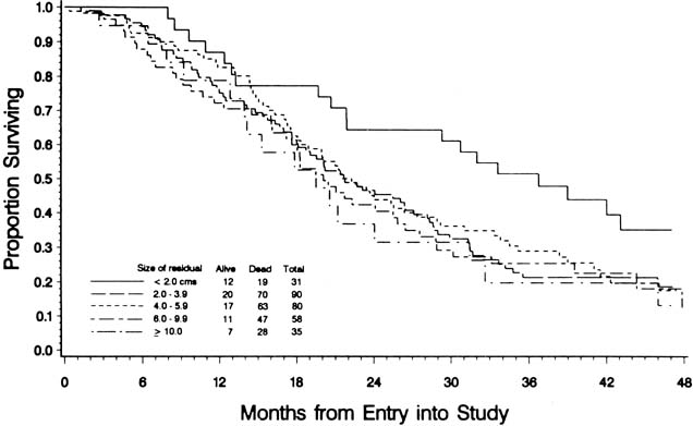
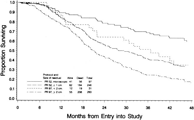
To evaluate the effect of residual disease on survival, Hoskins and colleagues65 reviewed data on 458 patients treated in accordance with a GOG protocol that evaluated standard versus high-dose chemotherapy with cisplatin and cyclophosphamide in suboptimal (residual disease greater than 1 cm) stage III and stage IV epithelial ovarian cancer. They found that patients with residual disease of 1 to 2 cm had better survival than patients with disease greater than 2 cm (Fig. 1). They found that cytoreduction that did not achieve residual disease of less than 2 cm had little effect on survival. When they combined the results of this analysis with an analysis of a similar GOG protocol (cisplatin and cyclophosphamide versus a cisplatin, cyclophosphamide, and doxorubicin combination) that enrolled patients with optimal (residual disease less than 1 cm) stage III epithelial ovarian cancer, they found that survival at 48 months was 58% for microscopic disease, 34% for residual gross disease up to 2 cm in diameter, and 16% for patients with residual disease greater than 2 cm (Fig. 2).

Fig. 1. Effect of residual disease on survival in 458 patients with stage III or stage IV ovarian cancer with suboptimal residual disease (greater than 1 cm in diameter) treated in accordance with GOG protocol 97. Patients with residual disease 1 to 2 cm in diameter have a better survival than patients with residual disease greater than 2 cm.

Fig. 2. Survival by size of residual disease in patients with stage III or stage IV ovarian cancer treated in accordance with GOG protocols 52 and 97.

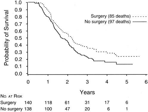
The finding of Hoskins and coworkers that disease that could not be cytoreduced to less than 2 cm in diameter was associated with uniformly poor survival and should be viewed in relation to a study of interval cytoreductive surgery by the European Organization for Research and Treatment of Cancer reported in 1995 by Van der Burg and associates.66

These authors randomized patients with suboptimal disease that could not be cytoreduced to six cycles of cisplatin and cyclophosphamide versus three cycles of the same chemotherapy followed by interval cytoreductive surgery. The surgical patients then received an additional three cycles of cisplatin and cyclophosphamide. They found a median survival of 26 months for patients undergoing interval cytoreduction versus 20 months for those who did not (p = .012) (Fig. 3).

Fig. 3. Improved survival after interval cytoreductive surgery in patients with advanced ovarian cancer with suboptimal residual tumor diameter after initial surgery.

Patients who present with suspected ovarian cancer should undergo the routine preoperative tests before any major operation, including a complete blood count, biochemical profile, chest radiograph, and electrocardiogram. In addition, special radiographic studies such as computerized tomography, ultrasonography, or magnetic resonance imaging may be helpful in confirming the presence of a mass and identifying ascites or other evidence of metastatic disease. Intestinal imaging studies or upper and lower endoscopy is performed if there is clinical suspicion of a gastrointestinal primary tumor. Most gynecologic oncologists use a regimen of mechanical and antibiotic bowel preparation to cleanse the intestine in case intestinal resection is necessary or the bowel is entered inadvertently. Many gynecologic oncologists also use a regimen of low-dose subcutaneous heparin or intermittent calf compression devices to prevent deep vein thrombosis and perioperative intravenous antibiotics to decrease infections.

Before surgery, the patient and her family should be counseled thoroughly about the risk of malignancy and the surgical procedures that might be necessary. If there is a suspicion of rectal involvement, the possibility of low rectal anastomosis should be mentioned. Although a colostomy is always possible, it is rarely performed during primary cytoreductive surgery. The patient should also understand that although every attempt will be made to remove as much tumor as possible, advanced ovarian cancer cannot be cured by surgery alone, and chemotherapy will be necessary.

The abdomen should be opened through a vertical incision extending from the pubic symphysis to well above the umbilicus to allow access to the upper abdominal structures. If ascites are present, a sample should be taken for cytologic analysis. If no ascites are present, saline washings may be obtained. Before undertaking any pelvic operation, a complete exploration of the upper abdomen should be performed, with particular attention to the hemidiaphragms and omentum. The stomach and the small and large intestines should be evaluated, as should the retroperitoneal structures, including the lymph nodes, pancreas, and kidneys. An estimate of the resectability of upper abdominal disease may be important in determining the appropriate procedure for dealing with the pelvic tumor. In evaluating the pelvis, the surgeon must consider whether the primary tumor in the ovaries can be completely removed, the extent to which hysterectomy would contribute to debulking, and the resectability of any areas of pelvic spread. An experienced gynecologic oncologist should be able to assess the extent of possible tumor removal and to decide which operative procedures would be most appropriate for cytoreduction.

In some cases, an omental tumor can be removed adequately by an infracolic omentectomy with division of the omental attachments to the transverse colon. If the tumor involves the gastrocolic omentum, the dissection will have to be carried upward to detach the omentum from the greater curvature of the stomach. Care must be taken to avoid injury to the mesentery of the transverse colon, to which the omentum is often adherent, and to the gastroepiploic vessels along the greater curvature of the stomach wall. Omental tumors often extend toward the spleen. An appropriately large incision usually allows resection of the tumor without the need for a splenectomy; however, a splenectomy is occasionally indicated. If the omental tumor infiltrates into the wall of the transverse colon, resection of the colon may be necessary to allow optimal debulking. Bulky tumor nodules on the diaphragm are often the limiting factor in achieving optimal cytoreduction, although to some extent they can be debulked sharply or with the use of the ultrasonic aspirator67 or argon beam coagulator.68 If resection of small areas of residual disease on the diaphragm will render the patient's disease optimal, small areas of the diaphragm can be resected with primary repair. It is usually necessary to mobilize the liver to perform this procedure. Although a chest tube may be required after resection of a portion of the diaphragm, one can often insert a catheter into the chest before tying the last stitch; with the end of the tube under water, the anesthesiologist can expel the air in the chest with a deep inspiration. Isolated nodules on the small intestine can be resected.

In the pelvis, bulky tumor often distorts or eliminates normal intraperitoneal tissue planes and anatomic landmarks. The retroperitoneal tissue planes, however, are usually well preserved and should be used as primary landmarks during the pelvic operation. The retroperitoneal space can usually be entered via incision of the peritoneum lateral to the infundibulopelvic ligament or the colon. After development of the space and direct identification of the ureter, the infundibulopelvic ligament can be ligated and transected. Continuing the retroperitoneal dissection, the uterine artery can also be ligated. After this is repeated contralaterally, mobilization of the ovarian tumors can proceed with less blood loss. In the case of a large tumor adherent to the pelvic sidewall, removal may require dissection of the ureter from the mass. If the tumor involves the lateral aspect of the uterus, one can perform the type of dissection used in a modified radical hysterectomy, unroofing the ureter as it passes through the parametrial tunnel and ligating the uterine vessels at this point. Extensive involvement of the rectosigmoid may require en bloc resection along with hysterectomy, as is performed in a posterior exenteration. Intestinal continuity can usually be reestablished by low anastomosis using intestinal stapling instruments. A colostomy is rarely indicated. Keeping in mind the observation first made by Griffiths that unless “optimal” cytoreduction can be accomplished the outcome is not improved, the surgeon should be reasonably certain that all bulky disease can be resected before undertaking an aggressive procedure.

Management of the pelvic and aortic lymph nodes in advanced ovarian cancer is not well defined. Although most patients with advanced ovarian cancer have involvement of pelvic or paraaortic nodes,69,70 most gynecologic oncologists agree that there is no need for routine diagnostic node sampling in patients with obvious intraperitoneal dissemination, because the information gained will have no bearing on treatment or prognosis. Whether an extensive lymphadenectomy has any effect on survival is unknown. One group has reported a retrospective series in which patients undergoing lymphadenectomy had an improved survival rate compared with historical controls.71 These data await confirmation. If the resection of bulky nodal disease allows optimal cytoreduction, it should be performed. It is our practice to remove enlarged lymph nodes in patients able to undergo optimal resection of intra-abdominal tumor.

The closure of the abdominal incision in advanced ovarian cancer patients deserves special attention. These patients are often elderly, obese, and nutritionally depleted, and they may have concomitant medical problems, such as diabetes, that interfere with wound healing and increase the risk of dehiscence. This closure may be performed with interrupted sutures or with a continuous suture using nonabsorbable or delayed absorbable material.72,73 Most studies show improved results with continuous mass closure techniques.

A number of centers have reported their experiences with the primary debulking of advanced ovarian cancer. Table 7 shows data from 11 reports,60,63,74,75,76,77,78,79,80,81 published since 1979, on a total of 2282 patients. The great variation among these individual studies in the ability to achieve optimal cytoreduction (range: 17%–87%) must in part be explained by patient selection, varying degrees of surgical skill and aggressiveness, and different definitions of optimal cytoreduction. In general, skilled and experienced gynecologic oncologists should be able to accomplish optimal debulking in at least one-half of their patients with advanced ovarian cancer.


Table 7. Optimal Cytoreduction at Primary Surgery in Stage III And Stage IV Ovarian Cancer


Patients Optimal Cytoreduction
First Author Year (N) Number Percentage
Smith 1979 792 190 24
Delgado 1984 75 13 17
Neijt 1984 186 76 41
Wharton 1984 395 154 39
Redman 1986 86 34 40
Heintz 1986 70 49 70
Neijt 1987 191 94 49
Piver 1988 40 35 87
Potter 1991 185 119 64
Eisenkop 1992 126 103 82
Baker 1994 136 113 83
Total   2282 980 43


Although some authors have questioned the value of debulking surgery in patients with stage IV ovarian cancer, recent reports from several institutions, including Memorial Sloan-Kettering and the University of Pennsylvania,82,83,84,85 indicate a significant survival benefit to optimal cytoreduction in stage IV patients. Data from these studies are summarized in Table 8. These authors concluded that attempted cytoreduction was indicated in all patients with advanced ovarian cancer, even in patients with stage IV disease.


Table 8. Effect of Debulking on Survival in Stage IV Ovarian Cancer


First Author Year Surgical Result Patients(N) Optimal (%) Median Survival P
Curtin 1997 Optimal (<2 cm) 41 45 40 .01
    Suboptimal 51   18  
Liu 1997 Optimal (<2 cm) 14 30 37 .02
    Suboptimal 33   17  
Munkarah 1997 Optimal (<2 cm) 31 34 25 .02
    Suboptimal 61   15  
Bristow 1998 Optimal (<1 cm) 25 30 38 .0004
    Suboptimal 59   10  


The morbidity of aggressive cytoreductive surgery appears to be within acceptable limits, based on publications from major centers. Heintz and coworkers78 reported that with increasing experience, improved cardiovascular monitoring, and the increased use of parenteral hyperalimentation, the risk of serious perioperative morbidity decreased to 10%, the most commonly encountered problem in their series being postoperative pneumonia.

Although patients with small-volume residual tumor at the initiation of primary chemotherapy have an improved prognosis, these patients may have had smaller, less aggressive tumors to begin with. Their improved prognosis may relate more to the biology of the tumor than to the extent of the surgery. Several authors have attempted to address this issue by retrospective studies that examine the prognostic significance of the extent of disease before and after cytoreduction. In a report by Hacker and coworkers,86 patients with large-volume metastatic disease (greater than 10 cm) were analyzed according to the success of cytoreduction. Of the patients with bulky disease, those for whom tumor was reduced to optimal status had a longer median survival than those for whom it was not. However, patients who started with smaller volume disease and achieved optimal status had an even longer survival. In this series, published in 1983, Hacker and associates used only a single alkylating agent as the chemotherapy after surgery. In a 1988 series, Heintz and colleagues87 reported on the effect of preoperative tumor size in patients treated with cisplatin-based combination chemotherapy. They observed that optimal tumor resection in patients with large-volume metastatic disease improved median survival by approximately 12 months. Once again, the survival of patients with optimal cytoreduction (smallest residual disease) who started with large-volume disease was not as good as that for their counterparts who started with smaller volume disease.

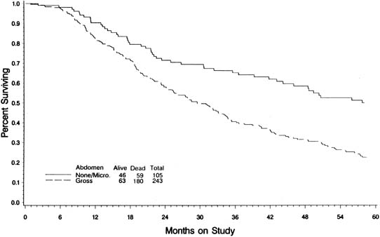
In an attempt to evaluate the relationship between cytoreductive surgery and tumor biology, Hoskins and colleagues88 reviewed 349 patients with stage III optimal (less than 1 cm) epithelial ovarian cancer. They divided patients into those found to have disease less than 1 cm in diameter on exploration and those who required cytoreductive surgery to achieve disease less than 1 cm. They theorized that if only surgery was important, survival would be equal between the two groups. They observed that patients found to have small-volume abdominal disease had a better survival than those who achieved small-volume residual disease via debulking (Fig. 4) (p = .0001). When evaluated by multivariate analysis, a variety of factors in addition to cytoreduction were found to be significant. The authors concluded that although cytoreduction was important, the biology of the tumor also influenced outcome. These studies suggest that although the finding of large-volume metastatic disease confers a poorer prognosis, and although the biology of the tumor is important, optimal tumor resection in these patients can improve median survival.

Fig. 4. Effect of tumor bulk found on initial exploration (excluding omental tumor) in patients with stage III ovarian cancer with optimal residual disease (less than 1 cm in diameter) treated in accordance with GOG protocol 52.

No prospective study randomizing ovarian cancer patients to aggressive cytoreduction versus less aggressive surgery has ever been performed, nor is such a study likely to be completed in the future. The GOG attempted such a trial with its protocol 80, but the trial came to a halt because of slow patient accrual; this failed attempt also was a reflection of the strong belief among gynecologic oncologists in the benefits of debulking.

One interesting concept that is emerging in gynecologic oncology is the possible use of neoadjuvant chemotherapy, in which patients initially undergo chemotherapy and then interval surgical cytoreduction. Retrospective analyses have demonstrated equivalent overall survivals when comparing patients treated with primary chemotherapy followed by interval debulking with conventionally treated patients.89,90 One prospective, phase II, nonrandomized trial conducted by Kuhn and associates91 noted the median survival of 31 patients with stage IIIC ovarian cancer with large volume ascites treated with three cycles of primary chemotherapy before surgical debulking to be 42 months versus only 23 months for 32 patients treated with initial surgery followed by chemotherapy (p = .007). Although promising, until these results can be validated by a large prospective randomized study, initial surgical cytoreduction followed by adjuvant chemotherapy remains the gold standard of treatment.

Few procedures offer as great a challenge to the skill and judgment of the surgeon as the cytoreduction of advanced ovarian cancer. Because optimal cytoreduction is such an important part of treatment, we believe that patients with suspected advanced ovarian cancer should be referred to a center where trained and experienced gynecologic oncologists are available.

Back to Top
REFERENCES

1. Jemal A, Murray T, Samuels A et al: Cancer statistics. CA Cancer J Clin 53:5, 2003

2. Young RA, Fuks Z, Hoskins WJ: Cancer of the ovary. In DeVita VT, Jr, Hillman SRosenberg SA, (eds): Principles and Practice of Oncology. Philadelphia, JB Lippincott, 1995

3. Scully RE: Tumors of the ovary and maldeveloped gonads. Washington, D.C., Armed Forces Institute of Pathology, 1979

4. Fuks Z: Patterns of spread of ovarian carcinoma: Relation to therapeutic strategies. Adv Biosci 26:39, 1980

5. Keettel WC, Pixley EE: Diagnostic value of peritoneal washings. Clin Obstet Gynecol 1:592, 1958

6. Feldman GB, Knapp RC: Lymphatic drainage of the peritoneal cavity and its significance in ovarian cancer. Am J Obstet Gynecol 119:991, 1974

7. Bagley CM, Jr, Young RC, Schein PS et al: Ovarian carcinoma metastatic to the diaphragm--frequently undiagnosed at laparotomy. A preliminary report Am J Obstet Gynecol 116:397, 1973

8. Piver MS, Barlow JJ, Lele SB: Incidence of subclinical metastasis in stage I and II ovarian carcinoma. Obstet Gynecol 52:100, 1978

9. Buchsbaum HJ, Brady MF, Delgado G et al: Surgical staging of carcinoma of the ovaries. Surg Gynecol Obstet 169:226, 1989

10. Eichner E, Bove E: In vivo studies on the lymphatic drainage of the human ovary. Obstet Gynecol 3:287, 1954

11. Plentyl A, Friedman E: Lymphatic System of the Female Genitalia. Philadelphia, WB Saunders, 1971

12. Musumeci R, De Palo G, Kenda R et al: Retroperitoneal metastases from ovarian carcinoma: Reassessment of 365 patients studied with lymphography. AJR Am J Roentgenol 134:449, 1980

13. Burghardt E, Pickel H, Stettner H: Management of advanced ovarian cancer. Eur J Gynaecol Oncol 5:155, 1984

14. Staging announcement: FIGO Cancer Committee. Gynecol Oncol 25:383, 1986

15. Pettersson F: Annual report on the results of treatment in gynecological cancer. Stockholm, International Federation of Gynecology and Obstetrics, 1991

16. Landis SH, Murray T, Bolden S et al: Cancer statistics 1998. CA Cancer J Clin 48:6, 1998

17. Young RC, Walton LA, Ellenberg SS et al: Adjuvant therapy in stage I and stage II epithelial ovarian cancer. Results of two prospective randomized trials N Engl J Med 322:1021, 1990

18. Schilder JM, Thompson AM, DePriest PD et al: Outcome of reproductive age women with stage IA or IC invasive epithelial ovarian cancer treated with fertility-sparing therapy. Gynecol Oncol 87:1, 2002

19. Zanetta G, Chiari S, Rota S et al: Conservative surgery for stage I ovarian carcinoma in women of childbearing age. Br J Obstet Gynaecol 104:1030, 1997

20. Querleu D: Laparoscopic paraaortic node sampling in gynecologic oncology: A preliminary experience. Gynecol Oncol 49:24, 1993

21. Scribner DR, Jr, Walker JL, Johnson GA et al: Laparoscopic pelvic and paraaortic lymph node dissection: Analysis of the first 100 cases. Gynecol Oncol 82:498, 2001

22. Hopkins MP, von Gruenigen V, Gaich S: Laparoscopic port site implantation with ovarian cancer. Am J Obstet Gynecol 182:735, 2000

23. Viala J, Morice P, Pautier P et al: CT findings in two cases of port-site metastasis after laparoscopy for ovarian cancer. Eur J Gynaecol Oncol 23:293, 2002

24. Boyd J, Rubin SC: Hereditary ovarian cancer: molecular genetics and clinical implications. Gynecol Oncol 64:196, 1997

25. Lynch HT, Bewtra CLynch JF: Familial ovarian carcinoma. Clinical nuances Am J Med 81:1073, 1986

26. Miki Y, Swensen J, Shattuck-Eidens D et al: A strong candidate for the breast and ovarian cancer susceptibility gene BRCA1. Science 266:66, 1994

27. Ford D, Easton DF, Bishop DT et al: Risks of cancer in BRCA1-mutation carriers. Breast Cancer Linkage Consortium Lancet 343:692, 1994

28. Brose MS, Rebbeck TR, Calzone KA et al: Cancer risk estimates for BRCA1 mutation carriers identified in a risk evaluation program. J Natl Cancer Inst 94:1365, 2002

29. Rebbeck TR, Lynch HT, Neuhausen SL et al: Prophylactic oophorectomy in carriers of BRCA1 or BRCA2 mutations. N Engl J Med 346:1616, 2002

30. Rubin SC, Benjamin I, Behbakht K et al: Clinical and pathological features of ovarian cancer in women with germ-line mutations of BRCA1. N Engl J Med 335:1413, 1996

31. Bast RC, Jr, Klug TL, St John E et al: A radioimmunoassay using a monoclonal antibody to monitor the course of epithelial ovarian cancer. N Engl J Med 309:883, 1983

32. Bourne T, Campbell S, Steer C et al: Transvaginal colour flow imaging: A possible new screening technique for ovarian cancer. BMJ 299:1367, 1989

33. Skipper HE: Thoughts on cancer chemotherapy and combination modality therapy. JAMA 230:1033, 1974

34. Skipper HE, Schabel FJ, Wilcox W: Experimental evaluation of potential anticancer agents XII: On the criteria and kinetics associated with “curability” of experimental leukemia. Cancer Chemother Rep 35:1, 1979

35. Goldie JH, Coldman AJ: A mathematic model for relating the drug sensitivity of tumors to their spontaneous mutation rate. Cancer Treat Rep 63:1727, 1979

36. Blythe JG, Wahl TP: Debulking surgery: does it increase the quality of survival. Gynecol Oncol 14:396, 1982

37. Morton DL: Changing concepts of cancer surgery: surgery as immunotherapy. Am J Surg 135:367, 1978

38. Munnell EW: The changing prognosis and treatment in cancer of the ovary. A report of 235 patients with primary ovarian carcinoma 1952-1961 Am J Obstet Gynecol 100:790, 1968

39. Elclos L, Quinlan EJ: Malignant tumors of the ovary managed with postoperative megavoltage irradiation. Radiology 93:659, 1969

40. Griffiths CT: Surgical resection of tumor bulk in the primary treatment of ovarian carcinoma. Natl Cancer Inst Monogr 42:101, 1975

41. Young RC, Chabner BA, Hubbard SP et al: Advanced ovarian adenocarcinoma. A prospective clinical trial of melphalan (L-PAM) versus combination chemotherapy N Engl J Med 299:1261, 1978

42. Omura GA, Bundy BN, Berek JS et al: Randomized trial of cyclophosphamide plus cisplatin with or without doxorubicin in ovarian carcinoma: A Gynecologic Oncology Group Study. J Clin Oncol 7:457, 1989

43. Rubin SC, Lewis JL, Jr: Second-look surgery in ovarian carcinoma. Crit Rev Oncol Hematol 8:75, 1988

44. Hoskins WJ: The influence of cytoreductive surgery on progression-free interval and survival in epithelial ovarian cancer. Baillieres Clin Obstet Gynaecol 3:59, 1989

45. Rubin SC, Hoskins WJ, Saigo PE et al: Prognostic factors for recurrence following negative second-look laparotomy in ovarian cancer patients treated with platinum-based chemotherapy. Gynecol Oncol 42:137, 1991

46. Barnhill DR, Hoskins WJ, Heller PB et al: The second-look surgical reassessment for epithelial ovarian carcinoma. Gynecol Oncol 19:148, 1984

47. Berek JS, Hacker NF, Lagasse LD et al: Second-look laparotomy in stage III epithelial ovarian cancer: Clinical variables associated with disease status. Obstet Gynecol 64:207, 1984

48. Cain JM, Saigo PE, Pierce VK et al: A review of second-look laparotomy for ovarian cancer. Gynecol Oncol 23:14, 1986

49. Carmichael JA, Shelley WE, Brown LB et al: A predictive index of cure versus no cure in advanced ovarian carcinoma patients—replacement of second-look laparotomy as a diagnostic test. Gynecol Oncol 27:269, 1987

50. Dauplat J, Ferriere JP, Gorbinet M et al: Second-look laparotomy in managing epithelial ovarian carcinoma. Cancer 57:1627, 1986

51. Gallup DG, Talledo OE, Dudzinski MR et al: Another look at the second-assessment procedure for ovarian epithelial carcinoma. Am J Obstet Gynecol 157:590, 1987

52. Lippman SM, Alberts DS, Slymen DJ et al: Second-look laparotomy in epithelial ovarian carcinoma. Prognostic factors associated with survival duration Cancer 61:2571, 1988

53. Lund B, Williamson P: Prognostic factors for outcome of and survival after second-look laparotomy in patients with advanced ovarian carcinoma. Obstet Gynecol 76:617, 1990

54. Podratz KC, Malkasian GD, Jr, Hilton JF et al: Second-look laparotomy in ovarian cancer: evaluation of pathologic variables. Am J Obstet Gynecol 152:230, 1985

55. Smirz LR, Stehman FB, Ulbright TM et al: Second-look laparotomy after chemotherapy in the management of ovarian malignancy. Am J Obstet Gynecol 152:661, 1985

56. Conte PF, Sertoli MR, Bruzzone M et al: Cisplatin, methotrexate, and 5-fluorouracil combination chemotherapy for advanced ovarian cancer. Gynecol Oncol 20:290, 1985

57. Hainsworth JD, Grosh WW, Burnett LS et al: Advanced ovarian cancer: Long-term results of treatment with intensive cisplatin-based chemotherapy of brief duration. Ann Intern Med 108:165, 1988

58. Louie KG, Ozols RF, Myers CE et al: Long-term results of a cisplatin-containing combination chemotherapy regimen for the treatment of advanced ovarian carcinoma. J Clin Oncol 4:1579, 1986

59. Neijt JP, ten Bokkel Huinink WW, van der Burg ME et al: Randomized trial comparing two combination chemotherapy regimens (CHAP-5 v CP) in advanced ovarian carcinoma. J Clin Oncol 5:1157, 1987

60. Piver MS, Lele SB, Marchetti DL et al: The impact of aggressive debulking surgery and cisplatin-based chemotherapy on progression-free survival in stage III and IV ovarian carcinoma. J Clin Oncol 6:983, 1988

61. Posada JG, Jr, Marantz AB, Yeung KY et al: The cyclophosphamide, hexamethylmelamine, 5 fluorouracil (CHF) regimen in the treatment of advanced and recurrent ovarian cancer. Gynecol Oncol 20:23, 1985

62. Pohl R, Dallenbach-Hellweg G, Plugge T et al: Prognostic parameters in patients with advanced ovarian malignant tumors. Eur J Gynaecol Oncol 5:160, 1984

63. Redman JR, Petroni GR, Saigo PE et al: Prognostic factors in advanced ovarian carcinoma. J Clin Oncol 4:515, 1986

64. Sutton GP, Stehman FB, Einhorn LH et al: Ten-year follow-up of patients receiving cisplatin, doxorubicin, and cyclophosphamide chemotherapy for advanced epithelial ovarian carcinoma. J Clin Oncol 7:223, 1989

65. Hoskins WJ, McGuire WP, Brady MF et al: The effect of diameter of largest residual disease on survival after primary cytoreductive surgery in patients with suboptimal residual epithelial ovarian carcinoma. Am J Obstet Gynecol 170:974, 1994

66. van der Burg ME, van Lent M, Buyse M et al: The effect of debulking surgery after induction chemotherapy on the prognosis in advanced epithelial ovarian cancer. Gynecological Cancer Cooperative Group of the European Organization for Research and Treatment of Cancer N Engl J Med 332:629, 1995

67. Adelson MD, Baggish MS, Seifer DB et al: Cytoreduction of ovarian cancer with the Cavitron ultrasonic surgical aspirator. Obstet Gynecol 72:140, 1988

68. Brand E, Pearlman N: Electrosurgical debulking of ovarian cancer: A new technique using the argon beam coagulator. Gynecol Oncol 39:115, 1990

69. Burghardt E, Pickel H, Lahousen M et al: Pelvic lymphadenectomy in operative treatment of ovarian cancer. Am J Obstet Gynecol 155:315, 1986

70. Wu PC, Qu JY, Lang JH et al: Lymph node metastasis of ovarian cancer: A preliminary survey of 74 cases of lymphadenectomy. Am J Obstet Gynecol 155:1103, 1986

71. Burghardt E, Lahousen M, Stettner H: The significance of pelvic and para-aortic lymphadenectomy in the operative treatment of ovarian cancer. Baillieres Clin Obstet Gynaecol 3:157, 1989

72. Archie JP, Feldtman RW: Primary abdominal wound closure with permanent, continuous running monofilament sutures. Surg Gynecol Obstet 153:721, 1981

73. Knight CD, Griffen FD: Abdominal wound closure with a continuous monofilament polypropylene suture. Experience with 1,000 consecutive cases Arch Surg 118:1305, 1983

74. Smith JP, Day TG, Jr: Review of ovarian cancer at the University of Texas Systems Cancer Center, M.D. Anderson Hospital and Tumor Institute Am J Obstet Gynecol 135:984, 1979

75. Baker T, Piver MS, Hempling RE: Improved long-term survival by cytoreductive surgery to less than 1 cm, induction cisplatin and monthly cisplatin, Adriamycin, cyclophosphamide in advanced ovarian adenocarcinoma. Proc Ann Meet Am Soc Clin Oncol 13:A861, 1994

76. Delgado G, Oram DH, Petrilli ES: Stage III epithelial ovarian cancer: The role of maximal surgical reduction. Gynecol Oncol 18:293, 1984

77. Eisenkop SM, Spirtos NM, Montag TW et al: The impact of subspecialty training on the management of advanced ovarian cancer. Gynecol Oncol 47:203, 1992

78. Heintz AP, Hacker NF, Berek JS et al: Cytoreductive surgery in ovarian carcinoma: Feasibility and morbidity. Obstet Gynecol 67:783, 1986

79. Neijt JP, ten Bokkel Huinink WW, van der Burg ME et al: Randomised trial comparing two combination chemotherapy regimens (Hexa-CAF vs CHAP-5) in advanced ovarian carcinoma. Lancet 2:594, 1984

80. Potter ME, Partridge EE, Hatch KD et al: Primary surgical therapy of ovarian cancer: How much and when. Gynecol Oncol 40:195, 1991

81. Wharton JT, Edwards CL: Cytoreductive surgery for common epithelial tumours of the ovary. Clin Obstet Gynaecol 10:235, 1983

82. Bristow RE, Montz FJ, Lagasse LD et al: Survival impact of surgical cytoreduction in stage IV epithelial ovarian cancer. Gynecol Oncol 72:278, 1999

83. Curtin JP, Malik R, Venkatraman ES et al: Stage IV ovarian cancer: Impact of surgical debulking. Gynecol Oncol 64:9, 1997

84. Liu PC, Benjamin I, Morgan MA et al: Effect of surgical debulking on survival in stage IV ovarian cancer. Gynecol Oncol 64:4, 1997

85. Munkarah AR, Hallum AV, 3rd, Morris M et al: Prognostic significance of residual disease in patients with stage IV epithelial ovarian cancer. Gynecol Oncol 64:13, 1997

86. Hacker NF, Berek JS, Lagasse LD et al: Primary cytoreductive surgery for epithelial ovarian cancer. Obstet Gynecol 61:413, 1983

87. Heintz AP: Surgery in advanced ovarian carcinoma: is there proof to show the benefit. Eur J Surg Oncol 14:91, 1988

88. Hoskins WJ, Bundy BN, Thigpen JT et al: The influence of cytoreductive surgery on recurrence-free interval and survival in small-volume stage III epithelial ovarian cancer: A Gynecologic Oncology Group study. Gynecol Oncol 47:159, 1992

89. Schwartz PE, Rutherford TJ, Chambers JT et al: Neoadjuvant chemotherapy for advanced ovarian cancer: long-term survival. Gynecol Oncol 72:93, 1999

90. Kayikcioglu F, Kose MF, Boran N et al: Neoadjuvant chemotherapy or primary surgery in advanced epithelial ovarian carcinoma. Int J Gynecol Cancer 11:466, 2001

91. Kuhn W, Rutke S, Spathe K et al: Neoadjuvant chemotherapy followed by tumor debulking prolongs survival for patients with poor prognosis in International Federation of Gynecology and Obstetrics Stage IIIC ovarian carcinoma. Cancer 92:2585, 2001

Back to Top