Chapter 40
The Histopathology of Vulvar Neoplasia
Edward J. Wilkinson and Heather M. Brown
Main Menu   Table Of Contents

Search

Edward J. Wilkinson, MD
Professor and Vice Chairman, Department of Pathology, Immunology & Laboratory Medicine, University of Florida College of Medicine, Gainesville, Florida (Vol 4, Chap 40)

Heather M. Brown, MD
Clinical Assistant Professor, Department of Pathology, Immunology and Laboratory Medicine, University of florida College of Medicine, Gainesville, Florida (Vol 4, Chap 40)

 
VULVAR CONDYLOMA ACUMINATUM
VULVAR INTRAEPITHELIAL NEOPLASIA
PAGET DISEASE
SQUAMOUS CELL CARCINOMA
VERRUCOUS CARCINOMA
BASAL CELL CARCINOMA
MELANOMA
BARTHOLIN GLAND TUMORS
SOFT TISSUE MALIGNANCY
OTHER SARCOMAS AND SOFT TISSUE TUMORS
OTHER PRIMARY TUMORS
METASTATIC TUMORS
PROCESSING OF SPECIMENS
ACKNOWLEDGMENTS
REFERENCES

VULVAR CONDYLOMA ACUMINATUM

Vulvar human papillomavirus (HPV) infection is commonly manifested by exophytic condyloma acuminatum with the typical appearance of a verrucoid, elevated, discreet lesion. Such lesions are commonly multifocal and may involve the vestibule and perianal areas. The lesions of HPV also may be flat and macular, especially when they involve the nonkeratinized epithelium of the vestibule, vaginal introitus, or urethra. The HPV types most commonly associated with typical genital warts are HPV 6 and 11; however, high-risk types may also be found. Fifty percent of women with vulvar condyloma acuminatum also have cervical intraepithelial neoplasia. Typical exophytic condyloma acuminatum have a fibrovascular stalk, and the epithelial surface is acanthotic and often hyperkeratotic. The fibrovascular stalk is lacking in the flat condyloma acuminatum. Both exhibit similar epithelial changes.

Microscopic features include a papillomatous lesion with some loss of epithelial maturation from the basal layer to the granular zone (Fig. 1). Basal and parabasal hyperplasia are usually present with accentuation of the intercellular bridges of the keratinocytes. Individual cell keratinization, dyskeratoses, and increased mitotic figures of normal morphology and location may all be present in varying degrees. Also seen are distinctive koilocytes, which are keratinocytes with distinctive perinuclear clearing and enlarged nuclei that may be hyperchromatic.

Fig. 1. Flat condyloma acuminatum/vulvar intraepithelial neoplasia (VIN) 1. There is lack of maturation in the lower one third of the epithelium and maturation with koilocytosis in the upper epithelium.

The differential diagnoses of vulvar condyloma acuminatum include verrucous carcinoma; vulvar intraepithelial neoplasia (VIN); condyloma lata of syphilis; and in children, other polypoid tumors, including botryoid rhabdomyosarcoma. The diagnosis can be made definitively on histologic examination of biopsy material. Their presence in children should raise the question of sexual activity or abuse, although HPV infection can be acquired at birth or may be due to cutaneous HPV types.

Back to Top
VULVAR INTRAEPITHELIAL NEOPLASIA

The International Society for the Study of Vulvovaginal Disease (ISSVD), in collaboration with the International Society of Gynecological Pathologists (ISGP), reviewed terminology for vulvar neoplastic disease and accepted the VIN terminology; however, the dysplasia/carcinoma in situ terms were placed by the World Health Organization (WHO) to develop terminology comparable to that for the cervix (Table 1).1–4 The biologic distinction between VIN 1 and flat condyloma acuminatum is unknown, and thus flat condyloma acuminatum is included in the VIN 1 category (Fig. 1). Exophytic condyloma acuminatum, in contrast, is not classified as VIN, although exophytic condyloma can occur in patients with VIN. VIN can also be classified by cellular type, including warty, basaloid, and differential types. There are corresponding invasive squamous cell carcinomas that correspond to these VIN cellular types.

Table 1. Classification of Vulvar Squamous Intraepithelial Neoplasia


VIN 1

Mild dysplasia

VIN 2

Moderate dysplasia

VIN 3

Severe dysplasia

VIN 3

Carcinoma in situ


VIN, vulvar intraepithelial neoplasia.

VIN can occur at any age starting from puberty. The median frequency appears to be among women in the 4th decade of life; this is far removed from the peak age incidence of invasive squamous carcinoma of the vulva. On clinical examination, VIN lesions may be pink, white, red, or brown and typically are slightly elevated (macular) above the level of the surrounding skin (Fig. 2). White or acetowhite lesions constitute approximately 70% of all cases, pigmented or red lesions being equally divided at approximately 15% each. Approximately 70% of the cases are multifocal, the remainder being unifocal.5 The most common sites of VIN are on the labia minora and the perineum. Perianal involvement occurs in approximately one third of cases of VIN and may extend into the anus. Pruritus is a frequent symptom but is not present in all patients.6

Fig. 2. Vulvar intraepithelial neoplasia (VIN) 3, multifocal. Note raised pale lesions on outer minora and across fourchette.

The natural history of VIN is poorly understood; however, VIN is one of the known precursors of vulvar squamous cell carcinoma. The frequency of progression of VIN to invasive squamous cell carcinoma appears to be low, with an estimated frequency from 0% to 18% of reported cases.7,8 In women with invasive squamous cell carcinoma, VIN is frequently found adjacent to the tumor. In deeply invasive tumors, VIN is seen in approximately 25% of the cases; however, in women with superficially invasive squamous carcinoma, the frequency of adjacent VIN approaches 85%. In women presenting with VIN, 2% to 28% have been found to have unsuspected invasive carcinoma with a lesion clinically appearing to be VIN.7 Women with VIN who are elderly, immunocompromised, or who have Fanconi's anemia are at increased risk for invasive disease occurring with a VIN lesion.7 Spontaneous regression of VIN lesions is well recognized, but long-term studies on these patients have not been reported, and the rates of recurrence among these women are unknown. When this disease arises during pregnancy, it may spontaneously regress in the postpartum period and require no therapy.9 These uncertainties should be kept in mind when the prognosis and therapy in any individual case of VIN are considered.

Invasive vulvar squamous cell carcinoma has adjacent warty or basaloid type of VIN in approximately 80% of cases. The prognosis in these cases is favorable because the majority of the carcinomas are superficial. In contrast, VIN 3 of the differentiated type is typically found associated with vulvar lichen sclerosus and is associated with well-differentiated squamous cell carcinoma; the mean age of these women is higher than for that of superficially invasive carcinoma (77 years versus 54 years, respectively).10,11 There is a high frequency of cervical or vaginal intraepithelial neoplasia or both (CIN, VAIN) among patients with VIN. In particular, VIN 3 is associated with a high frequency of synchronous or metachronous cervical neoplasia, with HPV infection as a common etiologic factor. Disease in these other areas may precede, coincide with, or occur after the onset of the vulvar lesion. Therefore, patients with VIN must be considered to be in a high-risk category for cervical and vaginal neoplasia and must receive appropriate follow-up, including cervical cytologic studies and colposcopy when the cytologic findings are abnormal. Because of the frequency with which vulvar VIN involves the anal canal and perianal skin, anoscopy is mandatory; sigmoidoscopy and barium enema may be helpful if anoscopy is abnormal.

Histopathologic Features

Vulvar intraepithelial neoplasia (VIN) is a term that encompasses Bowen's disease, erythroplasia of Queyrat, bowenoid papulosis, and carcinoma simplex. The clinical distinction of these lesions does not reflect biologic behavior. Pathologic distinctions have not been found to be biologically valid. The velvety-red lesions (erythroplastic) of erythroplasia of Queyrat are typically seen in the nonkeratinized squamous mucosa of the vulvar vestibule in a fashion similar to, and share many microscopic and biologic features with, those with white keratotic lesions (bowenoid) or those with hyperpigmented papules or plaques (bowenoid papulosis). These eponymic terms, therefore, have been superseded in favor of the more general term of vulvar intraepithelial neoplasia, which encompasses both clinical and pathologic findings.

In normal squamous epithelium, the basal cells divide and thereafter differentiate in a fixed postmitotic fashion. As maturation occurs, cytoplasm is acquired, granules of prekeratin are formed, and the cellular outline and nucleus eventually are lost in the superficial keratin layer. In VIN lesions, the epithelial cells continue to exhibit large nuclei with nuclear hyperchromasia as they progress upward. These cells are atypical. Mitotic figures are found elsewhere than in the basal layer and are often atypical in appearance. Multinucleation implies impairment of the cell division mechanism. Nuclear pleomorphism, hyperchromasia, and loss of maturation are evidence of abnormal development (Figs. 3 and 4). Finally, the rate of epithelial cell division and the number of cells undergoing cell division may be increased, resulting in an increased density of the cell population.

Fig. 3. Vulvar intraepithelial neoplasia (VIN) 3 (severe dysplasia), basaloid type. This intraepithelial lesion is composed of relatively uniform epithelial cells with little maturation and nuclear hyperchromasia.

Fig. 4. Vulvar intraepithelial neoplasia (VIN) 3 (severe dysplasia/carcinoma in situ ), warty type. Atypical cells extend throughout the full thickness of the epithelium. There is nuclear pleomorphism with koilocytes near the surface.

The histologic grading of VIN is determined by the degree of maturation loss of the epithelial keratinocytes (see Table 1) as follows1,2:

  VIN 1: Loss of maturation, with nuclear hyperchromasia and disordered maturation, confined to the lower one third of the epithelium (Fig. 1)
  VIN 2: Disordered maturation confined to the lower two thirds of the epithelium
  VIN 3: Disordered maturation involving more than two thirds of the epithelium and including lesions with full-thickness changes (carcinoma in situ)

The presence of a granular layer, or keratinized surface, is not an indication to downgrade a VIN lesion. Lesions having a keratin layer can be graded as VIN 3. Most lesions are VIN 2 or 3; VIN 1 lesions rarely occur in isolation, but rather usually are seen in association with higher grade VIN lesions. On the vulva, the superficial layers of cells are either keratinized or parakeratotic, so a “full-thickness” change, per se, rarely exists.

There may be a variety of microscopic features in VIN. Three histopathologic subtypes are currently recognized:

  Basaloid type: These are characterized by relatively uniform cells without significant maturation from the basal-parabasal cells (Fig. 3)
  Warty (condylomatous) type: These resemble condyloma acuminatum (Fig. 1) with koilocytosis, hyperkeratosis, and dyskeratosis (Fig. 4)
  Differentiated type (simplex type) (keratinized squamous cell): These have prominent eosinophilic cytoplasm in the parabasal layer and exhibit keratin formation. By convention, well-differentiated types of VIN are all considered VIN 3.11

HPV has been identified by Southern blot, in situ hybridization, and polymerase chain reaction in more than 80% of VIN lesions studied. The variety of HPV types found in preinvasive cervical lesions has not been observed in the vulvar intraepithelial lesions. HPV 16 is the most common type found in the vulva. Most of the warty and basaloid types of VIN and associated squamous carcinomas are HPV 16 related. The differentiated type of VIN is not usually HPV related but is associated with lichen sclerosus (see section on squamous cell carcinoma, microscopic features, below).10

In pigmented VIN lesions, there is a concentration of melanin in the dermal melanophages and basal keratinocytes. The presence or absence of pigmentation does not appear to be associated with clinical behavior but rather location on the vulva, with those VIN lesions involving the skin being more commonly pigmented than those involving the vulvar vestibule. Most lesions show an inflammatory response beneath the disordered epidermis.

Treatment

Vulvar intraepithelial neoplasia therapy should be administered on an individualized basis. Simple vulvectomy, as has been advocated in the past, constitutes overtreatment. Many patients with this disease are young and sexually active. Every attempt should be made to retain a functioning clitoris and to provide an acceptable functional and cosmetic result. VIN lesions are typically very superficial and confined to the epithelium.12 This permits sparing of the deeper tissues.

Lesions confined to the inner vulvar vestibule surfaces can be treated with superficial excision or laser ablation, provided that prior biopsy has excluded invasion. Wide superficial local excision with a 1-cm margin can be performed on localized lesions. Larger lesions can be treated with excision and laser ablation. Histologic evaluation of the specimen is necessary to ensure that there is no invasion and to define the status of the margins of resection. Frozen sections are not necessary and only add to the cost of treatment. With uninvolved margins of resection and absence of occult invasive foci within the lesion itself, the prognosis for patients treated in this manner is extremely good. Should new foci of disease occur, they can be handled in a similar fashion. The rate of local recurrence is approximately 6% for negative margins and approximately 20% for involved margins.6 Therefore, re-excision with involved margins is not always necessary; a follow-up examination determines whether VIN is present and further treatment necessary.

Extensive confluent multicentric lesions involving the entire vulva demand a careful treatment plan. Multicentric VIN lesions can be treated with superficial excision alone or with a combination of superficial excision and laser ablation.13 It is recognized that skin appendage involvement by VIN occurs and, in hair-bearing skin, may involve skin appendages to a depth of up to 2.9 mm. In nonhair-bearing skin (e.g., vulvar vestibule, labia minora, clitoris, perineal body), skin appendage involvement is not an issue, and in these areas, the VIN is typically less than 1-mm thick.12} Consequently, a plan of therapy should use a local superficial excision of the VIN lesions involving the hair-bearing skin and the skin with sebaceous glands without hair (e.g., medial labia majora, perianal and perineal areas peripheral to Hart's line).13 Superficial excision “skinning” procedures are one approach for such cases. As a rule, the surgical wound edges can be approximated primarily by mobilization of vaginal epithelium and creation of full-thickness flaps of adjacent skin. Some surgeons prefer the primary grafting of the denuded area with skin removed from a thigh donor site. Treatment with laser ablation or superficial excision of the VIN involving the clitoris, vestibule, labia minora, and perineal body proximal to Hart's line (i.e., the junction between the nonkeratinized epithelium of the vestibule and the skin) is an effective approach. In these nonhairbearing areas, it is essential that careful evaluation and selective biopsy of the VIN lesions be performed to exclude associated invasive carcinoma before treatment is initiated.7,14

Topical 5-fluorouracil has been advocated in the past for the treatment of VIN. Therapy consists of topical application of a 5% 5-fluorouracil cream applied three times per week for a period of 6 weeks. This therapy is not currently recommended in women of reproductive age because of changes in the U.S. Food and Drug Administration' s position regarding this medication and its potential side effects in pregnancy. Either technique gives a good cosmetic result and extremely low recurrence rate, provided the initial procedure is carried down to a level well below the epidermis and its adnexa and beyond the visible margins of disease. The clitoris usually can be spared, thus retaining the possibility of function. Irradiation and radical surgical excision have no place in the treatment of VIN.

Some studies have shown promising results using photodynamic therapy (PDT), particularly 5-aminolevulinic acid (5-ALA), as a nonsurgical approach to the treatment of VIN.15 In one study of 25 patients16 using topical sensitization of VIN lesions with 5-ALA and subsequent PDT, all VIN 1 lesions showed responsiveness, as did all monofocal and bifocal VIN lesions regardless of grade. However, multifocal VIN 3 disease did not respond well to the treatment. Use of topical Imiquimod, an immune response modifier, for VIN 3 lesions has also shown some promising preliminary results.17

Back to Top
PAGET DISEASE

Paget disease of the vulva is an intraepithelial neoplastic process; however, it is a unique and biologically distinct entity with specific morphologic characteristics and oncologic implications that warrant separate discussion. Paget disease of the skin was first described by Sir James Paget in 1874 in the nipple and areola of the breast and was associated with an underlying breast carcinoma (mammary Paget disease).

Extramammary Paget disease can arise in the female lower genital tract within the vulva or perianal area. Most cases of extramammary Paget disease of the vulva or perianal region are primary to these sites; that is, they arise from within the vulvar or perianal epithelium. However, not all cases of vulvar or perianal Paget disease are primary in origin, and a small percentage of cases are actually manifestations of an internal malignancy (most commonly anorectal adenocarcinoma or urothelial carcinoma from the bladder or urethra) that has spread or metastasized within the epithelium to secondarily involve the vulva or anus. Secondary Paget disease of the vulva constitutes approximately 12% of vulvar Paget cases.

To emphasize the importance of distinguishing primary from secondary vulvar Paget disease, because of the difference in treatment for the two entities, a recent classification scheme based on cell of origin has been proposed (Table 2). It is generally accepted that the Paget cells of primary cutaneous type (type 1) arise from an intraepidermal, pluripotent stem cell with apocrine gland differentiation.

Table 2. Subtypes of Vulvar Paget Disease


Type 1

Paget disease as a primary neoplasm of the skin


1a Paget disease as a primary intraepithelial neoplasm


1b Paget disease as an intraepithelial neoplasm with invasion


1c Paget disease as a manifestation of an underlying primary adenocarcinoma of skin, appendage, or subcutaneous gland

Type 2

Paget disease secondary to adenocarcinoma of nonskin


origin, including anal or rectal adenocarcinoma

Type 3

Paget disease secondary to urothelial neoplasia


(PUIN)


PUIN, Pagetoid urothelialintraepithelial neoplasia.

Virtually confined to postmenopausal white women, primary cutaneous vulvar Paget disease appears as a reddish-pink area interlaced with patches of hyperkeratotic white epithelium, usually on the lateral aspects of the labia majora (Fig. 5). The appearance is not unlike that of a severe cutaneous candidiasis or eczema, and the disease is often confused with these benign conditions. The reluctance to perform a simple office biopsy often leads to delay in diagnosis. The disease begins as a localized process, but it can involve the entire vulva, perianal skin, and inner thigh regions. It is not uncommon for the visible margins of the disease to not correspond to the extent of histologic involvement.

Fig. 5. Primary Paget disease (type 1). Note irregular patchy change present on lateral aspect of labium majus.

In contrast, vulvar Paget disease as a manifestation of anal or rectal adenocarcinoma (type 2) typically involves the perianal skin, as well as adjacent, contiguous vulvar skin, and as a manifestation of urothelial carcinoma (type 3) primarily involves the vulvar vestibule and periurethra. However, these cases of secondary Paget disease appear as eczematoid lesions, similar to primary vulvar Paget disease, and therefore, histologic examination is necessary to separate them. A summary of the clinical manifestations of Paget disease types 1, 2 and 3 is provided in Table 3.

Table 3. Clinical Manifestations of Subtypes of Vulvar Paget Disease


Type

Clinical Presentation

Type 1: Cutaneous vulvar Paget disease

Presents on vulvar skin, often on the lateral aspects of the labia majora

Type 2: Paget disease as a manifestation of anal or rectal adenocarcinoma

Typically involves the perianal skin, as well as adjacent, contiguous vulvar skin

Type 3: Paget disease as a manifestation of urothelial carcinoma

Primarily involves the vulvar vestibule and periurethral vulvar epithelium


From Wilkinson EJ, Brown HM: Pagetoid urothelial intra-epithelial neoplasia (PUIN): Differentiation from Paget disease of cutaneous or ano-rectal origin. Mod Pathol 14(1)147A, 2001.

Histopathologic Features

Microscopically, in cutaneous (type 1) Paget disease, the involved skin contains groups of large, pale cells that are round to oval (Fig. 6). In Paget disease secondary to spread from urothelial carcinoma, the cells tend to be smaller and more hyperchromatic than in type 1 (Fig. 7), but distinguishing the different types microscopically without the use of additional stains can be very difficult. The Paget cells, in cutaneous (type 1) or anorectal (type 2) Paget disease, have abundant cytoplasm rich in neutral and acidic mucopolysaccharides. Therefore, such cells have characteristic staining reactions, and their cytoplasm exhibits positive staining with periodic acid-Schiff (PAS, diastase-resistant) and mucicarmine. Paget cells are rich in carcinoembryonic antigen and cytokeratin, and immunohistochemical studies may be useful in distinguishing them from other intraepithelial neoplastic processes (Table 4).

Fig. 6. Primary Paget disease (type 1). Characteristic large, pale Paget cells are just above basal layer (magnification, ×500).

Fig. 7. Type 3 Paget disease of the vulva (pagetoid urothelial intraepithelial neoplasia [PUIN]). The vulvar epithelium is infiltrated by clusters of high-grade urothelial carcinoma. The nuclei are hyperchromatic with irregular contours.

Table 4. Summary of the Immunohistochemical Findings of Primary and Secondary Vulvar Paget Disease


CK 7

CK 20

GCDFP

CEA

UP-III

Primary

+

+/-

+/-

+

-

Secondary to anorectal

-(rare +)

+

-

+

-

 adenocarcinoma


Secondary to urothelial

+

+/-

-

+/-

+

 carcinoma


CK 7, cytokeratin 7; CK 20, cytokeratin 20; GCDFP, gross cystic disease fluid protein; CEA, carcinoembryonic antigen; UP-III, uroplakin-3.

The characteristic Paget cells usually are found singly or in nests along the basal layer of the epithelium, and they may extend to involve the sweat gland and hair shaft epithelia. This involvement of skin appendages should be distinguished from invasion. In most cases, Paget disease is entirely intraepithelial; however, there is a subgroup of cases in which associated invasive adenocarcinoma is found. Approximately 20% of patients with vulvar Paget disease have sweat gland or Bartholin' s gland adenocarcinoma beneath the clinically recognizable Paget disease.1

Clinical Behavior and Treatment of Vulvar Paget Disease

Vulvar Paget disease is a chronic, indolent process confined by the basement membrane and marked by recurrences. Invasive primary Paget disease has been observed contiguous to typical intraepithelial Paget disease in 10% to 20%. The prognosis in cases with underlying invasive disease is poor. Therefore, the primary approach to primary Paget disease should include surgical excision to the deep fascia to exclude underlying invasion.18 Excision should include all epithelium visibly involved with Paget disease, with a margin as wide as possible (a 1- to 2-cm border of apparently normal skin is not excessive). If the initial surgery shows underlying adenocarcinoma, radical vulvectomy with ipsilateral inguinal-femoral lymphadenectomy is indicated as a secondary procedure.

Despite generous surgical margins, in many cases the margins may prove to be microscopically positive, and recurrences along the lines of wound approximation are common. When the entire visible Paget lesion is excised, with a 1-cm margin, performing frozen sections for assessment of margins is not useful in that the presence or absence of intraepithelial Paget disease at the margins does not influence the risk of recurrence.20 Recurrences have been reported in skin grafts applied to the surgical defect, along and across suture lines, and in contiguous areas of “normal” skin. Because the danger of subjacent cancer already is ruled out by the primary surgery, the risk of invasive tumor in nonprimary clinically normal-appearing areas with Paget disease is remote, and radical excision of these areas has not been proved to reduce recurrence.21 The treatment of such recurrent lesions can be focused on the intraepithelial disease, and topical therapy is useful. The application of topical 5% 5-fluorouracil cream three times weekly for 6 consecutive weeks, as well as topical bleomycin, has been used to treat recurrence; however, laser ablation and local excision are both effective treatments in peripheral sites.13,22

Surgical treatment of secondary vulvar Paget disease should be different than that of primary vulvar disease. Vulvar Paget disease related to anal or rectal adenocarcinoma or urothelial carcinoma is usually entirely intraepithelial within the vulva. Therefore, the treatment for secondary vulvar Paget disease can be modified to address the primary neoplastic lesion and treat the intraepithelial vulvar disease as an intraepithelial, not an invasive, process.18 In most cases, the superficial involvement of the vulvar skin can be treated with superficial excision without having to extend to the fascia or with other superficial ablative techniques.18

In summary, vulvar Paget disease, from a surgical standpoint, should be viewed as either primary or secondary to the vulva because of the differences in treatment and tumor biology (Table 5).

Table 5. Etiologic Classification of Extramammary Genital Paget Disease

  Primary Paget Disease (Cutaneous Origin) (type 1)*
  Intraepithelial Paget disease (type 1a)
  Intraepithelial Paget disease with invasion (type 1b)
  Intraepithelial Paget disease with associated skin appendage adenocarcinoma (type 1c)
  Secondary Paget Disease (Noncutaneous Origin)
  A manifestation of associated adenocarcinoma (type 2), including:

  Anal/rectal/colonic
  Cervical
  Other


  A manifestation of urothelial carcinoma/PUIN (type 3)


PUIN, Pagetoid urothelial intraepithelial neoplasia.
* Other sites reported include axillary, eyelid, external ear canal, umbilical, inguinal femoral skin, etc.

Back to Top
SQUAMOUS CELL CARCINOMA

Squamous cell carcinoma is the most common malignant tumor of the vulva, constituting more than 90% of all vulvar malignancies and accounting for 4% of all genital malignancies in women. The frequency of this disease increases with age, and it is most common in women older than 75 years in whom its incidence is 19.9 per 100,000 women.14

Epidemiologic studies support the hypothesis that vulvar carcinoma has at least two origins: one HPV-related and one not. In younger patients, these lesions are frequently HPV-related with VIN as an apparent precursor. The VIN lesions are usually of the warty or basaloid type and are associated with basaloid or warty types of squamous cell carcinoma. These women are often heavy cigarette smokers as a group. Cigarette smoking alone appears to increase the odds ratio. The other group of women is older and does not appear to have HPV-related vulvar lesions, nor does there appear to be an association with cigarette smoking in this group. This older group of women may have associated vulvar dermatoses, usually lichen sclerosus. Other risk factors for vulvar carcinoma in all groups include carcinogen exposure, immunosuppression, chronic granulomatous disease, and prior cervical or vaginal carcinoma. The association between squamous cell carcinoma of the cervix or vagina and squamous carcinoma of the vulva is well established. As in VIN lesions, HPV infection is a common etiologic factor leading to multifocal genital neoplasia.23 Obesity and poor perineal hygiene also are implicated in many cases. Pregnancy and high parity do not appear to be associated.

Clinical Manifestations

Squamous carcinoma of the vulva usually appears as an ulcerated, exophytic, or indurated mass that may be associated with pruritus, local infection, and bleeding. Squamous cell carcinoma of the vulva is typically divided into two categories: (1) superficially invasive squamous cell carcinoma and (2) frankly invasive squamous cell carcinoma. VIN may be found adjacent to invasive carcinoma in approximately 60% of cases and is found more commonly among younger women and in those with more superficially invasive tumors. Conversely, superficially invasive vulvar carcinoma may be encountered in the treatment of VIN. Vulvar lichen sclerosus coexists with carcinoma in approximately 15% to 40% of cases.14 Although squamous cell hyperplasia has been found at the edges of invading tumor, it is most probable that the hyperplastic lesions seen immediately adjacent to squamous cell carcinomas are induced by factors associated with the carcinoma, or by the carcinoma itself, and may not necessarily be a precursor lesion. The tissue adjacent to the tumor may harbor VIN, lichen sclerosus, or nonspecific hyperplasia or may appear 
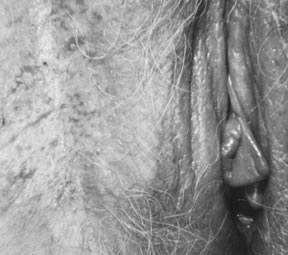
normal.24 In most cases of vulvar carcinoma, the tumor occurs within a single focus, usually within the clitoris, perineal body, or medial aspects of the labia majora or minora.

Squamous carcinoma of the vulva is generally slow growing and spreads by extension to contiguous skin, vagina, and rectum. The lesion may also invade deeper tissues and become fixed to the pelvic bones. Metastases to lymph nodes involve the femoral and inguinal nodes before they progress to the deep obturator, internal lilac, or periaortic group. When nodal metastases occur, the tumor typically involves the ipsilateral nodes and, in rare cases, the contralateral nodes. When the tumor is midline, bilateral nodal involvement may occur.25 The current clinical staging system for vulvar carcinoma was defined in 1995 by FIGO. The International TNM staging is also commonly used26 (Table 6). The system no longer relies on the clinical assessment of groin nodes, a procedure that can be quite inaccurate, based on a comparison of clinical findings with surgical and pathologic findings.27

Table 6. Vulvar Carcinoma Staging: Comparison of the AJCC and FIGO Nomenclatures, TNM Stage Groupings, and Correlation With FIGO Staging


TNM


FIGO

Stage 0Tis

N0

M0


Stage IT1

N0

M0


Stage I

Stage IA

T1a

N0

M0


Stage IB

T1b

N0

M0


Stage II

T2

N0

M0

Stage II

Stage III

T1

N1

M0

Stage III


T2

N1

M0


T3

N0,N1

M0


Stage IVA

T1

N2

M0

Stage IVA


T2

N2

M0


T3

N2

M0


T4

Any N

M0


Stage IVB

Any T

Any N

M1

Stage IVB


AJCC, American Joint Comission on Cancer.
FIGO, International Federation of Gynecology and Obstetrics.

The prognosis for vulvar carcinoma is primarily related to the size of the original lesion and the presence or absence of nodal metastases. Patients with tumors confined to the vulva with tumor-free groin nodes have a corrected 5-year survival rate of 90% after adequate excision of the tumor and usual ipsilateral inguinal-femoral lymphadenectomy. If the tumor has spread to the groin nodes, the corrected 5-year survival rate decreases to 65%. If the tumor has spread to involve the pelvic nodes as well, the survival rate is less than 25%.14,25,28 Large squamous carcinomas of the vulva have been reported in association with hypercalcemia, without bone metastasis.29 This is primarily mediated through the production of parathyroid hormone from the neoplastic keratinocytes.2,29 In addition, attempts at medical correction of the hypercalcemia have been unsuccessful. After excision of the tumor, the serum calcium level returns to normal but rises again with recurrent disease.

Superficially Invasive Carcinoma of the Vulva

The concept of superficially invasive carcinoma of the vulva implies that lesions with limited dermal invasion may constitute a separate prognostic category.28,30 The term microinvasive carcinoma has not been accepted for reference to vulvar carcinoma. Solitary tumors with a depth of invasion of 1 mm or less have essentially no risk of regional node metastasis, and node sampling or resection is not contributory in these.

HISTOPATHOLOGY

The FIGO staging of vulvar carcinoma has defined a stage IA subset of stage I carcinoma as originally set forth by the ISSVD, as a tumor with a diameter of 2 cm (20 mm) or less with a depth of invasion of 1 mm or less (Fig. 8). Measurement of tumor diameter alone is insufficient to define the risk of nodal metastasis. Most vulvar carcinomas with a depth of invasion of 1 mm or less are less than 2 cm in diameter. Clinically, VIN surrounding the tumor may be included erroneously in the measurement of the tumor diameter. In superficially invasive vulvar carcinoma, it is essential that the entire tumor be available for study before an attempt is made to determine the depth of invasion. Cases in which there is more than one tumor are not included in the stage IA group. The depth of invasion is defined as the measurement from the epithelial-stromal junction of the adjacent most superficial dermal papilla to the deepest point of invasion, as described by Wilkinson and colleagues.1,30,31 The ISGP and the WHO have accepted this definition of the “depth of invasion” in the vulva.1,2 This measurement should be distinguished from the “thickness of the tumor,” which is defined as the measurement from the surface, or bottom of the granular layer of keratin, if present, to the deepest point of invasion (Fig. 9A, B). The ISGP and WHO recommend that the pathologist report both measurements whenever possible. The stage IA group includes patients whose tumors involve capillary-like spaces, provided skin measurements are available. A protocol for the evaluation of vulvar specimens drafted by the senior author (Wilkinson) has been published by the College of American
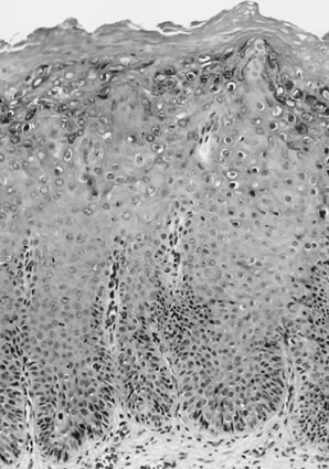
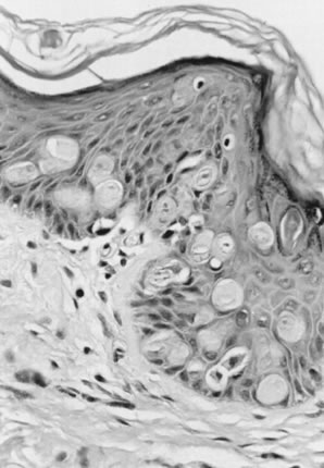
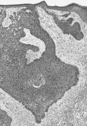
 Pathologists.26

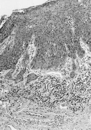
Fig. 8. Superficially invasive squamous cell carcinoma with vulvar intraepithelial neoplasia (VIN) 3. There is a marked inflammatory response.

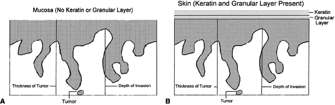
Fig. 9. Measurement methods of squamous cell carcinoma of the vulva. Depth of invasion is defined as the distance from the epithelial-stromal junction of the adjacent most-superficial dermal papilla to the deepest point of invasion. Tumor thickness is defined as the distance from the surface (mucosa) or bottom of the granular layer of the keratin (skin) to the deepest point of invasion.

TREATMENT

Stage IA carcinomas, as defined in Table 7, can be treated by wide and deep local excision without lymphadenectomy. In excising a squamous carcinoma, the entire tumor should be included in the excision so that accurate measurements of the diameter, depth of invasion, and thickness of tumor can be assessed. An excision with margins of approximately 2 cm, extending to the fascia (partial deep vulvectomy), is recommended.25 If the tumor is then found to be deeper than 1 mm (no longer a stage IA) on pathologic examination, ipsilateral lymphadenectomy is indicated unless the tumor is midline, in which case bilateral inguinal-femoral lymphadenectomy is necessary. Total or more complete partial deep vulvectomy may be necessary if the tumor extends to a margin of resection or is more than 3-mm deep.

Table 7. Suggestions for Sampling Tissue Removed for Diagnosis or Treatment of Vulvar Cancer

CLINICAL FOLLOW-UP

Superficially invasive vulvar squamous cell carcinomas that meet the definition of stage IA have essentially no risk of nodal metastases. In contrast, with tumors 3-mm deep, the risk of nodal metastasis is approximately 12%. With a 5-mm depth of invasion, or thickness, there is a reported risk of nodal metastases of approximately 15%.1,25

Follow-up of women with vulvar superficially invasive carcinoma is essential because it is recognized that these women, although they have a relatively low risk of local recurrence, are at risk of having a “reoccurrence” of a new primary tumor or the vulva, independent of the original tumor site. Although this risk is low, awareness and observation, with biopsy when indicated, can reduce the risk of metastases or death from a recurrent or new “reoccurrent” vulvar carcinoma.25,31

Frankly Invasive Squamous Cell Carcinoma of the Vulva

Frankly invasive squamous cell carcinomas include all of those that invade to a depth beyond the limit used to define superficially invasive carcinoma (1 mm).

HISTOPATHOLOGY

Histologic subtypes of squamous cell carcinomas are listed in Table 8. Squamous cell carcinomas with keratin pearls and obvious squamous cellular characteristics are considered well differentiated (Fig. 10). This pattern is usually the most frequent histologic type encountered. Other terms that have been suggested for the keratin pearl-forming carcinomas include large cell keratinizing, keratinizing, and epidermoid carcinoma. These tumors are more common in older women and are usually not associated with HPV.1,10 The less well-differentiated squamous cell carcinomas are less obviously squamous in origin without prominent intercellular bridges or keratinization (Fig. 11).

Table 8. Histologic Subtypes of Vulvar Squamous Cell Carcinoma Including Basal Cell Carcinomas

  Squamous cell carcinoma, well-differentiated (not otherwise specified)
  Basalold carcinoma
  Warty (condylomatous) carcinoma
  Verrucous carcinoma
  Giant cell squamous carcinoma
  Spindle cell squamous carcinoma
  Acantholytic squamous cell carcinoma (adenoid squamous carcinoma)
  Lymphoepithelioma-like carcinoma
  Basal cell carcinoma

  Metatypical basal cell carcinoma (basosquamous carcinoma)
  Adenoid basal cell carcinoma
  Sebaceous cell carcinoma


From Wilkinson EJ: Premalignant and malignant tumors of the vulva. In Kurman RJ: Blaustein's Pathology of the Female Genital Tract, 5th ed. New York, Springer-Verlag, 2001.

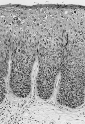
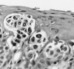
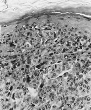
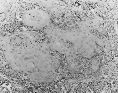
Fig. 10. Well-differentiated squamous carcinoma. Large cells with abundant cytoplasm form keratin pearls (magnification, ×80).

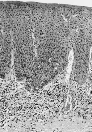
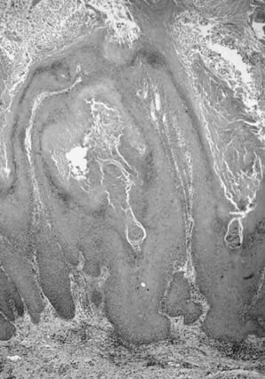
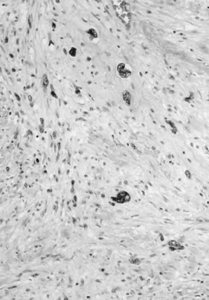
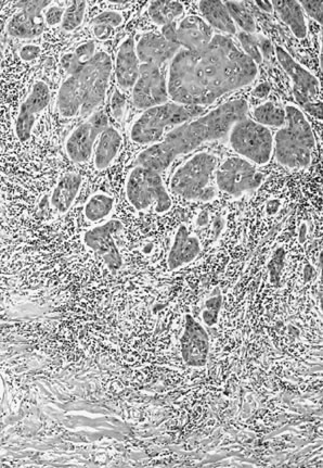
Fig. 11. Poorly differentiated squamous cell carcinoma of the vulva. The tumor cells are nonkeratinized, without prominent intercellular bridges. The tumor has a “finger-like” pattern of invasion.

Basaloid carcinomas are described as squamous tumors that do not form keratin and are composed of smaller cells with an increased nuclear-to-cytoplasmic ratio. These are less common than the well-differentiated tumors and are associated with HPV, especially type 16.10

Warty (condylomatous) carcinomas have superficial features of condyloma acuminatum. Their prognosis appears to be intermediate between well-differentiated squamous cell carcinomas and verrucous carcinomas. They also are associated with HPV, especially type 16.10

The giant cell forms of vulvar carcinoma are characterized by multinucleated tumor giant cells and may have a poorer prognosis than the well-differentiated carcinoma. These tumors must be distinguished from malignant melanoma, which also may have tumor giant cells. An analysis of cases classified as giant cell carcinoma of the vulva showed that criteria described for giant cell carcinoma may be observed in vulvar malignant melanoma and that the use of immunohistochemical studies is essential to distinguish between the two (see section on melanoma, below).32

In a study of 50 squamous cell carcinomas, Lasser and coworkers33 described two patients in whom the tumor showed primarily an adenoid-squamous cell pattern. In 15 other patients, loci of adenoid-squamous change were evident in isolated areas. The adenoid-squamous pattern of growth is characterized by the formation of pseudoglandular spaces lined with a single layer of squamous cells (Fig. 12). Within the spaces, dyskeratotic and acantholytic cells may be present. Mucin stains do not show evidence of secretion. These adenosquamous areas are usually found in tumors that would otherwise be considered well-differentiated carcinomas with keratin pearl formation, and this pattern of growth has not been observed in metastases or in recurrent tumors. However, neoplasms with a predominantly adenoid pattern are poorly differentiated and have a poor prognosis. One study noted a 6% 5-year survival for such patients compared with a 77% survival for patients 
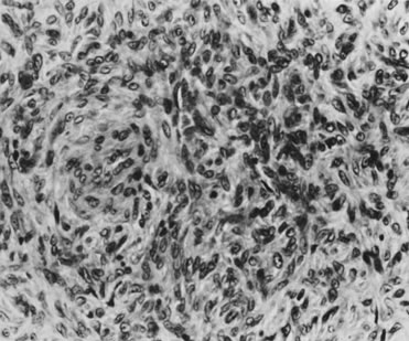
with conventional squamous cell carcinoma.34

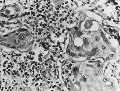
Fig. 12. Adenoid-squamous pattern with pseudoglandular spaces lined by squamous cells (magnification, ×500).

Other Types of Vulvar Carcinoma

Other primary carcinomas that have been reported arising within the vulva include verrucous carcinoma, basal cell carcinoma, small cell,35 Merkel cell carcinoma,36–38 and adenocarcinoma.39 The ISGP has recommended that pathologists report the following information for partial or total vulvectomy specimens1:

  1. Depth of tumor invasion (in millimeters)
  2. Thickness of tumor (in millimeters)
  3. Whether there is vascular space involvement by tumor
  4. Diameter of the tumor (in millimeters) measured in the fresh or fixed state
  5. Clinical measurement of the diameter of the tumor

TREATMENT

The treatment for vulva squamous cell carcinoma is directed toward sparing as much of the vulva as possible with wide local excision (partial deep vulvectomy) and associated ipsilateral regional lymph node dissection (inguinal-femoral lymphadenectomy). A recent study has shown that limited lymphadenectomy may be adequate in assessing the lymph node status in patients with vulvar squamous carcinoma.40 If the tumor is midline or extends near the midline, bilateral inguinal-femoral lymphadenectomy is usually performed.41 If the superficial lymph nodes are involved with tumor, radiation of the deep pelvic nodes is usually recommended.41

Back to Top
VERRUCOUS CARCINOMA

Verrucous carcinomas are an uncommon, special type of tumor with characteristic gross and microscopic features.42 Clinically, they are slow-growing, papillary, cauliflower-like lesions that eventually may become necrotic, ulcerated, and fungating.1,42 They may be locally destructive, and when surgically excised, they commonly recur but rarely metastasize. On histologic examination, verrucous carcinomas show a wartlike character similar to condylomata acuminata, demonstrating numerous branching papillary stalks composed of extremely well-differentiated squamous epithelium. Verrucous carcinomas are characterized by a “pushing” border with a well-defined tumor-dermal interface, without apparent infiltration.42 Cellular atypia is minimal and always restricted to the basal layer.42 Atypical mitotic figures are not identified and dyskeratosis and koilocytosis are absent. A chronic inflammatory cell infiltrate within the dermis is usually present. The lesion, called “giant condyloma of Buschke Lowenstein,” is now considered verrucous carcinoma.1 Verrucous carcinomas have been found to be associated with HPV, types 6 and 11 being the most common.43 These tumors are typically DNA diploid.

The main differential diagnoses for verrucous carcinomas include condyloma acuminatum and squamous carcinoma with verrucous features (warty carcinoma). Condylomata are typically not infiltrative, have prominent vascular dermal papillae separating the acanthotic epithelial elements, and have koilocytosis and dyskeratosis. Occasionally, VIN may occur within a condyloma, which can be distinguished by the nuclear pleomorphism and hyperchromasia, abnormal mitoses, and junction with the typical condyloma acuminatum. Warty squamous carcinomas have infiltrating tumors at the tumor-dermal interface (Fig. 13), rather than the pushing margins of verrucous carcinoma. Warty carcinomas usually have adjacent warty VIN, with koilocytosis, dyskeratosis, multinucleation, nuclear pleomorphism, and hyperchromasia. Usually abnormal mitotic figures are evident unlike verrucous carcinoma.

Fig. 13. Squamous cell carcinoma, warty or condylomatous type. The epithelium shows little cellular atypia and the tumor-dermal interface is infiltrative rather than pushing in appearance.

Verrucous carcinoma is a slowly growing tumor, but left untreated, it can be deeply infiltrative and locally destructive. The treatment of verrucous carcinoma is partial deep vulvectomy (wide local excision to the fascia). Lymphadenectomy is not necessary if the lymph nodes are not clinically suspicious. If they are suspicious, lymph node biopsy or fine-needle aspiration is recommended to exclude metastasis rather than lymphadenectomy since metastases to regional lymph nodes are not generally encountered.42 In the majority of cases, lymphadenopathy, if present, is related to an inflammatory reaction evoked by the tumor or secondary to infection. Recurrence is uncommon; however, any new lesions require re-excision. Radiation therapy should be contraindicated because of the possibility that the tumor can undergo anaplastic change with subsequent aggressive behavior after radiation.42 Local application of podophyllin, bleomycin therapy, and cryosurgery are considered ineffective methods of treatment.44 Recently, there has been a report of control of tumor progression using acitretin in a case of verrucous carcinoma not amenable to surgery.45

Back to Top
BASAL CELL CARCINOMA

Basal cell carcinomas constitute 2% to 3% of all vulvar malignancies, but make up approximately 65% of all nonvulvar cutaneous malignancies. Vulvar basal cell carcinoma can be found in any woman older than 30 years of age, but is most commonly encountered in older women.46

The clinical presentation of basal cell carcinoma has been divided into three basic types. The first type is the nodulo-ulcerative lesion, which may appear as a deep ulcer with raised margins. The second presentation is a more superficial, flat lesion with a waxy surface that is macular and slightly erythematous. The third type appears as a polypoid excrescence with an intact surface. These three clinical patterns may occur in any combination and carry no particular significance with regard to prognosis, but rarely is the diagnosis made on clinical appearance alone. The tumor is typically solitary and confined to the vulva, usually involving the labia majora. Multiple basal cell carcinomas can be seen in the autosomal-dominant nevoid basal cell carcinoma (or Gorlin) syndrome and may be associated with other anomalies, including jaw cysts and skeletal abnormalities.47 The finding of a basal cell carcinoma should prompt a search for basal cell lesions elsewhere on the skin, although the majority of basal cell carcinomas are not syndrome-associated. In a recent small study of basal cell carcinoma of the genitalia, no predisposing risk factors or evidence of HPV infection was identified.48

Microscopic features and behavior of basal cell carcinomas of the vulva are the same as those of basal cell carcinomas elsewhere in the skin. Basal cell carcinoma derives its name from the similarity of the tumor cells to the normal basal cells of the epidermis and is composed of relatively uniform cells with prominent palisading without keratinization. The tumor usually has a well-defined infiltrating margin. A desmoplastic reaction is common in the stroma (Fig. 14). Keratinization may be noted with a basal cell carcinoma, but this does not suggest a transformation to squamous carcinoma. Such tumors are termed metatypical basal cell carcinomas. Pigmented basal cell carcinomas have pigment in the dermal macrophages rather than within the tumor cells themselves. These variations do not imply a difference in the prognosis of the lesion.

Fig. 14. Basal cell carcinoma. The tumor is composed of small, uniform hyperchromatic cells with peripheral palisading of the nuclei. Central necrosis is present focally.

A distinction must be made between basal cell carcinoma and adenoid cystic carcinoma of the vestibular glands of the vulva. Adenoid cystic carcinoma of the vestibular glands is very rare and characterized by cribriform glandlike spaces filled with eosinophilic material. This distinction is necessary because, unlike the basal cell carcinoma, the adenoid cystic carcinoma is an aggressive lesion prone to producing distant metastases.49

The treatment of choice for basal cell carcinoma is local excision.50 Vulvectomy is not indicated nor is removal of nodes necessary. The frequency of nodal metastasis in vulvar basal cell carcinoma is extremely low and is largely restricted to the deeply invasive lesions.51 Local recurrence on the vulva has been reported and, therefore, long-term follow-up is important.52

Back to Top
MELANOMA

Melanoma (malignant melanoma) is the second most common malignant tumor of the vulva, but it is much less frequent than squamous cell carcinoma. Melanoma accounts for approximately 9% of all malignant neoplasms of the vulva. Patients with melanoma of the vulva reportedly vary in age from 17 to 80 years of age and older (mean, 55 years). Such tumors may arise either in a pre-existing pigmented nevus or from apparently normal skin. They should be distinguished from benign nevomelanocytic nevi and atypical or dysplastic nevi. Consultation with an expert dermatopathologist with skill in this area is essential in questionable cases. Chung and coworkers53 observed that 54.5% of vulvar melanomas involved mucosal sites, whereas 45.5% were of cutaneous origin. Melanomas are relatively rare in persons with heavily pigmented skin. They are unrelated to parity. The most common presenting complaint is bleeding or a lump noted by the patient.54 Additional symptoms include pruritus and change in color, shape, or size in a pre-existing pigmented lesion.54 Melanomas clinically appear pigmented in approximately 70% of cases; approximately 30% are macroscopically amelanotic.54 Malignant melanomas of the vulva occur with nearly equal frequency on the clitoris, labia minora, or labia majora and are typically solitary lesions.54 The microscopic appearance is similar to that of melanoma of the skin.

The histopathologic types of melanoma include nodular melanoma, superficial spreading melanoma, and acral lentiginous melanoma.55,56 Nodular and superficial spreading melanomas tend to predominate in most series, with acral lentiginous tumors comprising approximately 14% of the cases. Variations in histopathologic criteria to separate nodular from superficial spreading melanoma may account for some of the observed variations in reported frequency. Nodular melanomas have a predominantly vertical growth pattern, involving fewer than four rete ridges adjacent to the tumor. If the radial growth of the adjacent superficial component of the melanoma involves four or more adjacent rete ridges, the tumor can be classified as a superficial spreading melanoma regardless of the shape or nodularity of the vertical, or invasive, component of the tumor.57

Cell types of melanomas include epithelioid, spindle, nevoid (dendritic), and mixed varieties (Fig. 15). With lesions of the vulva, there is no significant correlation between survival rates and cell type, the presence or absence of pigment, or epithelial or inflammatory cell reaction. The prognosis for patients with melanoma depends on the thickness of the tumor and the depth of invasion. In determining the level of invasion of a melanoma, Clark and coworkers divided melanomas into five groups (modified for the vulva by Chung and associates53):

Fig. 15. Malignant melanoma. Large nevoid cells with prominent nucleoli extend down from dermoepidermal junction. Some contain pigment (magnification, ×400).

  Level I: Lesion confined to the epidermis (in situ). These have no measurable thickness.
  Level II: Lesion penetrates the papillary dermis below the basement membrane. These are usually 0.75 mm or less in thickness.
  Level III: Tumor cells aggregate at the junction of the papillary and reticular dermis. These are more than 1 mm but not more than 2 mm in thickness.
  Level IV: Tumor cells infiltrate the reticular dermis. These exceed 2 mm in thickness, but do not invade fat or deeper dermal structures.
  Level V: Tumor invades the underlying fat.

Breslow58 showed that the depth of invasion measured in millimeters from the granular layer, or its estimated position in the epidermis, correlated well with the levels of Clark and associates53 and was a more readily learned technique. In addition, excellent precision was noted when pathologists compared measurements. In melanomas that involved the nonkeratinized vulvar mucous membrane, the most superficial layer of squamous cells corresponded to the granular layer. When this leveling system was used, nodular melanomas were found to have deeper penetration than the other types, a fact that most probably accounted for their poorer prognosis. It was concluded that both the thickness and the level of invasion have an impact on prognosis.

Prognosis and Treatment

The prognosis for patients with vulvar melanoma is worse than that for patients with cutaneous melanoma.54 The number of positive lymph nodes seems to represent the strongest prognostic factor in melanoma of the vulva.59 Level of thickness of the primary tumor is also an important prognostic parameter.60 The prognosis of vulvar melanoma is excellent with thin tumors at Clark level II or less or with a thickness measuring 1.49 mm or less.61 Most melanomas of the vulva are advanced (Clark's level III or IV) at the time of diagnosis.62 The usual treatment for vulvar melanomas with a thickness of 0.75 mm or less is wide local excision with a 1.0-cm circumferential and 1.0- to 2.0-cm-deep margin. Melanomas with a thickness of 1–4 mm thick require 2.0-cm surgical margins with deep margins of at least 1.0 to 2.0 cm.63 Melanomas with a thickness greater than 4 mm are usually treated by wide excision to the fascia, or partial or total vulvectomy.64 Depending on the tumor size, the surgical procedure may include bilateral inguinal lymphadenectomy.65 Radical vulvectomy does not appear to improve survival when compared with that of deep local excision with bilateral groin lymphadenectomy. Lymphatic mapping, to sample the sentinel lymph node, is currently being used in some centers. The sentinel node is identified using peritumoral injection of a blue dye or technetium 99 (Tc99) colloid or both and identification of the primary draining nodes. These are evaluated. If tumor is present, total lymphadenectomy is done; if the sentinel lymph node or nodes are negative, no further node dissection is done.66

Recurrence of melanoma may be local or metastatic. Prognosis after recurrence is poor, with a reported 5-year survival of 5%.55 Metastatic sites from the vulva include the regional lymph nodes as well as the vagina, cervix, bladder, urethra, and rectum.5–7 Lymphatic and vascular spread to the lungs, brain, and bone marrow is well documented.67

Cutaneous melanomas tend to spread initially via lymphatics, and metastases are usually first noted in the node groups draining the tumor site. Thus, metastatic disease from the vulva usually involves the groin nodes; however, metastases can occur in almost any organ as a result of hematogenous spread. Early detection is, therefore, particularly important. Because it is not possible to differentiate all dark lesions of the vulva on clinical examination alone, all elevated pigmented lesions 4 mm or larger in diameter on the vulva should be excised. Although the majority of such lesions will prove to be vulvar melanosis, lentigines, nevi, seborrheic keratosis, pigmented condyloma acuminatum, or VIN, these lesions are sometimes clinically indistinguishable from early superficial spreading melanoma.68 If a lesion is suspected of being a melanoma on the basis of clinical appearance, the optimal approach is to excise the lesion locally, widely, and deeply, providing generous margins. It is necessary for the physician to think in three dimensions when approaching such lesions and to appreciate the fact that the tumor may well be as deep as it is wide. The value of this type of approach (compared with partial excision, or shave biopsy) is twofold:

  1. To establish the level of the melanoma, the entire depth of the lesion must be studied. Local biopsy will not allow assessment of the depth or level of infiltration. In fact, a superficial biopsy is not adequate for this purpose because the lesions may be misinterpreted as active junctional nevi or atypical nevi misinterpreted as melanoma.
  2. The deep excisional biopsy may serve as the definitive treatment for the patient. With this approach, the problem of attempting to excise residual tumor buried by a prior biopsy, necessitating excessive surgery, can be avoided.

The differential diagnosis of melanoma is extensive and includes typical junctional and congenital nevi, atypical vulvar nevi, squamous cell carcinoma, extramammary Paget disease, and soft tissue tumors.69 Immunohistochemistry can be very useful in distinguishing melanomas from nonmelanocytic tumors in that melanomas are usually immunoreactive for S-100 and melanoma-specific antigen (HMB-45), Melan-A, and other melanoma markers but nonimmunoreactive for cytokeratin.70

Back to Top
BARTHOLIN GLAND TUMORS

Bartholin gland malignancy is a relatively rare lesion, accounting for approximately 1% to 2% of all carcinomas of the vulva.14,49 Bartholin gland malignancy is usually a disease of the older age groups, and primary tumors generally present as enlargement of the Bartholin gland. However, because of common benign causes of Bartholin gland enlargement, this finding initially may be misinterpreted clinically as a cyst or abscess. Therefore, any bleeding from a Bartholin duct or a persistent nodule in the Bartholin gland area in women older than 50 years warrants excision of the entire gland. Even in younger women, a Bartholin mass that persists after marsupialization of a Bartholin cyst requires histologic evaluation if no other explanation is evident to rule out malignancy.

Criteria for establishing a tumor as a primary Bartholin gland carcinoma1 include that the tumor (1) is histologically consistent with a Bartholin gland carcinoma; (2) is connected to the Bartholin gland duct; (3) contains a transitional area from benign to malignant in the gland; and (4) is not metastatic, as recognized by histologic features consonant with a known primary. In many cases, these criteria cannot be fulfilled, usually because of the inability to define any normal adjacent Bartholin gland tissue.

The histologic type of Bartholin gland carcinoma reflects the origin of the tumor. The Bartholin gland and its duct are composed of three types of epithelium: the secretory cells of the gland acini, transitional cells lining the duct, and squamous cells lining the vestibular orifice. In the glandular or acinar secretory portions of the gland, adenocarcinomas arise, accounting for approximately 40% of the cases. Squamous cell carcinomas account for another 40% and may arise from squamous metaplasia of the ductal epithelium. Such squamous carcinomas are generally nonkeratinizing, and a pattern of basal cell type may be seen. Adenoid cystic carcinomas comprise approximately 15% of the cases, with transitional cell carcinoma, adenosquamous carcinoma, and undifferentiated carcinomas each comprising 5% or less of the total.1,14,49,71 Bartholin gland adenocarcinomas, adenosquamous carcinomas, and squamous cell carcinomas are usually immunoreactive for carcinoembryonic antigen. The adenocarcinoma and adenosquamous carcinomas typically contain mucin.34 Adenoid cystic carcinomas are characterized by their acinic architecture, in which acellular eosinophilic material is seen within the glandlike acini. This material has been characterized as a basement membrane-like material. These tumors are keratin immunoreactive and contain S-100 antigen,72 which may reflect a myoepithelial component. Occasionally, mixed tumors that show both squamous and adenomatous elements are found. The prognosis is probably not influenced by cell type but rather by the presence or absence of lymph node involvement. Lymphatic spread to the superficial groin nodes and deep pelvic nodes is common, and early nodal involvement most probably accounts for the relatively poor (30%) 5-year survival rate.

Bartholin gland adenocarcinomas must be distinguished from hidradenomas, which are benign primary neoplasms of the skin appendages.1 Hidradenomas may exhibit bizarre glandular architecture on low-power microscopy, but the component cells are arranged in a typical epithelial and myoepithelial cell fashion and lack the characteristics of malignancy. Complete excision shows the well-defined relationship of this benign tumor within the underlying dermis.

Bartholin gland carcinomas occur deep beneath the inferior labia minora where there is access to lymphatics. The surgical treatment is wide and deep partial vulvectomy with ipsilateral inguinal-femoral lymphadenectomy. If the inguinal lymph nodes are involved with tumor, pelvic radiation therapy is the usual treatment.

The prognosis correlates with the involvement of the inguinal lymph nodes by metastatic tumor. In approximately one fifth of the cases, the inguinal nodes are involved at the time of diagnosis. When this occurs, the 5-year survival rate is less than 20%. When the nodes are not involved, the survival is approximately 50% at 5 years. Unlike adenocarcinoma or squamous carcinoma of Bartholin gland, adenoid cystic carcinomas have a predilection for local recurrence and only infrequently metastasize.

Although most adenocarcinomas of the vulva are of Bartholin's gland origin, primary adenocarcinomas arising from cloacal remnants, Paget disease, skin appendages, and Skene glands have been reported.39,73,74 Tumors, including undifferentiated sweat gland adenocarcinoma, eccrine porocarcinoma, hidradenocarcinoma,74 and ductal eccrine carcinoma,74 as well as clear cell hidradenocarcinoma, apocrine carcinoma, and variants of adenocarcinoma, have all been reported.

Back to Top
SOFT TISSUE MALIGNANCY

Soft tissue malignancies involving the vulva include a broad range of lesions of diverse origin. Although the superficial appearance of such lesions may be unremarkable, observations regarding fixation of the tumor to the overlying epidermis and the presence or absence of fixation to the deeper fascia may prove most helpful in later diagnostic analysis.

Sarcoma of the vulva is relatively rare and accounts for 1.1% to 3% of all vulvar malignancies.75,76 Sarcomas may occur in any age group but are seen most frequently among women between the ages of 30 and 50 years. Most sarcomas arise from the labia majora.77

Leiomyosarcoma is the most common type encountered in the vulva.78 These tumors are usually not ulcerated and appear as a firm nodule in the deeper subcutaneous tissues, possibly fixed to the underlying fascia.79 When the lesions are large, an incisional biopsy can be performed; when the lesions are small, total excision is preferred. Microscopically, leiomyosarcomas are characterized by interlacing bundles of smooth muscle cells with anaplastic-appearing nuclei and numerous mitotic figures (Fig. 16). The criterion used to distinguish leiomyosarcoma from leiomyoma is a mitotic count of at least 10 per 10 high-power fields (hpf) or evidence of invasion or metastasis, or both.77 Necrosis is a common and important finding in leiomyosarcoma. There is an indeterminate group of smooth muscle tumors of the vulva that have an uncertain risk of recurrence. They have infiltrative margins and a mitotic rate between 5 and 9/10 hpf without significant nuclear atypia. If three or more of the following four criteria are fulfilled, the lesion is considered a leiomyosarcoma: significant nuclear atypia is present, the mitotic rate is 5/10 hpf or higher, the greatest tumor diameter is 5.0 cm or more, and the margin is infiltrative.80 Tumors with only one of these features are considered leiomyomas and tumors with only two of these features may be classified as atypical leiomyomas, which are still considered benign or indeterminate.80 Because the indeterminate and malignant smooth muscle tumors infiltrate the adjacent fat and fascia, wide margins should be included in the excision. Leiomyosarcomas may carry a good prognosis if adequately and completely excised. Wide and deep local excision, without regional lymphadenectomy, is the preferred approach for a leiomyosarcoma confined to the vulva. If metastasis occurs, it may be remote, with pulmonary or hepatic involvement common.

Fig. 16. Leiomyosarcoma of the vulva. The tumor is composed of fascicles of smooth muscle with nuclear atypia, characterized by enlarged, irregular, and hyperchromatic nuclei.

Fibrous histiocytomas can be malignant or benign. Those confined to the dermis, without infiltration into fat or subcutaneous fascia, are generally considered benign lesions, although they have the potential to recur locally. Wide local excision with an adequate deep margin is the treatment of choice. Careful pathologic examination of the deep margins is mandatory because lesions that involve the underlying fat, fascia, or muscle must be considered malignant fibrous histiocytomas (MFHs) until proved otherwise.1 MFH is rare on the vulva, but it is the second most common sarcoma of this site. This tumor typically presents as a solitary deep subcutaneous nodule or an ulcerated mass in middle-aged women.81,82 It is an aggressive tumor that freely infiltrates subcutaneous fat, fascia, and muscle. The tumor is thought to arise from histocytes that have undergone fibroblastic differentiation. MFH is characterized by marked pleomorphism with giant and multinucleated cells. The treatment is wide local excision or radical vulvectomy. Lymphadenectomy is reserved for those cases with clinical evidence of regional node involvement. Postoperative radiation therapy is believed to be of value in reducing local recurrence. The prognosis in patients with primary MFH is guarded.1

Dermatofibrosarcoma protuberans (DFSP) is believed by some pathologists to be an intermediate form between the benign and the MFH. They are typically superficial subcutaneous nodules that may infiltrate subcutaneous fat, but do not infiltrate into underlying fascia. Local recurrence may be seen, but metastases are rare.83,84

Microscopically, these three neoplasms (benign and MFHs and DFSP) show a similarity of cell type and tissue organization. The cells are spindle shaped and often have a foamy cytoplasm (Fig. 17). They are usually arranged in haphazard bundles, resembling straw cast on the ground, the so-called storiform pattern.

Fig. 17. Fibrous histiocytoma. Spindle-shaped cells with elongated nuclei form swirling patterns (magnification, ×400).

Rhabdomyosarcoma (alveolar and embryonal types) is an uncommon tumor in the vulva of striated muscle origin. They are rapidly growing neoplasms with the capacity to metastasize early. These tumors have a variety of histologic patterns, and careful search of the tumor for striated muscle cells is required. The embryonal subtype (also known as sarcoma botryoides) typically occurs in children, generally younger than 10 years of age. They grow in a distinctive polypoid manner, most commonly arising within the vagina and involving the labia minora. Rhabdomyosarcoma arising in the vulva are most often of the alveolar rather than the embryonal type. Phosphotungstic acid stain of the tissue may be helpful because it selectively stains the muscle striations. They are immunoreactive for myosin, actin, and desmin. Electron microscopy may also prove useful and can be helpful in soft tissue tumors in which there is a question of diagnosis.1,77

Back to Top
OTHER SARCOMAS AND SOFT TISSUE TUMORS

Epithelioid sarcoma has been reported on the vulva, although it is most common on the extremities. Epithelioid sarcoma of the vulva tends to be superficially located and tends to run a more aggressive course than in its usual location in the distal extremities. This may be in part because this tumor, when located in the vulva, often exhibits rhabdoid features, known to be associated with a more aggressive course.85 When rhabdoid features are prominent, it may be difficult to distinguish it from a malignant rhabdoid tumor. Microscopically, the lesion has a granuloma-like appearance, composed of nodules of epithelial-appearing tumor cells with areas of necrosis. The tumor cells resemble nonkeratinizing squamous carcinoma cells; however, they do not connect with the adjacent squamous epithelium and have relatively abundant eosinophilic cytoplasm. The histogenesis of this tumor remains obscure, although it clearly shows features of epithelial differentiation both by electron microscopy and immunohistochemistry. By immunohistochemistry, these tumors are immunoreactive for keratin and epithelial membrane antigen. Local excision is the primary treatment of choice.

Malignant rhabdoid tumor is a rare soft tissue malignant neoplasm of uncertain origin that has been reported to present as a labial subcutaneous mass resembling a Bartholin gland abscess in young women of reproductive age.85 These tumors appear as deep subcutaneous, multinodular lesions with poorly defined margins. The polygonal tumor cells resemble rhabdomyocytes, as the name implies; however, the tumor cells are of uncertain origin. Similar to the epithelioid sarcoma, these tumors are immunoreactive for keratins by immunohistochemistry. Unlike epithelioid sarcomas, from which they must be differentiated, the malignant rhabdoid tumor is aggressive and infiltrative, rather than indolent, and extended wide local excision to the fascia or total vulvectomy with bilateral inguinal-femoral lymphadenectomy is the treatment of choice.

Aggressive angiomyxoma is a soft tissue tumor that is not considered to be malignant in that it has not been reported to metastasize, but is described here because of its relatively aggressive local behavior and because it occurs in the vulva, vagina, and perineum. In the vulva, it usually presents as a subcutaneous mass, most often in the areas of the Bartholin gland . On clinical examination, the mass is typically firm and “rubbery” and may clinically simulate a Bartholin gland cyst. The tumor is very paucicellular and composed of fibroblasts and myofibroblasts within a myxoid stroma and prominent clustered muscular arterioles. Wide local excision is the treatment of choice, and lymphadenectomy is not required. Local recurrence is a significant problem in some cases,86 often occurring many years after initial resection (35% to 72% of cases). These tumors have not been found to metastasize.77,87,88

Angiomyofibroblastoma, also not a malignant tumor, is included here because of its potential confusion with the aggressive angiomyxoma. Angiomyofibroblastoma, similar to the aggressive angiomyxoma, has a predilection for the vulva and, to a lesser extent, the vagina.89 It typically presents as a polypoid submucosal mass, ranging from 0.5 to 12 cm in size, and grossly appears well circumscribed. Microscopically, the tumor is more cellular than the aggressive angiomyxoma, with areas of hypercellularity alternating with areas of hypocellularity. These tumors are microscopically sharply circumscribed and do not recur after local excision.90 Additional lesions included in the differential diagnosis of aggressive angiomyxoma and angiomyofibroblastoma are fibroepithelial stromal polyps and cellular angiofibroma.91

Hemangiopericytoma has been reported in the labia majora and fourchette, presenting as a subcutaneous cystic mass.92 Hemangiopericytomas can be locally aggressive and may metastasize. Patients with larger tumors with high cellularity, mitotic counts of four per 10 hpf or higher, and associated tumor necrosis or hemorrhage are considered to be at high risk for recurrence or metastasis.77 Wide local excision is the primary treatment of choice.

Angiosarcoma of the vulva has been described.93Lymphangiosarcoma of the thigh after radiation therapy also has been described.94Kaposi sarcoma,77,95alveolar soft part sarcoma,96malignant schwannoma,97 and liposarcoma98–100 have all been documented within the vulva.

Back to Top
OTHER PRIMARY TUMORS

Endodermal sinus tumor (yolk sac tumor) is of germ cell origin and primarily occurs in the ovary and testis. It has been rarely reported as a primary tumor of the vulva, vagina, or pelvis primarily in children and young women.101 The tumor presents as a deep subcutaneous mass, but may be polypoid in advanced cases. Microscopic features include Schiller-Duval bodies, but a variety of microscopic patterns have been observed. These tumors produce alpha-fetoprotein, which can be shown by immunohistochemical techniques as well as by elevated serum levels. The treatment of choice is wide, deep local excision with adjuvant chemotherapy. Platinum-based chemotherapy has markedly improved survival rates in patients with this tumor.101

Primary vulvar malignant lymphoma is rare. Vulvar malignant lymphoma usually occurs in women of reproductive age as a Bartholin, clitoral, or vulvar mass.102 A variety of lymphoma types have been described, but they are usually of diffuse large B-cell type. Lymphomas must be distinguished from lymphoepithelial carcinoma, which is a rare variant of vulvar squamous carcinoma associated with a prominent chronic inflammatory infiltrate.

Merkel cell tumors are rare but highly aggressive with a high predilection for metastasis. They are of neuroendocrine origin, and in the vulva, they usually present as intradermal nodules. They are often associated with an inflammatory reaction that clinically may mimic an infection or abscess.103,104 Three histopathologic subtypes are seen: small cell (oat-cell), intermediate, and carcinoid-like (trabecular). The cells are relatively small with hyperchromatic chromatin without nucleoli. Extensive infiltration of the dermis is typical. Cytokeratin stain shows a distinctive perinuclear cytoplastic immunoreactive “dot.” The cells are usually neuron-specific-enolase immunoreactive; however, chromogranin is variable in reactivity and may be negative. The treatment of Merkel cell tumor is wide, deep local excision. If tumor has extended beyond where it is locally resectable or if it has metastasized, chemotherapy protocols as used for small cell carcinoma of the lung may be applicable; however, patients with metastatic Merkel cell tumor have a guarded prognosis.

Back to Top
METASTATIC TUMORS

Metastatic tumors account for approximately 8% of all vulvar malignancies. Carcinoma of the cervix is the primary tumor in nearly half of the cases reported. Often, the vulva is involved as the tumor spreads distally along the vagina. Less often, metastatic vulvar nodules are noted without vaginal extension and represent hematogenous or lymphatic emboli. Adenocarcinoma of the endometrium or ovary also may spread to the vulva via lymphatic channels, and they are the second and third most common sites of origin of metastasis. Additional metastases from primary tumors of the vagina, bladder, and urethra have been recorded.105,106 Other less-common metastatic tumors arise from the breast, lung, kidney, stomach, colon, cutaneous melanoma, gestational choriocarcinoma, neuroblastoma, and lymphoma.105,107 Tumors of the cervix, vagina, urethra, bladder, and rectum may involve the vulva by direct extension.

Back to Top
PROCESSING OF SPECIMENS

Vulvar specimens submitted to a pathology laboratory range from simple punch biopsy of the skin to radical vulvectomy dissections. Proper identification of all tissues with relevant clinical information, exact site of the lesion, and name of the physician is mandatory. When surgical excision is performed on intraepithelial or invasive neoplasms, the surgical margins should be marked with a suture, India ink, or tattoo dye when they are not apparent from gross examination of the specimen. Vulvar biopsy specimens should be specified by site and submitted in separate designated containers. A diagram of the vulva specifying the site of the biopsy specimen is very useful. Discussion with the pathologist is essential in complex cases. The orientation and fixation of the tissue are critical. Small biopsy specimens are first placed on a small piece of absorbent paper or other material to which they will adhere, epidermal side at right angles to the adherent surface. They are then allowed to float (tissue down) on the surface of the fixative (usually 10% buffered formaldehyde solution). This ensures flat fixation and proper orientation so that tangential cutting can be avoided. Larger specimens should be sent to the laboratory in the fresh state, wrapped in moist gauze. When received, such specimens should be promptly laid flat on a corkboard and pinned along the periphery of the tissue. The surgical margins must be identified with India ink or tattoo dye, after which the specimen (tissue down) can be immersed in fixative solution.1 Specimens of total or partial vulvectomy, with or without inguinal-femoral groin dissection, deserve further attention.1

When lymph nodes are submitted, the identification, excision, and proper sampling of all lymph nodes are crucial to the planning of future management. Photography is often useful to document the full extent of tumor in the specimen. Radiographic techniques can be used to assist in the identification of nodes in en bloc specimens. Once all the nodes have been separated from fat, they are sectioned at 2-mm intervals, and alternate-cut surfaces are placed down into a cassette. When lymph nodes are processed in this manner, compared with the usual method of simple bisection, the probability of finding nodal metastasis is enhanced.108

All nodal tissue is submitted for processing with the exception of nodes that grossly contain tumor, which are less extensively sectioned with fewer sections submitted for microscopic examination. After the tissue sections are placed in cassettes, they are submitted by the pathologist to be embedded in paraffin and sectioned for histologic examination.108

The correct diagnosis and treatment of vulvar malignancy depend on close cooperation between the clinician and the pathologist as accurate communication is essential and often facilitates patient treatment. Consultation between the pathologist and surgeon provides the flow of information necessary to make an effective decision; the result of such cooperation is an improved quality of patient care.

Back to Top
ACKNOWLEDGMENTS

The authors dedicate this chapter to Dr. Eduard G. Friedrich Jr, a colleague and a previous coauthor of this chapter. The authors thank Robin M. Foss, P.A., Associate in Surgical Pathology, for the drawing of Figure 9.

Back to Top
REFERENCES

1. Kurman RJ, Norris HJ, Wilkinson EJ: Tumors of the cervix, vagina and vulva. In Atlas of Tumor Pathology, Fascicle 3. Washington, DC, Armed Forces Institute of Pathology, 1992

2. Scully RE, Bonfiglio TA, Kurman RJ et al: World Health Organization International Histological Classification of Tumors. In Histological Typing of Female Genital Tract Tumors, 2nd ed. New York, Springer-Verlag, 1994

3. Wilkinson EJ, Kneale B, Lynch PJ: Report of ISSVD Terminology Committee. J Reprod Med 31: 973, 1986

4. Ridley CM, Frankman OF, Jones ISC et al: New nomenclature for vulvar disease: International Society for the Study of Vulvar Disease. Human Pathol 20: 495, 1989

5. Wilkinson EJ, Friedrich EG Jr, Fu YS: Multicentric nature of vulvar carcinoma in situ. Obstet Gynecol 58: 59, 1981

6. Friedrich EG Jr, Wilkinson EJ, Fu YS: Carcinoma in situ of the vulva: A continuing challenge. Am J Obstet Gynecol 136: 830, 1980

7. Chafe W, Richards A, Morgan LS et al: Unrecognized invasive carcinoma in vulvar intraepithelial neoplasia (VIN). Gynecol Oncol 31; 154, 1988

8. Jones RW, McLean MR: Carcinoma in situ of the vulva: A review of 31 treated and 5 untreated cases. Obstet Gynecol 68: 499, 1986

9. Friedrich EG: Reversible vulvar atypia. Obstet Gynecol 39: 173, 1972

10. Toki T, Kurman RJ, Park JS et al: Probable nonpapillomavirus etiology of squamous cell carcinoma of the vulva in older women: A clinicopathologic study using in situ hybridization and polymerase chain reaction. Int J Gynecol Pathol 10: 107, 1991

11. Hart WR: Vulvar intraepithelial neoplasia: Historical aspects and current status. Int J Gynecol Pathol 20: 16, 2001

12. Shatz P, Bergeron C, Wilkinson EJ et al: Vulvar intraepithelial neoplasia and skin appendage involvement. Obstet Gynecol 74: 769, 1989

13. Wilkinson EJ, Stone IK: Atlas of Vulvar Disease. Baltimore, Williams and Wilkins, 1995

14. Zaino RJ: Carcinoma of the vulva, urethra and Bartholin's gland. In Wilkinson EJ (ed): Contemporary Issues in Surgical Pathology: Pathology of the Vulva and Vagina, pp 119–153, Vol 9. New York, Churchill Livingstone, 1987

15. Martin-Hirsch PL, Whitehurst C, Buckley CH et al: Photodynamic treatment for lower genital tract intraepithelial neoplasia. Lancet 351: 1403, 1998

16. Hillemanns P, Untch M, Dannecker C et al: Photodynamic therapy of vulvar intraepithelial neoplasia using 5-aminolevulinic acid. Int J Cancer 85: 659, 2000

17. Davis G, Wentworth J, Richard J: Self-administered topical Imiquimod treatment of vulvar intraepithelial neoplasia: A report of four cases. J Reprod Med 45: 619, 2000

18. Wilkinson EJ, Brown HM: Pagetoid urothelial intraepithelial neoplasia (PUIN): Differentiation from Paget disease of cutaneous or ano-rectal origin. Mod Pathol 14: 147A, 2001

19. Brown HM, Wilkinson EJ: Uroplakin-III to distinguish vulvar Paget disease secondary to urothelial carcinoma Hum Pathol (in press)

20. Fishman DA, Chambers SK, Schwartz PE et al: Extramammary Paget's disease of the vulva. Gynecol Oncol Feb;56:266, 1995

21. Ewing TL: Paget's disease of the vulva treated by combined surgery and laser. Gynecol Oncol 43: 137, 1991

22. Ridley CM: The Vulva, New York, Churchill Livingstone, 1988

23. Hording U, Daugaard S, Junge J et al: Human papillomaviruses and multifocal genital neoplasia. Int J Gynecol Pathol 15: 230, 1996

24. Rodke G, Friedrich EG Jr, Wilkinson EJ: Malignant potential of mixed vulvar dystrophy (lichen sclerosus associated with squamous cell hyperplasia). J Reprod Med 33: 545, 1988

25. Hacker NF, Van der Velden J: Conservative management of early vulvar cancer. Cancer 71 (Suppl 4): 1673, 1993

26. Wilkinson EJ: Protocol for the examination of specimens from patients with carcinomas and malignant melanomas of the vulva. Arch Pathol Lab Med 124: 51, 2000

27. Creasman WT: Editorial: New gynecologic cancer staging. Gynecol Oncol 58: 157, 1995

28. Kneale BL: Microinvasive cancer of the vulva: Report of the International Society for the Study of Vulvar Disease Task Force, VIIth Congress. J Reprod Med 29: 454, 1984

29. Niebyl JR, Genadry R, Friedrich EG et al: Vulvar carcinoma with hypercalcemia. Obstet Gynecol 45: 343, 1975

30. Wilkinson EJ, Rico JM, Pierson KK: Microinvasive carcinoma of the vulva. Int J Gynecol Pathol 1: 29, 1982

31. Wilkinson EJ: Superficially invasive carcinoma of the vulva. In Wilkinson EJ (ed): Contemporary Issues in Surgical Pathology: Pathology of the Vulva and Vagina, p 103, Vol 9. New York, Churchill Livingstone, 1987

32. Wilkinson EJ, Croker BP, Friedrich EG Jr et al: Two distinct pathologic types of giant cell tumor of the vulva: A report of two cases. J Reprod Med 33: 519, 1988

33. Lasser A, Cornog JL, Morris JM: Adenoid squamous cell carcinoma of the vulva. Cancer 33: 224, 1974

34. Underwood JW, Adcock LL, Okagahi T: Adenosquamous carcinoma of skin appendages (adenoid squamous cell carcinoma, pseudoglandular squamous cell carcinoma, adenoacanthoma of sweat gland of Lever) of the vulva. A clinical and ultrastructural study. Cancer 42: 1851, 1978

35. Jones MA, Mann EW, Caldwell CL et al: Small cell neuroendocrine carcinoma of Bartholin's gland. Am J Clin Pathol 94: 439, 1990

36. Husseinzadeh N, Wesseler T, Newman N et al: Neuroendocrine (Merkel cell) carcinoma of the vulva. Gynecol Oncol 29: 105, 1988

37. Gil-Moreno A, Garcia-Jimenez A, Gonzalez-Bosquet J et al: Merkel cell carcinoma of the vulva. Gynecol Oncol 64: 526, 1997

38. Chen KTK: Merkel's cell (neuroendocrine) carcinoma of the vulva. Cancer 73: 2186, 1994

39. Tiltman AJ, Knutzen VK: Primary adenocarcinoma of the vulva originating in misplaced cloacal tissue. Obstet Gynecol 51: 330, 1978

40. DeLasCasas LE, Kurtycz DF, Caya JG: Inguinal lymph node dissections in patients with vulvar carcinoma. How many lymph nodes is enough? Mod Pathol 14: 135A, 2001

41. Burke TW, Eifel PJ, McGuire WP et al: Vulva. In Principles and Practice of Gynecologic Oncology, pp 775–810, 3rd ed. Philadelphia, Lippincott Williams Wilkins, 2000

42. Brisigotti M, Moreno A, Murcia C et al: Verrucous carcinoma of the vulva. A clinicopathologic and immunohistochemical study of five cases. Int J Gynecol Pathol 8: 1, 1989

43. Kondi-Paphitis A, Deligeorgi-Politi H, Liapis A et al: Human papilloma virus in verrucous carcinoma of the vulva: An immunopathological study of three cases. Eur J Gynaecol Oncol 19: 319, 1998

44. Andersen ES, Sorensen IM: Verrucous carcinoma of the female genital tract: Report of a case and review of the literature. Gynecol Oncol 30: 427, 1988

45. Mehta RK, Rytima E, Sterling JC: Treatment of verrucous carcinoma of vulva with acitretin. Br J Dermatol 142: 1195, 2000

46. Feakins RM, Lowe DG: Basal cell carcinoma of the vulva: A clinicopathologic study of 45 cases. Int J Gynecol Pathol 16: 319, 1997

47. Gorlin RJ: Nevoid basal cell carcinoma syndrome. Derm Clin No Am 13: 113, 1995

48. Nehal KS, Levine VJ, Ashinoff R: Basal cell carcinoma of the genitalia. Dermatol Surg Dec 24: 1361, 1998

49. Morita Y, Hikage S, Oginom: Adenoid cystic carcinoma of Bartholin's gland. Int J Gynaecol Obstet Sep; 54: 279, 1996

50. Benedet JL, Miller DM, Ehlen TG: Basal cell carcinoma of the vulva: clinical features and treatment results in 28 patients. Obstet Gynecol Nov; 90: 765, 1997

51. Perrone T, Twiggs LB, Adcock LL et al: vulvar basal cell carcinoma. An infrequently metastasizing neoplasm. Int J Gynecol Pathol 6: 152, 1987

52. Piura B, Rabinovich A, Dgani R: Basal cell carcinoma of the vulva. J Surg Oncol Mar; 70: 172, 1999

53. Chung AF, Woodruff JM, Lewis JL: Malignant melanoma of the vulva. Obstet Gynecol 45: 638, 1975

54. Ragnarsson-Olding BK, Kanter-Lewensohn LR, Lagerlof B et al: Malignant melanoma of the vulva in a nationwide, 25-year study of 219 Swedish females. Cancer 86: 1273, 1999

55. Podratz KC, Gaffey TA, Symmonds RE et al: Melanoma of the vulva: An update. Gynecol Oncol 16: 153, 1983

56. Tasseron EW, van der Esch EP, Hart AA et al: A clinicopathological study of 30 melanomas of the vulva. Gynecol Oncol 46: 170, 1992

57. Benda JA, Platz CE, Anderson B: Malignant melanoma of the vulva: A clinical-pathologic review of 16 cases. Int J Gynecol Pathol 5: 202, 1986

58. Breslow A: Tumor thickness, level of invasion and node dissection in stage I cutaneous melanoma. Ann Surg 182; 572, 1975

59. Raspagliesi F, Ditto A, Paladini D et al: Prognostic indicators in melanoma of the vulva. Ann Surg Oncol Dec; 7: 738, 2000

60. Ragnarsson-Olding BK, Nilsson BR, Kanter-Lewensohn LR et al: Malignant melanoma of the vulva in a nationwide, 25-year study of 219 Swedish females-predictors of survival. Cancer 86: 1285, 1999

61. Beller U, Demopoulos RI, Beckman EM: Vulvovaginal melanoma: A clinicopathologic study. J Reprod Med 31: 315, 1986

62. Ronan SG, Eng AM, Briele HA et al: Malignant melanoma of the female genitalia. J Am Acad Dermatol 22: 428, 1990

63. Trimble EL: Melanomas of the vulva and vagina. Oncology 10: 1017, 1996

64. Trimble EL, Lewis JL, Jr, Williams LL et al: Management of vulvar melanoma. Gynecol Oncol 45: 254, 1992

65. Woolcott RJ, Henry RJ, Houghton CR: Malignant melanoma of the vulva. J Reprod Med 33: 699, 1988

66. Maffioli L, Sturm E, Roselli M et al: State of the art of sentinel node biopsy in oncology. Tumori Jul-Aug; 86: 263, 2000

67. Johnson TC, Kumar NB, White CD et al: Prognostic features of vulvar melanoma: A clinicopathologic analysis. Int J Gynecol Pathol 5: 110, 1986

68. Rock B: Pigmented lesions of the vulva. Dermatol Clin 10: 361, 1992

69. Blickstein I, Feldberg E, Dgani R et al: Dysplastic vulvar nevi. Obstet Gynecol 78: 968, 1991

70. Banerjee SS, Harris M: Morphologic and immunophenotypic variations in malignant melanoma. Histopathology May; 36: 387, 2000

71. Obermair A, Koller S, Crandon AJ: Primary Bartholin's gland carcinoma: A report of seven cases. Aust NZ J Obstet Gynaecol Feb 41: 78, 2001

72. Nadji M, Ganjei P: The application of immunoperoxidase techniques in the evaluation of vulvar and vaginal disease. In Wilkinson EJ (ed): Contemporary Issues in Surgical Pathology: Pathology of the Vulva and Vagina, pp 239–248, Vol 9. New York, Churchill Livingstone, 1987

73. DiBonito L, Patriarca S, Falconieri G: Aggressive “breast-like” adenocarcinoma of vulva. Pathol Res Pract 188: 211, 1992

74. Wick MR, Goellner JR, Wolfe IT III et al: Vulvar sweat gland carcinoma. Arch Pathol Lab Med 109: 43, 1985

75. DiSaia PJ, Rutledge F, Smith JP: Sarcoma of the vulva. Obstet Gynecol 38; 180, 1971

76. Davos I, Abell MR: Soft tissue sarcomas of vulva. Gynecol Oncol 4: 70, 1976

77. LiVolsi VA, Brooks JJ: Soft tissue tumors of the vulva. In Wilkinson EJ (ed): Contemporary Issues in Surgical Pathology: Pathology of the Vulva and Vagina, pp 209–238, Vol 9. New York, Churchill Livingstone, 1987

78. Curtin JP, Saigo P, Slucher B et al: Soft-tissue sarcoma of the vagina and vulva: A clinicopathologic study. Obstet Gynecol 86: 269, 1995

79. Tavassoli FA, Norris HI: Smooth muscle tumors of the vulva. Obstet Gynecol 53: 213, 1979

80. Nielsen GP, Rosenberg AE, Koerner F et al: Smooth muscle tumors of the vulva. A clinicopathologic study of 25 cases and review of the literature. Am J Surg Pathol 20: 779, 1996

81. Taylor RN, Bottles K, Miller TR et al: Malignant fibrous histiocytoma of the vulva. Obstet Gynecol 66: 145, 1985

82. Santala M, Suonio S, Syrjanen K et al: Malignant fibrous histiocytoma of the vulva. Gynecol Oncol May; 27: 121, 1987

83. Soergel TM, Doering DL, O'Connor D: Metastatic DFSP of the vulva. Gynecol Oncol 71: 320, 1998

84. Bock JE, Andreason B, Thorn A et al: Dermatofibrosarcoma protuberans of the vulva. Gynecol Oncol 20: 129, 1985

85. Weissmann D, Amenta PS, Kantor GR: Vulvar epithelioid sarcoma metastatic to the scalp. A case report and review of the literature. Am J Dermatopathol 12: 462, 1990

86. Fetsch JF, Laskin WB, Lefkowitz M et al: Aggressive angiomyxoma. A clinicopathologic study of 29 female patients. Cancer 78: 79, 1996

87. Begin LR, Clement PB, Kirk ME et al: Aggressive angiomyxoma of pelvic soft parts: A clinicopathologic study of nine cases. Hum Pathol 16: 621, 1985

88. Steeper T, Rosal J: Aggressive angiomyxoma of the female pelvis and perineum: Report of nine cases. Am J Surg Pathol 7: 463, 1983

89. Laskin WB, Fetsch JF, Tavassoli FA: Angiomyofibroblastoma of the female genital tract: Analysis of 17 cases including a lipomatous variant. Hum Pathol 28: 1046, 1997

90. Ockner DM, Sayadi H, Swanson PE et al: Genital angiomyofibroblastoma. Comparison with aggressive angiomyxoma and other myxoid neoplasms of skin and soft tissue. Am J Clin Pathol 107: 36, 1997

91. Nucci MR, Fletcher CDM: Vulvovaginal soft tissue tumors: Update and review. Histopathology 36: 97, 2000

92. ZaKut H, Lotan M, Lipnilsky M: Vulvar hemangiopericytoma: A case report and review of previous cases. Acta Obstet Gynecol Scand 64: 619, 1985

93. Nirenberg A, Ostor AG, Slavin J et al: Primary vulvar sarcomas. Int J Gynecol Pathol Jan;14:55, 1995

94. Huey GR, Stehman FB, Roth LM et al: Lymphangiosarcoma of the edematous thigh after radiation therapy for carcinoma of the vulva. Gynecol Oncol 20: 394, 1985

95. Agarossi A, Vago L, Lazzarin A et al: Letter: Vulvar Kaposi's sarcoma: A case report. Ann Oncol 2: 609, 1991

96. Shen IT, D'Ablaing G, Morrow CP: Alveolar soft part sarcoma of the vulva: Report of first case and review of literature. Gynecol Oncol 13: 120, 1982

97. Terada KY, Schmidt TW, Roberts JA: Malignant schwannoma of the vulva: A case report. J Reprod Med 33: 969, 1988

98. Brooks JJ, LiVolsi VA: Liposarcoma presenting on the vulva. Am J Obstet Gynecol 156: 73, 1987

99. Genton DY, Maroni ES: Vulval liposarcoma. Arch Gynecol 240: 63, 1987

100. Nucci MR, Fletcher CD: Liposarcoma (atypical lipomatous tumors) of the vulva: A clinicopathologic study of six cases. Int J Gynecol Pathol 17: 17, 1998

101. Flanagan CW, Parker JR, Mannel RS et al: Primary endodermal sinus tumor of the vulva: A case report and review of the literature. Gynecol Oncol 66: 515, 1997

102. Tuder RM: Vulvar destruction by malignant lymphoma. Gynecol Oncol 45: 52, 1992

103. Husseinzadeh N, Whesseler T, Newman N et al: Neuroendocrine (Merkel cell) carcinoma of the vulva. Gynecol Oncol 29: 105, 1988

104. Cliby W, Soisson AP, Berchuck A et al: Stage I small cell carcinoma of the vulva treated with vulvectomy, lymphadenectomy, and adjuvant chemotherapy. Cancer 67: 2415, 1991

105. Mazur MT, Hsueh S, Gersell DJ: Metastases to the female genital tract: Analysis of 325 cases. Cancer 53: 1978, 1984

106. Powell CS, Jones PA: Carcinoma of the bladder with a metastasis in the clitoris. Br J Obstet Gynecol 90: 380, 1983

107. Radman HM: Metastatic melanoma of the vulva. MD State Med J 30: 60, 1981

108. Wilkinson EJ, Hause LL, Hoffman RG et al: Occult axillary lymph node metastases in invasive breast carcinoma: Characteristics of the primary tumor and significance of the metastases. Pathol Annu 17: 67, 1982

Back to Top