Chapter 48
Genetic and Nongenetic Causes of Pregnancy Loss
Joe Leigh Simpson and Sandra Ann Carson
Main Menu   Table Of Contents

Search

Joe Leigh Simpson, MD
Ernst W. Bertner Chairman and Professor, Department of Obstetrics and Gynecology, Baylor College of Medicine, Houston, Texas (Vol 2, Chap 6; Vol 3, Chaps 110, 112; Vol 4, Chap 66; Vol 5, Chaps 48, 75, 80, 83, 84, 85, 86, 87, 90, 95; Vol 6, Chap 37)

Saundra Ann Carson, MD
Department of Obstetrics and Gynecology, Baylor College of Medicine, Houston, Texas (Vol 1, Chap 22; Vol 5, Chap 90)

 
INTRODUCTION
FREQUENCY AND TIMING OF PREGNANCY LOSSES
LIKELIHOOD OF CLINICAL PREGNANCY LOSS
PREIMPLANTATION LOSSES DUE TO CHROMOSOMAL ABNORMALITIES
CLINICALLY RECOGNIZED LOSSES ASSOCIATED WITH CHROMOSOMAL ABNORMALITIES
CLINICALLY RECOGNIZED PREGNANCY LOSSES NOT ASSOCIATED WITH CHROMOSOMAL ABNORMALITIES
ROLE OF (UNRECOGNIZED) MENDELIAN AND POLYGENIC FACTORS
CLINICAL EVALUATION
REFERENCES

INTRODUCTION

A large proportion of embryos never implant, and many that do are lost without clinical recognition of pregnancy. Pregnancy losses are repetitive. Of married women in the United States, 4% have experienced two clinically recognized losses and 3% three or more losses.1 By far the most common etiology for pregnancy loss is genetic, especially cytogenetic. This chapter will discuss the frequency and timing of pregnancy loss throughout gestation, the likelihood of recurrence, and the relative likelihood of pregnancy due to genetic and nongenetic causes.

Back to Top
FREQUENCY AND TIMING OF PREGNANCY LOSSES

Embryos implant 6 days after conception but are not generally recognized clinically until 5 to 6 weeks after the last menstrual period. Before this time, β-chorionic gonadotropin (hCG) assays can detect preclinical pregnancies. To determine the frequency of losses before clinical recognition, Wilcox and colleagues2 performed daily urinary hCG assays beginning around the expected time of implantation (day 20 of gestation). Of pregnancies detected in this fashion, 31% (61/198) were lost; the preclinical loss rate was 22% (43/198). The clinically recognized loss rate in this cohort was 12% (19/155). These rates are consistent with data gathered by us and colleagues3 in a National Institute of Child Health and Human Development (NICHD) collaborative study using serum β-hCG assays performed 28 to 35 days after the previous menses. That cohort was ascertained approximately 10 days later than the date of ascertainment in the sample of Wilcox and colleagues.3 The total loss rate (preclinical and clinical) in the NICHD cohort was lower, at 16%.

In couples with prior losses, abortion rates seem lowest when conception occurs in days other than the date of ovulation or the day before (optimal interval).4 To assess this question further, Wilcox and associates5 reanalyzed their cohort of 221 pregnancies. Of 189 pregnancies in which sufficient data existed, 141 lasted beyond 6 weeks. Assuming implantation to correspond with first appearance of hCG, 84% of implantations leading to a clinical pregnancy did so on day 8, 9, or 10 after ovulation. None occurred beyond day 12. The loss rate was 13% if implantation occurred by day 9, rising to 26%, 52%, and 82% on the next 3 days, respectively. The above data relate to natural ovulatory cycles. Successful implantation has occurred up to 14 days after ovulation in in vitro fertilization cycles.6 Clinically recognized first-trimester fetal loss rates of 10% to 12% are well documented in both retrospective and prospective cohort studies.7 Higher clinical loss rates reported in some older studies may have reflected misclassification, unwittingly including surreptitious illicit abortions.

Timing of pregnancy loss is clinically relevant. Information in older studies was based on clinical pregnancy losses that traditionally were not appreciated until 9 to 12 weeks' gestation, at which time bleeding and passage of tissue (products of conception) occurred. After ultrasonography became widely available, it was shown that fetal demise actually occurred weeks before the time overt clinical signs are manifested. This conclusion was first reached on the basis of cohort studies showing that only 3% of viable pregnancies are lost after 8 weeks' gestation.8 Studies involving obstetric registrants were very similar.8,9,10,11,12 Given the accepted clinical loss rate of 10% to 12%, fetal viability must cease weeks before maternal symptoms appear; thus, most fetuses aborting clinically at 9 to 12 weeks have died weeks previously. That almost all losses are retained in utero for an interval before clinical recognition means most losses are “missed abortions.” This term is probably archaic.

Most pregnancy losses after 8 weeks occur in the next 2 gestational months. This can be deduced from loss rates being only 1% in women confirmed by ultrasound to have viable pregnancies at 16 weeks. One calculation of the declining loss rate with increasing week of gestation is illustrated in Figure 1.

Fig. 1. Probability of subsequent fetal loss after confirmed viability at given gestational ages. Rates of declining probability are shown on the right ordinate and visualized on the descending curve( darkened diamonds ). Histographic bases show probability of loss in the next week (left ordinate).(Hoesli IM et al: Spontaneous fetal loss rates in a non-selected population. Am J Med Genet 100:106, 2001)

Back to Top
LIKELIHOOD OF CLINICAL PREGNANCY LOSS

Clinical loss rates reflect many factors, but two associations are worth emphasizing. First, maternal age is positively correlated with pregnancy loss rates. A 40-year-old woman has twice the risk of a 20-year-old woman. This increase occurs in euploid as well as aneuploid pregnancies.13 Second, prior pregnancy history is important. Among nulliparous women who have never experienced a loss, the rate is low: 5% (4/87) in primiparas and 4% (3/73) in multiparas14 (Table 1). The loss rate increases to 25% to 30% for women with three or more losses.14,15 Whitley and coworkers16 derived an odds ratio of 3.19 for subsequent loss in women with two prior losses. Parazzini and colleagues17 reached similar findings. These risks apply not only to women whose losses were recognized at 9 to 12 weeks' gestation but also to those whose pregnancies were ascertained in the fifth week of gestation.18 Although loss rates are increased among women who have experienced previous losses, they are not nearly as high as once thought. For decades, many obstetricians fervently believed in the concept of “habitual abortion.” After three losses, the risk of subsequent losses was thought to rise sharply. Such beliefs were based on theoretical calculations made in 1938 by Malpus,19 who concluded that after three abortions the likelihood of a subsequent one was 80% to 90%. The occurrence of three consecutive spontaneous abortions was thus said for decades to confer on a woman the designation of “habitual aborter.” These theoretically derived risk figures not only proved incorrect but also and unfortunately were used as the background expectation in clinical studies evaluating various treatment regimens. This practice led to unwarranted acceptance of certain interventions to prevent spontaneous abortion, the most famous of which was diethylstilbestrol (DES).

Table 1. Approximate Recurrence Risk Figures Useful for Counseling Women With Repeated Spontaneous Abortions


Prior Abortions

Risk (%)

Women with live-born infants

0

5–10


1

20–25


2

25


3

30


4

30

Women without live-born infants

3

30–40


Recurrence risks are slightly higher for older women, for those who smoke cigarettes or drink alcohol, and for those exposed to high levels of selected chemical toxins.
(Based on data from Warburton D, Fraser FC: Spontaneous abortion risks in man. AM J Hum Genet 16:1, 1964;Poland BJ, Miller JP, Jones DC et al: Reproductive counseling in patients who had a spontaneous abortion. Am J obstet Gynecol 127:685, 1977; Regan L: A prospective study on early abortion. In Beard RW, Sharp F [eds]: Early Pregnancy Loss:Mechanisms and Treatment, p 22. London, Royal College of Obstetricians and Gynaecologists, 1988.)

In 1964, Warburton and Fraser20 showed that the likelihood of recurrent abortion was not nearly as high as Malpus had calculated. The risk increased to only 25% to 30% irrespective of whether a woman had previously experienced one, two, three, or even four spontaneous abortions. This concept has been confirmed in many subsequent studies, with the additional observation that if no previous live-borns have occurred, the likelihood of fetal loss is somewhat higher.21 Lowest risks (5%)are observed in nulliparous women with no prior losses.22 Women who smoke cigarettes or drink alcohol moderately are probably at slightly higher risk.23 Recurrence risks are higher if the abortus is cytogenetically normal than cytogenetically abnormal.24 Taking all the above into account, the prognosis is reasonably good even without therapy. The predicted success rate is 70% despite two or three prior losses. This favorable success rate has been confirmed often in cohort trials. Vlaanderen and Treffers25 reported pregnancies in each of 21 women having unexplained prior repetitive losses but subjected to no intervention. Similar findings were reported by Liddell and associates26 and Houwert-de Jong and coworkers.27 Of 325 consecutive British women with idiopathic recurrent abortions followed up by Brigham and colleagues,28 70% conceived (n = 222), with 167 pregnancies persisting beyond 24 weeks. Most of the 55 losses occurred before 6 to 8 weeks. In a National Institutes of Health collaborative immunotherapy trial involving women with a history of losses, the success rate in 92 untreated women (placebo) was 65%.29

To be judged efficacious in preventing spontaneous abortions, therapeutic regimens must therefore show success rates significantly greater than 70%, adjusted for maternal age and other confounding variables. Essentially no therapeutic regimen can make this claim, indicating that almost never should a proposed therapy be promised as efficacious in treating women with two or three first-trimester losses. The situation could well differ if five or more losses occur, but this is unproved.

Back to Top
PREIMPLANTATION LOSSES DUE TO CHROMOSOMAL ABNORMALITIES

Morphologic Abnormalities

Establishing an etiology for preimplantation and preclinical losses is not easy, but the one proven explanation is morphologic abnormalities in the early embryo. It is presumed that most are due to chromosomal abnormalities.

Initial advances were made decades ago by Hertig and associates30,31,32 who examined the fallopian tubes, uterine cavities, and endometria of women undergoing elective hysterectomy. Women studied were of proven fertility, with a mean age of 33.6 years. Coital times were recorded before hysterectomy. Eight preimplantation embryos (less than 6 days from conception) were recovered. Four of these eight embryos were morphologically abnormal. The four abnormal embryos presumably would not have implanted, or, if implanted, would not have survived long thereafter. Nine of 26 implanted embryos (6 to 14 embryonic days) were morphologically abnormal (Fig. 2).

Fig. 2. A. Cross-section of endometrium containing an abnormal 14-day-old embryo compared to a normal 11-day-old embryo.(Hertig AT, Rock J: A series of potentially abortive ova recovered from fertile women prior to the first missed menstrual period. Am J Obstet Gynecol 58:968, 1949.) B. In the abnormal embryo no embryonic disc is present and only syncytiotrophoblasts are identifiable.(Hertig AT, Rock J: On the development of the early human ovum, with special reference to the trophoblast of the previllous stage: A description of seven normal and 5 pathological human ova. Am J Obstet Gynecol 47:149, 1944.)

That morphologically abnormal embryos in mammals were likely to result from genetic causes was first shown in elegant mouse studies performed in the 1970s and 1980s by Gropp33 in Germany. Mice heterozygous for a variety of Robertsonian translocations were selectively mated to generate monosomies and trisomies for each chromosome. By sacrifice of pregnant animals at varying gestational ages, survival and phenotypic characteristics of the abnormal complements could be determined. In mice, as in humans, autosomal monosomy proved inviable. Monosomes aborted around the time of implantation (4 to 5 days after conception) (Fig. 3). Trisomies usually survived longer but rarely to term. These findings are analogous to those observed in aneuploid human fetuses.

Fig. 3. Timing for loss of murine autosomal monosomy and murine autosomal trisomy.(Gropp A: Chromosomal animal model of human disease: Fetal trisomy and development failure. In Berry L, Poswillo DE[eds]: Teratology, pp 17–35. Berlin, Springer-Verlag, 1975.)

Chromosomal Abnormalities in Preimplantation Embryos (Six to Eight Cells)

In humans, a high frequency of chromosomal abnormalities began to be observed as soon as it became possible to perform cytogenetic studies on human preimplantation embryos. Embryos were initially available from in vitro fertilization programs in which embryos fertilized were neither transferred nor cryopreserved.34,35 (Successful cryopreservation techniques were not developed until several years later.) Of morphologically normal embryos, the consensus arose that 25% show an abnormal chromosomal number as determined by metaphase analysis. An aneuploidy rate of this magnitude in morphologically normal embryos is consistent with aneuploidy occurring in 6% of sperm from ostensibly normal men36 and in perhaps 20% of oocytes.37,38

Morphologically abnormal embryos are even more frequently abnormal. Plachot and associates34 found chromosomal abnormalities in 78% of fragmented embryos, compared with 12.5% of morphologically normal embryos. Pellestor and coworkers39 reported 90% abnormalities in 118 poor-quality embryos. Given the above, it has been accepted for more than a decade that chromosomal abnormalities are the explanation for most of the morphologically abnormal embryos recovered by Hertig and associates.30,31,32 Given difficulties in obtaining an interpretable metaphase from preimplantation embryos, fluorescent in situ hybridization (FISH) with chromosome-specific probes began to be used (Fig. 4). Probes are now available for all chromosomes, and with innovative hybridization schedules as many as 9 or 10 probes can be done on a single cell. A major caveat when comparing results of FISH studies to results of earlier studies using metaphase analysis is the differing composition of the study sample embryos available for study. Embryos available in the 1980s, before modern cryopreservation techniques, are relatively more normal morphologically than most available in the 1990s. The latter tend to be those not transferred and, hence, more likely to be morphologically abnormal. FISH studies on available preimplantation embryos first showed the predictable positive correlation between aneuploidy and maternal age.40 Also as expected, morphologically abnormal embryos were more likely to be chromosomally abnormal (50% to 75%) than morphologically normal embryos(25% to 50%). Of fragmented embryos, 57% were shown to be chromosomally abnormal by Munné and colleagues.40 This total was contributed by aneuploidy (23%), mosaicism (22%), and less often polyploidy (13%). More than half of slow-growing embryos at the two- to four-cell stage or arrested embryos are aneuploid.41

Fig. 4. Aneuploid preimplantation embryos. Numbers refer to chromosome-specific probes (13, 21; X, 13, 18, 21).Left, trisomy 21 (three signals); right, normal female (46, XX).(Courtesy of Farideh Bischoff, PhD, Baylor College of Medicine.)

Another line of evidence showing a very high frequency of chromosomal abnormalities in preimplantation embryos was gathered by Delhanty and associates.42 This group assessed aneuploidy by performing FISH for chromosomes X, Y, and 17 in disaggregated embryos that were not transferred because preimplantation genetic diagnosis (PGD) had shown a Mendelian mutation. In contrast to previous data sets used, this study involved women of relatively young maternal age, more closely approximating the general population. Morphologically normal but nontransferable embryos were disaggregated so that individual cells could be studied by FISH (X, Y, 17). Of 95 disaggregated embryos, only 48% proved chromosomally normal(diploid). Nonmosaic aneuploidy was present in 2%, and the remaining 50%were mosaic; half of the latter showed only a single nonmodal cell(mosaicism), whereas the other half showed a variety of different chromosomal abnormalities in a single embryo. These findings have been confirmed,43,44 albeit with different percentages of abnormalities. Table 2 estimates the distribution of cytogenetic abnormalities in preimplantation embryos.

Table 2. Chromosomal Abnormalities in Normal and Abnormal Preimplantation Embryos at Various Stages


Morulae (8 cells)


Chromosomal

Normal

Abnormal


Complement

Morphology

Morphology

Blastocysts

Normal

55%

30%

70% (defined as >70% diploid)

Mosaicism

25%

25%

20%

Nonmosaic

2%

30%

5–10%

 aneuploidy


Polyploidy

-

15%

Rare

Chaotic

18%

?

5–10%

Chromosomal Abnormalities in Blastocysts

Given the trend toward blastocyst transfer, it is of clinical relevance that the “background” mosaicism noted above persists to the blastocyst stage. Thus, a clinical strategy of waiting until the blastocyst stage (5 to 6 days) for embryo transfer is not sound reasoning for assuring no chromosomally abnormal embryos. Of 50 blastocysts studied by Sandalinas and coworkers,45 17 (13%) were cytogenetically abnormal when studied with FISH probes for chromosomes X, Y, 13, 15, 16, 18, 21, and 22. Detected were 14 trisomies, 2 cases of monosomy 21, and 1 case of monosomy X. Coonen and colleagues46 disaggregated blastocysts and found mosaicism to be frequent in ostensibly normal embryos. In fact, the mean percentage of normal diploid cells (2n = 46) in blastocysts was only 72% ± 17% in 199 specimens. The authors defined a normal blastocyst as one consisting of 70% to 90% diploid cells. Still, 31% were considered abnormal on the basis of a majority of cells not being diploid. These included 15% mosaic, 11% chaotic, and 5% monosomic.

Significance of Preimplantation Chromosomal Abnormalities

Two phenomena seem to be accounting for the high rate of mosaicism seen in preimplantation embryos. First, true low-grade mosaicism must sometimes exist, perhaps reminiscent of that in chorionic villus or amniotic fluid cells. As for chorionic villi, only one of six to eight cells failing to show the modal count (2n = 46) may have no lasting significance. The developing embryo may simply be manifesting inevitable and not unexpected errors of cell division, aberrant cells being eliminated without deleterious effect. Single cell aneuploidy could even be programmed (apoptosis). Otherwise, the ineluctable conclusion is that virtually every surviving embryo would be abnormal chromosomally, given a rate of 25% observed in only three chromosomes (X, Y, 17) studied by Delhanty and colleagues.42 If data were available for all chromosomes, the rate of mosaicism would approximate 100%. By the above reasoning, the significance of a single aneuploid cell might be less biologic than diagnostic, confusion being generated in single-cell preimplantation genetic diagnosis.

The second cytogenetic phenomenon encountered in preimplantation development is chaotic mosaicism, which unequivocally affects outcome. In these preimplantation embryos, almost every cell is cytogenetically abnormal. Unlike the types of mosaicism encountered in prenatal cytogenetic diagnosis of chorionic villi (2n/2n+1 or 2n/2n-1),47 not just one but many different chromosomes are involved. There may be little consistency or pattern; among cells from the same embryo, trisomy for different chromosomes may be observed. This situation presumably reflects generalized abnormalities of cell division, perhaps involving faulty cell cycle checkpoints.

Conclusion

In summary, the frequency of chromosomal abnormalities in preimplantation embryos is very high, as shown initially by metaphase analysis and more recently by larger studies using FISH with chromosome-specific probes. Nearly 50% of morphologically normal cleavage-stage embryos are chromosomally abnormal, showing either single-cell (30%) mosaicism, chaotic multiple-cell mosaicism (15% to 20%), or nonmosaic aneuploidy (2%). The frequency of aneuploidy exceeds 50% in fragmented, arrested, tripronuclear, multinucleated, and morphologically abnormal embryos.

Back to Top
CLINICALLY RECOGNIZED LOSSES ASSOCIATED WITH CHROMOSOMAL ABNORMALITIES

Frequency, Distribution, and Cytologic Origin

The major cause of clinically recognized pregnancy losses is chromosomal abnormalities, just as it is in preimplantation embryos that are either morphologically normal or morphologically abnormal. For decades it has been accepted that at least 50% of clinically recognized abortuses result from a chromosomal abnormality.48,49,50 In fact, the proportion is probably even higher. If one analyzes chorionic villi after ultrasound diagnosis of fetal demise rather than relying on recovery of spontaneously expelled products, the frequency of chromosomal abnormalities is 75% to 90%.51,52,53,54 Sánchez and colleagues55 found an incidence of chromosomal abnormalities of 66%, even when including abortuses up to 14 weeks' gestation. Even given that maternal age is somewhat advanced in the above samples, the frequencies of first-trimester chromosomal abnormalities are probably 70% or more.

Autosomal trisomies represent the largest (approximately 50%) single group of chromosomal complements in cytogenetically abnormal spontaneous abortions. Monosomy X is the most common single chromosomal abnormality, accounting for 20% of all abnormalities or 10% of all abortions. Trisomy for every chromosome except number 1 has been reported, and trisomy has been observed in an eight-cell embryo.56 Polyploidy as a group accounts for 25% to 30%;unbalanced chromosomal rearrangements occur in 5% or less. Table 3 lists relative frequencies of the various chromosomal abnormalities found in abortions.

Table 3. Chromosomal Complements in Spontaneous Abortions Recognized Clinically in the First Trimester


Complement

Frequency

(%)

Normal 46,XX or 46,XY


54.1

Triploidy


7.7

 69,XXX

2.7


 69,XYX

0.2


 69,XXY

4.0


 Others

0.8


Tetraploidy


2.6

 92,XXX

1.5


 92,XXYY

0.55


 Not stated

0.55


Monosomy X


8.6

Structural abnormalities


1.5

Sex chromosomal polysomy


0.2

 47,XXX

0.05


 47,XXY

0.15


Autosomal monosomy (G)


0.1

Autosomal trisomy for chromosomes


22.3

 1

0


 2

1.11


 3

0.25


 4

0.64


 5

0.04


 6

0.14


 7

0.89


 8

0.79


 9

0.72


 10

0.36


 11

0.04


 12

0.18


 13

1.07


 14

0.82


 15

1.68


 16

7.27


 17

0.18


 18

1.15


 19

0.01


 20

0.61


 21

2.11


 22

2.26


Double trisomy


0.7

Mosaic trisomy


1.3

Other abnormalities or not specified


0.9


100.0

(Pooled data from several series, as referenced by Simpson JL, Bombard AT: Chromosomal abnormalities in spontaneous abortion. In Edwards K, Benett MJ [eds]: Spontaneous Abortions, p 51. London, Blackwell, 1987.)

Attempts to correlate morphologic abnormalities with specific trisomies have met with limited success.57,58 Schimdt-Saroli and associates59 concluded that empty gestational sacs characterize trisomies 2, 4, 7, 9, 14, 15, 20, and 22; discernible embryonic tissue was found in monosomy X and in abortuses trisomic for 12, 13, 15, 18, 20, and 22. Trisomies incompatible with life predictably show slower growth than trisomies compatible with life. The mean crown-to-rump length in abortuses trisomic for chromosomes 13, 18, or 21 is 20.65 mm, compared with 10.66 mm for trisomies that never survive until term (e.g., trisomy 10 or 16).57 Either the former survive longer, the latter show more intrauterine growth restriction, or both. Potentially viable trisomies are more likely to show anomalies reminiscent of those found in full-term live-born trisomic infants.57,58 Malformations in trisomic abortuses have been said to be more severe than those found in abortuses induced after prenatal genetic diagnosis.

Among second-trimester losses, chromosomal abnormalities observed become more similar to those in live-born infants: trisomies 13, 18, and 21;monosomy X; and sex chromosome polysomies. Among third-trimester losses(stillborn infants), the frequency of chromosomal abnormalities is approximately 5%. This frequency is far less than observed in earlier abortuses but still much higher than among live-borns (0.6%).

Autosomal Trisomy

Most trisomies show a maternal age effect, but the effect varies among chromosomes. Maternal age effect is especially impressive for double trisomies, the mean maternal age being 36 years.60 As predicted given the maternal age effect, autosomal trisomies are predictably more likely to arise cytologically in maternal meiosis than in paternal meiosis. Most autosomal trisomies (90%) arise during maternal meiosis.

Maternal meiotic I errors constitute 75% to 90% of all maternal meiotic errors. This generalization is well studied in trisomy 1561 and trisomy 21 and in trisomy 16, where almost all cases arise in maternal meiosis I.62 A notable exception is trisomy 18, in which two thirds of the 90% maternal meiotic cases arise at meiosis II.63,64 The association of maternal meiosis I errors with advanced maternal age is correlated in turn with decreased to absent meiotic recombination between the homologous chromosomes that undergo nondisjunction.63,64,65 Cytologic hypotheses invoked to explain this relationship can generally be collapsed into these two general explanations: the product-line hypothesis, which states that oocytes ovulated earlier in life are characterized by more recombinants and hence less nondisjunction;66 and nonspecific cytologic entanglement, which assumes disturbance secondary to generalized chiasmatic disruption.62 This could be more likely at advanced maternal age for reasons related to oocyte selection.

It has more recently become clear that cytologic errors in maternal meiosis II are also associated with advanced maternal age effect.67 Errors of recombination thus constitute the primary explanation for aneuploidy arising in either meiosis I or II. The clinical significance of this finding for meiosis II errors is that periconceptional events such as exposure to toxins or fertilization involving gametes aged in vivo (delayed fertilization) are unlikely to play pivotal roles in trisomies. Failing to find an increased frequency of Down syndrome in pregnancies conceived on days other than the day of ovulation or the day before is in retrospect not surprising.68,69,70 Errors in paternal meiosis account for approximately 10% of acrocentric trisomies 13, 14, 15, 21, and 22.71,72,73 In trisomy 21, paternal meiotic errors are equally likely to arise from meiosis I or II.74 This contrasts with the meiosis I predominance in maternal meiotic errors. Among nonacrocentric chromosomes, the paternal contribution is even less common, virtually unobserved in trisomy 16.62

Polyploidy

Triploidy (3n = 69) and tetraploidy (4n = 92) account for 30% of chromosomally abnormal spontaneous abortuses. Triploid abortuses are usually 69,XXY or 69,XXX, the result of dispermy. An association exists between triploidy and hydatidiform mole, a “partial mole” said to exist if molar tissue and fetal parts coexist. “Complete” hydatidiform moles are most often 46,XX and of androgenic origin.

Pathologic findings in triploid placentas include a disproportionately large gestational sac, cystic degeneration of placental villi, hemorrhage, and hydropic trophoblasts (pseudomolar degeneration).57,58 Malformations include neural tube defects and omphalocele, anomalies reminiscent of those observed in triploid conspectuses progressing to term. Facial dysmorphia and limb abnormalities have also been reported.

Tetraploidy rarely progresses beyond 4 to 5 weeks of gestation.

Monosomy X

Monosomy X is the single most common chromosomal abnormality among spontaneous abortions, accounting for 15% to 20% of all abortuses. Monosomy X usually (80%) occurs as a result of paternal sex chromosome loss.75,76 If the remaining X is paternal in origin (Xp), the mean maternal age is 23.8 ± 6.1 years; if the remaining X is maternal (Xm), the mean maternal age is 29.6 ± 5.5 years.76 In other words, maternal age is decreased only if 45,X arises due to a paternal error (45,Xm). Spontaneously aborted monosomy X embryos may display only an umbilical cord stump or may show anomalies characteristic of the Turner syndrome. Later in gestation, cystic hygromas and generalized edema are typical. Liveborn 45,X individuals usually lack germ cells, but 45,X abortuses show germ cells; however, these germ cells rarely proceed beyond primordial follicles. Thus, the pathogenesis of 45,X germ cell failure involves not failure of germ cell development but more rapid attrition in 45,X than in 46,XX embryos.77,78 This observation helps makes plausible the rare but well-documented pregnancies occurring in 45,X individuals.79

Structural Chromosomal Rearrangement

Structural chromosomal rearrangements are an important cause of repetitive spontaneous abortions but account for only 1.5% of abortuses (see Table 3). Rearrangements (e.g., translocation) may either arise de novo during gametogenesis or be inherited from a parent carrying a “balanced” translocation or inversion. Phenotypic consequences depend on the chromosomal segments duplicated or deficient.

Sex Chromosome Polysomy

X and Y polysomies are only slightly more common in abortuses than in live-borns. The complements 47,XXY and 47,XYY each occur in about 1 per 800 live-born male births; 47,XXX occurs in 1 per 800 female births. In 47,XXX the cytologic origin is 59% maternal meiosis I, 16% maternal meiosis II, 6% paternal meiosis I or II, and 19% postzygotic.80 In 47,XXY, half (46%) are paternal and half maternal in origin; the latter usually originate at meiosis I.80 Almost all cases of 47,XYY are paternal in origin.81,82

Recurrent Aneuploidy

Successive abortuses in a given family are more likely to have either recurrently normal chromosomal complements or recurrently abnormal complements81 (Table 4). If the complement of the first abortus is abnormal,81,83 successive abortuses are likely also to be abnormal. The recurrent abnormality usually is trisomy. It follows that numeric chromosomal abnormalities (aneuploidy) may be responsible for both recurrent and sporadic losses. Although it has been argued that adjustments for maternal age actually render the ostensibly nonrandom distribution marginally nonsignificant (p = 0.06),83 it seems more likely to us that some couples are genuinely predisposed toward chromosomally abnormal conceptions.

Table 4. Recurrent Aneuploidy


Complement of Second Abortus


Complement of


De novo

 First Abortus

Normal

Trisomy

Monosomy

Triploidy

Tetraploidy

Rearrangement

Normal


Trisomy

142

18

5

7

3

2

Monosomy X

31

30

1

4

3

1

Triploidy

7

5

3

3

0

0

Tetraploidy

7

4

1

4

0

0

De novo

3

1

0

2

0

0

Rearrangement

1

3

0

0

0

0


The relationship between karyotypes of successive abortuses.
(Warburton D, Kline Stein Z et al: Does the kamotype of a spontaneous abortion predict the kamotype of a subsequent abortion?AM J Hum Genet 41: 465, 1987.)

If recurrent aneuploidy is a genuine phenomenon, couples might logically be at increased risk not only for aneuploid abortuses but also for aneuploid live-borns. Consistent with this hypothesis, Stern and colleagues84 found the same frequency of chromosomal abnormalities (57%) in abortuses from repetitively aborting women versus sporadically aborting women. Ogasawara and associates85 found the frequency among repetitively aborting women to be 51%, and Carp and coworkers86 33% (38/116). In the last study, ascertainment of aborters included women with losses up to 20 weeks, perhaps accounting for the relatively lower rate of chromosomal abnormalities in aborters.

The recurrent trisomic autosome might not always confer lethality but rather might be compatible with life (e.g., trisomy 21). The live-born recurrence risk of trisomy 21 after an aneuploidy is about 1%, based on ascertainment through a live-born.87 Recurrence risks more relevant to aneuploid abortuses would be based on ascertainment after detection of trisomy during pregnancy. This would occur after abnormal nuchal translucency or maternal serum analyte screening. In 750 women with a prior trisomy 18 pregnancy, the increased risk was also 0.75%.

Limited cytogenetic data from preimplantation embryos is consistent with the phenomenon of recurrent aneuploidy being operative at that stage of development as well. We described above the studies of disaggregated embryos by Delhanty and colleagues.42 Chaotic embryos were found in successive cycles in certain women; other women in the same sample failed to show chaotic embryos in successive cycles. Similarly, embryos of women experiencing repetitive abortions reveal consistently higher aneuploidy rates than women not having that problem. In 19 PGD cycles generating 136 embryos from 14 women, Rubio and associates88 found the percentage of chromosomal abnormalities to be 19.8%, 24.8%, 11.0%, 23.1%, and 20.0% for chromosomes 13, 16, 18, 21, and 22, respectively: PGD in 17 cycles generating 106 embryos from 10 control women yielded 2.1%, 7.1%, 4.6%, 7.8%, and 4.5% aneuploidies, respectively. Women with unexplained abortions were thus far more likely to show chromosomally abnormal preimplantation embryos.

Cryptic parental gonadal mosaicism is another possible explanation for recurrent aneuploidy. Parental gametes could be abnormal (24 chromosomes) by secondary nondisjunction.

If no cytogenetic information exists on prior abortuses, analysis of archival specimens (products of conception) fixed in paraffin could still be informative. Comparative genome hybridization and FISH analysis for selected chromosome probes could be applied.89

Imprinting and Novel Cytogenetic Mechanisms Contributing to Pregnancy Loss

Mosaicism may be restricted to the placenta, the embryo itself being normal. This phenomenon is called “confined placental mosaicism” (CPM). Actually, losses due to this mechanism may already have been reflected in existing data (e.g., Table 3) because cytogenetic studies of abortuses may involve analysis only of villus material. A relationship between CPM and intrauterine growth restriction (IUGR) also exists; this probably furnishes an explanation for few if any early abortions.

Another phenomenon that could explain increased loss with CPM is uniparental disomy. In uniparental disomy, both homologues of a given chromosome are derived from a single parent, probably as a result of expulsion of a chromosome from a trisomic zygote. If the expelled chromosome were from the parent contributing only the one chromosome, the karyotype would be normal(46,XX or 46,XY), but the embryo would lack any genetic contribution for genes on the chromosome from one parent. Both chromosomes for a given autosome would be derived from the parent contributing the disomic (n = 24)gamete. The likelihood that expulsion of a chromosome from a trisomic embryo would lead to uniparental disomy is one in three.

Uniparental disomy is deleterious for some chromosomes but not others. Uniparental disomy was detected in an abortus with chromosome 21.90 However, it was not found in 18 fetal losses studied by a genome-based approach.91 The consensus is that uniparental disomy is not responsible for substantive numbers of abortions and need not be necessarily sought.

Chromosomal Translocations

Among couples experiencing repeated losses, the most common structural rearrangement is a translocation.79,92,93 Individuals with balanced translocations are phenotypically normal, but chromosomal duplications or deficiencies may arise as a result of normal meiotic segregation. Imbalance can be manifested either solely as spontaneous abortuses or as a combination of abortuses and abnormal live-borns in a single kindred. About 60% of translocations found in couples experiencing repetitive abortions are reciprocal; 40% are Robertsonian. Women are about twice as likely as men to show a balanced translocation.79

If a child has Down syndrome as a result of a translocation, the rearrangement will have originated de novo in 50% to 75% of cases;that is, neither parent will have a balanced translocation. The likelihood of Down syndrome offspring recurring to such a couple is minimal. On the other hand, the risk is substantive in the 25% to 50% of families in which individuals have Down syndrome as a result of a balanced parental translocation [e.g. 45,XX, -14, -21, +(14q;21q)]. Among live-borns, the theoretical risk for a child with Down syndrome is 33%, but empiric risks are much lower. The likelihood is 10% if the mother carries the translocation and only 2% if the father carries the translocation.94,95 In these cases half of all products would be lethal (Fig. 5). If Robertsonian (centric-fusion) translocations involve chromosomes other than numbers 14 and 21, empiric risks are lower. In t(13q;14q), the risk for live-born trisomy 13 is 1% or less.

Fig. 5. Possible gametes and progeny of a phenotypically normal individual heterozygous for a translocation between chromosomes 14 and 21. Three of the six possible gametes are incompatible with life. The likelihood that an individual with such a translocation would have a child with Down syndrome is thus 33%. However, the empiric risk is considerably less.(Gerbie AB, Simpson JL: Antenatal detection of genetic disorders. Postgrad Med J 59:129, 1976.)

Reciprocal translocations are those that do not involve centromeric fusion. Empiric data are available for few specific translocations, but a few generalizations can be made on the basis of data pooled from different translocations. Theoretical risks for abnormal offspring (unbalanced reciprocal translocations) are far greater than empiric risks. The risk is 12% for offspring of either female heterozygotes or male heterozygotes.94,95 The frequency of unbalanced fetuses is lower if the parental balanced translocation was ascertained through repetitive abortions (3%) than if ascertained from an anomalous live-born infant(nearly 20%).94 In theory, studies using sperm chromosomes or fluorescent in situ hybridization (FISH) with chromosome-specific probes should provide data relevant for a translocation in a specific family. This would allow determination of the relative proportion of balanced and unbalanced gametes (spermatozoa), a result that should reflect the respective expected number of normal fertilizable gametes and, hence, embryos. Specifically, the likelihood of alternate versus adjacent 1 or 2 segregation can be estimated (Fig. 6). However, meiotic studies are laborious and expensive and are available only on an investigational basis.

Fig. 6. Meiotic segregation in a reciprocal translocation( top ). Alignment of two translocation chromosomes and two normal chromosomes at pachytene ( bottom ). Segregation is said to be alternate if centromeres T1 and T2 pass to one pole, with N1 and N2 passing to the opposite pole. Adjacent I segregation occurs if T1 and N1 pass to the same pole and T2 and N2 to the other. If T1 and N2 pass to the one pole and T2 and N1 to the other, adjacent II segregation occurs.(Modified from Simpson JL: Pregnancies in women with chromosomal abnormalities. In Schulman JD, Simpson JL [eds]: Genetic Diseases in Pregnancy, p 439. New York, Academic Press, 1981.)

Occasionally a parental translocation precludes the possibility of a normal live-born infant. This occurs when translocations involve homologous chromosomes [e.g., t(13q13q) or t(21q21q)]. If the father carries such a homologous translocation, artificial insemination may be appropriate. If the mother carries the translocation, donor oocytes or donor embryos (assisted reproductive technologies) should be considered.

Chromosomal Inversions

A common chromosomal rearrangement responsible for repetitive pregnancy is an inversion, in which the order of the genes is reversed. There are two types of inversions: paracentric and pericentric (Fig. 7). Individuals heterozygous for an inversion should be clinically normal if their genes are merely rearranged and do not interrupt the coding sequence of the genes present at the breakpoints; however, adverse reproductive consequences could arise as a result of normal meiotic phenomena. Pericentric inversions are present in perhaps 0.1% of women and 0.1% of men experiencing repeated spontaneous abortions. Some inversions (e.g., centromeric region of chromosome 9) are merely variants without effect, whereas others may not be detected by routine cytogenetic studies using fewer than 500 to 600 metaphase bands. Prometaphase banding (1200 bands) is more sensitive.

Fig. 7. Origin of paracentric (top) and pericentric(bottom) inversions.(Simpson JL: Disorders of Sexual Differentiation: Etiology and Clinical Delineation, p 25. New York, Academic Press, 1976.)

The cytologic basis of unbalanced gametes in inversions involves crossing over within the inverted region (Fig. 8). Homologous chromosomes can pair in inversions only if a loop is formed. Crossing-over may or may not occur within an inversion loop, but it is likely to do so if the loop encompasses a relatively long length of chromosome. (Recombination normally occurs at least once per chromosome, providing a mechanism to retain homologues in close proximity in preparation for orderly disjunction). A single crossover at any site within a pericentric loop yields four types of gametes. Two of the four gametes have the parental genetic composition (one normal, one inverted); the other two have combinations of genes present in neither parent (recombinants). In both the recombinants, crossing-over will have resulted in duplication for genes distal to one breakpoint and deficiency for other genes.

Fig. 8. Recombination in an inv (18) heterozygote. Pericentric inversion for number 18. Crossing over at meiosis I would be expected to produce the four types of gametes shown. Two of these would be genetically unbalanced. The chromosomal region outside the inverted segment appears as duplications or deficiencies in the two types of unbalanced gametes. Thus, the smaller the inversion, the greater the genetic imbalance in recombinant gametes and the more severe the expected phenotypic effect. Only two of the four strands are shown.(Martin AO, Simpson JL, Deddish DB et al: Clinical implications of the chromosomal inversions: A pericentric inversion in no. 18 segregating in a family ascertained through an abnormal proband. Am J Perinatol 1:81, 1983.)

The unbalanced segments (genes) in inversion recombinants are those in the chromosomal region outside the inverted segment. As result, the paradox exists that when crossing-over occurs within the inverted segment, the smaller inversion produces the greater genetic imbalance and, hence, the more deleterious phenotype. In other words, crossing-over within an inversion characterized by only a small portion of the total chromosomal length results in large duplications and deficiencies. The paradox is that imbalances may be so great that lethality occurs, the clinical consequences thus being almost nil.

Counseling an individual couple having an inversion is complex. Inversions involving a very small portion of the total chromosomal length may be of little significance because the likelihood of crossing-over is low. Conversely, inversion involving a large portion of the chromosome may not be significant because the large duplications or large deficiencies resulting from crossing-over are lethal. Inversions involving 30% to 60% of the total chromosomal length are most likely to be characterized by duplications or deficiencies compatible with survival.96,97 Risks also reflect gender and mode of ascertainment, as for translocations. Women having a pericentric inversion carry a 7% risk of abnormal live-born infants; men carry a 5% risk. Also analogous to translocations, pericentric inversions ascertained through phenotypically normal probands are less likely to result in abnormal live-born infants.

Fewer data are available for paracentric inversions. These inversions should show less risk for unbalanced products than pericentric inversions because recombinants in the former should more often be lethal. However, both abortions and abnormal live-born infants have been observed when a parent has a paracentric inversion. The risk for viable recombinant offspring has been estimated to be 4%.98

Back to Top
CLINICALLY RECOGNIZED PREGNANCY LOSSES NOT ASSOCIATED WITH CHROMOSOMAL ABNORMALITIES

Luteal Phase Defects

DIAGNOSIS AND VALIDATION.

Implantation in an inhospitable endometrium is an entirely plausible explanation for spontaneous abortions. Progesterone deficiency in particular could result in the estrogen-primed endometrium being unable to sustain implantation. Luteal phase deficiency (LPD) describes the condition in which the endometrium manifests an inadequate response to progesterone, irrespective of reason. Progesterone secreted by the corpus luteum is necessary to support the endometrium until the trophoblast produces sufficient progesterone. The pathogenic mechanisms postulated as explanations for LPD include decreased secretion of gonadotrophin-releasing hormone (GnRH), decreased follicle-stimulating hormone (FSH), decreased luteinizing hormone (LH), inadequate ovarian steroidogenesis, endometrial receptor defects, or deficiencies of any of the gene products induced by progesterone (e.g., glycodelin, integrins, or MMPs).

Once almost universally accepted by gynecologists as a common cause for fetal wastage, LPD is now generally considered an uncommon explanation for clinical pregnancy loss. One major problem is lack of reproducible diagnostic criteria. Another is that endometrial histology identical to that observed with luteal phase “defects” exists in fertile women. When regularly menstruating fertile women having no history of abortions underwent endometrial biopsies in serial cycles, LPD was found in 51.4% in any single cycle and 26.7% in sequential cycles.99 Not only is the diagnosis of LPD not specific, but interobserver variation in reading endometrial biopsies is considerable. Biopsies read by five different pathologists resulted in one third of patients having differences of interpretation sufficient to alter management.100 Pathologists reading coded endometrial biopsy slides a second time agreed with their own initial diagnosis in only 25% of cases.101 With the possible exception of daily serial progesterone assays in research settings, measuring serum hormone levels shows little improvement in sensitivity and specificity over that of endometrial biopsy. A single low serum progesterone value in the luteal phase is only 71% predictive of a luteal phase defect as defined on the basis of an abnormal endometrial biopsy.102 Soules and colleagues103,104 have long attempted to characterize gonadotropin and progesterone secretion in LPD. This group believes that diagnosis is best made on the basis of a single assay of three pooled blood samples; a level of more than 10 ng/mL defines LPD.105 Combining Doppler ultrasound and hormone assays has been considered as an alternative to endometrial biopsy, but this approach was not verified as reliable by Sterzik and associates.106 Li and coworkers107 showed a correlation between low plasma progesterone levels (less than 30 nmol/L) and endometrial dating 2 days behind that expected on the basis of LH surge; the biopsy was taken 7 days after the LH surge. Of 24 women with recurrent abortions whose plasma progesterone level was less than 30 nmol/L, 8 showed endometrial delay; of 62 with a progesterone level of more than 30 nmol/L, only 7 showed a delay. The same study further concluded that the prevalence of endometrial defects was higher (27%) than in a control group of 22 fertile women with no prior abortions (11%). Like similar studies, voluminous endocrine data were gathered, but relatively few other potential confounding variables (e.g., maternal age) were taken into account.

Endometrial abnormalities due to hormonal deficiency could be the explanation 
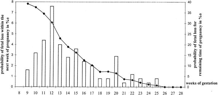
for the finding by Wilcox and coworkers5 of progressively increased loss rates beginning on day 9 after ovulation; no pregnancies occurred beyond day 12. The other explanation could be late implantation of slower-growing genetically abnormal embryos.

Molecular analysis of markers such as endometrial receptors or factors genes one day might prove useful, provided their perturbation is indeed the explanation for LPD. However, no such explanation for LPD has been proved. It is possible that LPD is a secondary effect of abnormal oocytes and nature's method of preventing abnormal concepti from developing into abnormal babies.

TREATMENT.

The efficacy of treatment is unproved, principally because no randomized studies have validated LPD as a genuine entity. Studies by Tho and associates108 and by Daya and Ward109 have been cited as providing some evidence of efficacy, but their experimental designs can be criticized because concurrent control groups were not recruited. Li and coworkers110 identified 21 women with three or more consecutive first-trimester losses. Following their previously published protocol,111 they obtained blood or urine daily for 9 days until the LH surge; 7 days later, timed endometrial biopsy was obtained to detect LPD, diagnosed as more than 2 days behind chronologic dating. Among 25 subsequent pregnancies by the 21, 13 conceived without and 12 conceived with ovarian stimulation (hMG followed by hCG). The two groups were apparently not randomized. Of 13 pregnancies conceiving with ovarian stimulation, 11 continued to at least 24 weeks; of 12 conceiving without ovarian stimulation, only 5 continued to at least 24 weeks. Results were statistically significant by chi-square testing but there was no correction (Yates) for small numbers and no adjustment for potential confounding variables (e.g., maternal age). Meta-analysis by Karamardian and Grimes112 showed no beneficial effect of progesterone treatment. The consensus is that LPD is either an arguable entity or cannot be proved to be treated successfully with progesterone or progestational therapy.

Using the St. Mary's (London) Early Pregnancy Assessment unit cohort, Rai and colleagues113 found that 11% (53/486) of unexplained recurrent aborters had elevated LH levels. However, neither Carp and coworkers114 nor Tulppala and associates115 observed elevated LH levels in women experiencing recurred losses. Bussen and colleagues116 failed to find elevated LH levels among 42 women with recurrent losses, despite such women showing prolactin and androstenedione levels 40% higher than those in 15 other women who showed normal LH and two losses. We conclude that LH elevations by themselves are not significant but could point to other underlying problems.

Thyroid Abnormalities

Decreased conception rates and increased fetal losses are associated with overt hypothyroidism or hyperthyroidism. Subclinical thyroid dysfunction has not generally been considered an explanation for repeated losses.117 However, thyroid perturbation could play a role. Bussen and Steck118 studied 22 women with recurrent abortions in comparison to a like number of nulligravid and multigravid controls. Thyroid antibodies were increased in the former. Stagnaro-Green and associates119 and Glinoer and coworkers120 concluded that antithyroid antibodies and mild thyroid disease were associated with spontaneous abortions, and Singh and colleagues121 opined that thyroid antibodies were a useful marker for clinical losses in the assisted reproductive technology population. Earlier, Pratt and associates122 reported increased antithyroid antibodies in euthyroid women experiencing first-trimester losses, but the same group123 concluded that the ostensible association was secondary to nonspecific organ antibodies.

Rushworth and colleagues124 prospectively studied 870 women in the St. Mary's (London) Early Pregnancy Assessment Unit; all women had at least three spontaneous abortions. Of these 1621, 19% had antibodies against thyroglobulin or thyroid microsomal factors. However, subsequent outcomes did not significantly differ whether women were positive or negative for thyroid antibody.

Overall, asymptomatic thyroid antibodies would not seem to be a major cause of early pregnancy loss.

Diabetes Mellitus

Women with poorly controlled diabetes mellitus clearly show an increased risk for fetal loss. In the cohort best studied to address this question, women whose glycosylated hemoglobin level was greater than four standard deviations above the mean showed higher pregnancy loss rates than either diabetic women showing lower glycosylated hemoglobin levels or euglycemic controls.3 Total pregnancy loss rates were 16.1% (62/386) compared with 16.2% (70/432) in controls. Almost all these losses were early in pregnancy, by 8 weeks. Similar conclusions have been repeatedly reached in retrospective studies.125 Poorly controlled diabetes mellitus should thus be considered one cause for early pregnancy loss. However, subclinical or gestational diabetes is probably not a major etiologic factor because the number of insulin-dependent diabetic women who experience pregnancy loss and have poor control is too small to exert a large attributable effect.

Intrauterine Adhesions (Synechiae)

Intrauterine adhesions could interfere with implantation or with early embryonic development. Most often these adhesions arise after overzealous uterine curettage during the puerperium or intrauterine surgery (e.g., myomectomy). Adhesions are most likely to develop if curettage is performed 3 or 4 weeks postpartum. Women with uterine synechiae usually manifest hypomenorrhea or amenorrhea, but 25% to 30% show repeated abortions. Adhesions surely cause early pregnancy failure in rare individuals, but the overall contribution to pregnancy loss is probably very small.

Incomplete Müllerian Fusion

Müllerian fusion defects are well-accepted causes of second-trimester losses and pregnancy complications. Low birthweight, breech presentation, and uterine bleeding are also commonly accepted correlates. However, the rate of incomplete Müllerian fusion in early (first-trimester) losses is probably overstated. Most studies lack controls126,127,128,129,130,131,132 or uncritically pool early and later pregnancy losses.

One major problem in attributing second-trimester losses to uterine anomalies is that both phenomena occur so frequently that concurrent adverse outcomes could be coincidental. Stampe-Sorenson126 found unsuspected bicornuate uteri in 2 of 167 (1.2%) women undergoing laparoscopic sterilization; another 3.6% had a septate uterus and 15.3%showed fundal anomalies. Simon and associates133 found Müllerian defects in 3.2% (22/679) of fertile women; 20 of the 22 defects were septate. A more unbiased figure was found by Byrne and colleagues,134 who performed ultrasound in women not undergoing imaging for gynecologic reasons. The frequency was estimated to be 0.4%(8/2065).

Some studies claim an increased rate of spontaneous abortions in women with septate uteri132 or T-shaped uteri.129 In septate uteri, an increased risk of pregnancy loss could plausibly reflect implantation occurring on an inhospitable fibrous surface. There is less reason to believe early losses would be increased with bicornuate uteri or T-shaped uteri, but poor vascularization could still play a role. Other studies show no discernible differences among various subtypes.128 Grimbizis and coworkers135 reviewed several series to divide abortion rate by subtype. Overall, the authors concluded that uterine malformations were found in 13% of women with recurrent losses and 5% of fertile women. The highest rates were for untreated septate uterus (44.3%); rates were for bicornuate uterus 36.0%, untreated arcuate uterus 25.7%, didelphys 32.2%, and unicornuate uterus 36.5%.

Losses later in pregnancy can more confidently be attributed to uterine anomalies. Losses occurring in the first trimester after 8 weeks but lacking confirmation of prior fetal viability are statistically more likely to represent missed abortions in which fetal demise actually occurred weeks earlier. Losses occurring before 8 weeks should in general be attributed to other causes, although doubtless some are due to implantation on septi. Quantitative estimates of the role that uterine anomalies play in first-trimester losses cannot be cited with confidence.

Leiomyomas

Leiomyomas occur frequently and produce clinical problems well recognized by gynecologists. Leiomyomas plausibly could also cause early pregnancy loss, but analogous to uterine fusion anomalies, the coexistence of uterine leiomyomas and reproductive losses need not necessarily imply a casual relationship.

The location of leiomyomas is more important than size, submucous leiomyomas being most likely to cause abortion. Plausible mechanisms increasing pregnancy loss rates might include thinning of the endometrium over the surface of a submucous leiomyoma, predisposing to implantation in a poorly decidualized site. Rapid growth of leiomyomas could occur due to the hormonal milieu of pregnancy, compromising blood supply and resulting in necrosis (“red degeneration”) that in turn leads to uterine contractions or infections that eventually lead to fetal expulsion.

Clinically, it is best initially to assume that leiomyomas have no etiologic relationship to pregnancy loss. Surgery for this indication alone should thus be undertaken with reticence.

Incompetent Internal Cervical Os

A functionally intact cervix and lower uterine cavity are obvious prerequisites for a successful intrauterine pregnancy. Characterized by painless dilation and effacement, cervical incompetence usually occurs during the middle of the second trimester or the early part of the third trimester. This condition frequently follows traumatic events such as cervical amputation, cervical laceration, forceful cervical dilatation, or conization. There is little reason to postulate a relationship to first-trimester losses.

Intermittently the question arises as to whether prior induced abortion is associated with subsequent loss. The long-term consensus is that little if any relationship exists;136 however, controversy persists.137,138 Not all studies have taken into account obvious potential confounding variables such as increasing maternal age in subsequent pregnancies. Indeed, an increased loss rate is observed as the number of prior terminations increases,139 but this increase merely parallels that with increasing numbers of spontaneous losses.

Infections

Infections are accepted causes of late fetal losses and logically could be responsible for early fetal losses as well. Among the many microorganisms reported to have been associated with spontaneous abortion are variola, vaccinia, Salmonella typhi, Vibrio fetus, malaria, cytomegalovirus, Brucella, toxoplasmosis, Mycoplasma hominis, Chlamydia trachomatis, and Ureaplasma urealyticum. Transplacental infection doubtless occurs with each of these microorganisms, and sporadic losses could logically be caused by any.

Proof that infections truly cause repetitive losses has been less forthcoming. One line of indirect evidence is that certain organisms (e.g., U. urealyticum and M. hominis) have been isolated in midtrimester placentas and abortuses, but only rarely from induced (control)midtrimester abortions.140 Other evidence consists of studies in which empiric antibiotic therapy ostensibly benefits couples experiencing repeated losses. For example, repetitive aborters treated for 4 weeks with tetracycline before pregnancy showed a subsequent fetal loss in only 10%;141 aborters who chose not to take tetracycline showed a 38% loss rate. However, the two groups (treated and untreated) were not randomized and therefore were not necessarily comparable. Other studies have not found any difference in outcome between women treated or not treated with antibiotics.142

BACTERIAL VAGINOSIS.

Some studies have suggested a relationship between bacterial vaginosis, presumed due to Gardnerella vaginalis, and abortion. There are few data specific to early losses. Most literature in this field has focused on the relationship to pregnancy complications in the second and third trimesters. For example, Hay and associates143 found a 5.5-fold increased risk for losses at 16 to 24 weeks, and McGregor and colleagues144 found an increased risk for losses before 22 weeks. Another study involved Belgian women seen at obstetric registration earlier than 14 weeks.145 At the initial visit, an investigator knowledgeable in bacterial vaginosis performed vaginal fluid microscopy, searching for clue cells and leukocytes; vaginal and cervical cultures were initiated for G. vaginalis, U. urealyticum, and M. hominis, various other bacterial species, Chlamydia trachomatis, and herpes simplex. Of 218 pregnancies, 21 (10%) aborted, and in this group bacterial vaginosis was five times more common (relative risk 5.5, 95%confidence interval 2.9–10); gestational age at pregnancy loss was usually less than 14 weeks (mean 11.3 ± 2.9 weeks). Relative risk for pregnancy loss was also increased for U. urealyticum (1.58) and M. hominis (1.25) but not for herpes, chlamydia, or enteric bacteria. Regression analysis led the authors to conclude that bacterial vaginosis was the likely explanation. It is uncertain whether the above study took into account key confounding variables such as maternal age, prior pregnancy history, or gestational age. The magnitude of the attributable risk found in this study also is at considerable odds with other studies. Findings could be applicable only to this particular sample population, could be explained on the basis of failing to take into account confounding variables, or could be applicable only to pregnancies of later gestational age (late first and early second trimester). Recall that all but 3% of pregnancy losses have occurred by 8 weeks and all but 1% by 16 weeks.

U. urealyticum AND M. hominis.

Of the organisms implicated in repetitive abortion, these seem most plausibly related to repetitive spontaneous abortions because they fulfill several prerequisites: the putative organism can exist in an asymptomatic state, and virulence is not universally severe enough to cause infertility due to fallopian tube occlusion and, hence, preclude the opportunity for pregnancy in which spontaneous abortions might occur. Kundsin and associates146,147 have long contended that Ureaplasma is associated with recurrent abortions. From 46 women with histories of three or more consecutive losses of unknown etiology, Stray-Pedersen and colleagues148 recovered mycoplasma significantly more often among women with repetitive abortions (28%) than among controls (7%). Infected women and their husbands (n = 43) were then treated with doxycycline, with subsequent cultures confirming eradication of mycoplasma. Nineteen of the 43 women became pregnant; of the 19, 3 experienced another spontaneous abortion, whereas 16 had normal full-term infants. Among 18 women with untreated mycoplasma, there were only five full-term pregnancies. A recent study showing a positive correlation between pregnancy loss earlier than 14 weeks and U. urealyticumM. hominis has been discussed above.145

HERPES.

Data are less compelling than for other organisms. Women with a history of herpesvirus have shown a higher spontaneous abortion rate than controls, 34% versus 10.6% in an early study by Nahmias and coworkers.149 Other reports substantiated this potential relationship.150 However, potential confounding variables were not taken into account in the above studies. Donders and colleagues145 found no correlation, despite doing so for several other organisms.

C. trachomatis.

Chlamydial antibodies have been sought in the sera of women who experienced repeated losses, and an association was claimed on the basis of high antibody titers.151,152 Gronroos and associates150 studied a population of women with threatened abortions and concluded that cervical IgA (but not IgG) antibody liters were increased in women who actually aborted. Other data show no relationship.153,154 Rae and colleagues154 found no significant difference between the frequency of IgG anti-chlamydial antibodies in the sera of women with recurrent abortion (n = 106) versus controls (n = 3890; antipodal gravidas), 24.5% versus 20.3%, respectively. Paukku and associates155 reported no differences in the frequencies of C. trachomatis (IgG or IgA antibodies) in 70 Finnish women with histories of spontaneous abortions, compared with 40 parous women and 94 asymptomatic sexually active women. Donders and associates145 also found no relationship in their prospective Belgian study.

In conclusion, most studies indicate no significant role for C. trachomatis. A caveat is the possibility that only certain strains of chlamydia could confer embryotoxicity. The power to detect such an effect would be limited.

TOXOPLASMOSIS.

Toxoplasmosis antibodies have been observed in Mexican and in Egyptian women having repetitive losses.156,157 However, the ubiquitous nature of this organism makes it unclear whether antibody frequencies are higher than in the general Mexican or Egyptian populations in which toxoplasma is endemic. Most recent studies have tended to conclude that toxoplasmosis is not a significant cause of reproductive loss.158

IS INFECTION AN EPIPHENOMENON IN PREGNANCY LOSS?

Do the infectious agents discussed above actually cause fetal losses, or do they merely arise after fetal demise due to other etiologies? To what extent do the two phenomena overlap? Cohort surveillance for infections beginning in early pregnancy can help shed light on the true role of infections in pregnancy loss. To this end, the frequency of infections in pregnant women was prospectively determined using the multicenter United States NICHD Diabetes in Early Pregnancy study alluded to earlier.159 Data were collected prospectively on the frequency of clinical infections in 386 diabetic subjects and 432 control subjects seen frequently (weekly or every other week) during the first trimester. Infection occurred no more often in 112 subjects experiencing pregnancy loss than in 706 having successful pregnancies. This held true both for the 2-week interval in which a given loss was recognized clinically as well as in the prior 2-week interval. Similar findings were observed in both control and diabetic subjects and further held true when data were stratified into ascending genital infection only versus systemic infection only.

CONCLUSION.

Infections surely explain some pregnancy losses, especially later in the first trimester and subsequently throughout pregnancy. However, the attributable risk is probably low for an early loss, even in sporadic cases, and lower still among women experiencing repetitive abortions.

Antiphospholipid and Anticardiolipin Antibodies

An association between second- and third-trimester pregnancy loss and certain autoimmune phenomena is accepted.160,161 Antibodies found in women with pregnancy loss are diverse, encompassing nonspecific antinuclear antibodies (ANA) as well as antibodies against such specific cellular components as phospholipids, histones, and double- or single-stranded DNA. Antiphospholipid antibodies (aPL) in turn represent a broad category that encompasses lupus anticoagulant (LAC) antibodies and anticardiolipin antibodies (aCL). The consensus has long been that midtrimester fetal death rates are increased in women with LAC or aCL, perhaps dramatically so.162 Controversy centers on the role these antibodies play in first-trimester losses.

Descriptive studies initially seemed to show increased aCL levels in women with first-trimester pregnancy losses. However, frequencies of various antiphospholipid antibodies (LAC, aCL, aPL) soon were shown to be similar in women who experienced and who did not experience first-trimester abortions.163,164,165,166 A major pitfall in assessing the role these antibodies play in first-trimester losses is the unavoidable selection bias in ascertaining and studying couples only after they have presented with spontaneous abortions. That antibodies did not arise until after the pregnancy loss cannot readily be excluded. To address this pitfall, the multicenter NICHD collaborative study cohort (Diabetes in Early Pregnancy study) was again used, analyzing sera prospectively obtained from insulin-dependent and nondiabetic women within 21 days of conception. A total of 93 women who later experienced pregnancy loss (48 diabetic, 45 nondiabetic) were matched 2:1 with 190 controls (93 diabetic, 97 nondiabetic) who subsequently had a normal live-born offspring.167 No association was observed between pregnancy loss and the presence of either aPL or aCL. Neither aCL nor aPL would thus seem to contribute greatly, if at all, to first-trimester pregnancy losses in the general population.

The issue is, however, not closed in the opinion of some. It has been stated that results would be different if more specific assays were performed, such as antiphosphatidylethanolamine.168 The marked variation in antiphospholipid antibodies occurring during pregnancy is another confusing factor.169 Could antibodies connote significance only in selected clinical subsets, such as women attempting to achieve pregnancy by in vitro fertilization? This is plausible, given that some reports show an increased prevalence of aCL170 in women requiring in vitro fertilization. The possibility of an effect restricted to the group experiencing only many repetitive abortions cannot be excluded. Power calculations are not adequate to exclude categorically an effect operative only in the 1% to 3% of women with three or more repetitive losses, or especially in the few having five or more. However, one third of women in the cohort study by Simpson171 experienced at least one prior loss, and in that group the frequency of aCL and aPL was still no greater than among women without prior losses.172

It has also been claimed that even if aCL and aPL are not present, antibodies to β2 glycoprotein (aβ2 GP-1) might still be increased and relevant to repetitive aborters. However, Balasch and coworkers173 found no such increase. Only 1 of 100 women having repetitive abortions and not having LAC or aCL showed a β2 GP-1. Any possible association between a β2 GP-1 and spontaneous abortions is also unlikely to be independent of the association with aPL and aCL. No relationship is likely to exist between a β2 GP-1 and implantation failure in the in vitro population, despite earlier claims by Stern and associates.84

In conclusion, a relationship between first-trimester loss and aPL and aCL seems unlikely to be generally applicable. Treatment regimens should thus be embarked on with reticence and initiated only with relatively nontoxic agents (e.g., aspirin and heparin, but not steroids). Specifically giving aspirin to women having only prior first-trimester abortions shows no improvement in outcome compared with those not given aspirin (68% [251/367]vs. 64% [278/438])113 These results contrast with the beneficial effect observed in women having a previous second-trimester loss (65% vs. 49%). Administering intravenous immunoglobulin is not recommended.174,175

Anti-sperm Antibodies

Anti-sperm antibodies (ASA) are another group of antibodies in which a relationship to fetal loss was once claimed. After vasectomy, approximately 50% of men show ASA. In men, the presence of these antibodies connotes difficulty in impregnating women even after vasectomy is surgically reversed. Women manifesting ASA could have their fertilization adversely affected. Several studies show an increased frequency of ASA among women experiencing repeated abortions.176,177,178,179 Others reached opposite conclusions.180,181 The biologic basis for an association might be cross-reaction with paternally derived whole-body antigens essential for embryonic survival.

Again seeking to obtain prospective data on the relationship between presence of ASA in maternal sera and first-trimester pregnancy losses, first-trimester sera from the NICHD DIEP cohort were studied. Recruited within 21 days of conception, a total of 111 women who experienced pregnancy loss (55 diabetic, 56 nondiabetic) were matched 2:1 with 104 diabetic and 116 nondiabetic women (controls) who subsequently had a normal live-born infant.182 No differences were observed with respect to IgG, IgA, or IgM binding when a positive ASA test was defined as 50% of sperm showing antibody binding. At 20% binding, no association was found to IgG and IgM ASA antibodies, although a significant difference was observed for IgA ASA. This single positive finding probably reflects multiple comparisons.

In conclusion, the presence of ASA contributes little to pregnancy loss in the general population. A role in the selective subset of couples experiencing repeated losses is not excluded.

Hypercoagulopathies

Any relationship between fetal loss and aCL or aPL could reflect placental thrombosis. If so, other maternal hypercoagulable states could be associated with increased fetal losses. Currently observed associations include factor V Leiden (Q1691G→A), prothrombin 20210G→A, and homozygosity for the 677C→T polymorphism in the methylene tetrahydrofolate reductase gene(MTHFR).

Studying sera from women previously experiencing repetitive losses, Rai and associates183 found an association between activated protein C resistance and second-trimester losses. The prevalence of factor V Leiden was 7.1% in women with abortions184 versus 4% to 5% in the general population; no association was observed between factor V Leiden and aPL or aCL. Women with histories of repetitive abortions who had factor V Leiden and then became pregnant had a lower likelihood (4/11 [30%]) of a live birth than women who lacked factor V Leiden (77/177[60%]).185 This group also found thromboelastographic abnormalities to be more common in repetitive aborters than in controls,186 offering a way to identify high-risk women. In this group's latest contribution,187 they showed that acquired activated protein C resistance but not congenital (factor V Leiden) activated protein C resistance was more common in aborters. To elevate acquired activated protein C resistance, they studied 904 women with three or more consecutive losses at less than 12 weeks and 207 women with at least one loss after 12 weeks. Acquired activated protein C resistance Leiden mutation was more common in both groups (8.8% each, 87% late) than in 150 controls (3.3%). By contrast, the rate of factor V Leiden (the congenital form of activated protein C resistance) was similar among the three groups (3.3% of 1808 women with recurrent early loss, 3.9% of 414 women with late loss, and 4.0% of 300 controls). In Brazilian women, Souza and coworkers188 found factor V Leiden in 4 of 56 (7.1%) aborters versus 6 of 384 (1.6%)controls; factor II G20210A (prothrombin) was found in 2 of the 56 (3.6%) aborters versus 4 of 384 (1%) controls.

Not all authors accept a relationship. Dizon-Townsend and associates,189 Preston and colleagues,190 Pauer and coworkers,191 and Balasch and associates192 failed to find a relationship between early losses and the presence of factor V Leiden. Deficiencies of antithrombin, protein C, or protein S were found by Kutteh and colleagues193 to be no more frequent in 50 women with three or more losses than in 50 controls. Coumans and associates194 assessed 52 women with two or more losses before 12 weeks for markers and found no relationship to any of these same hemostatic markers. An increased frequency of hyperhomocystinemia was observed (6 of 35 patients tested [17.1%])compared with controls (4.5%). Such a relationship was also shown by Grandone and colleagues195 and Ridker and associates.196

In conclusion, hypercoagulable states, especially those confirmed by factor V Leiden or prothrombin 20120G→A, seem plausible causes of repetitive losses. Additional studies would be helpful.

Anti-fetal Antibodies, Embryotoxic Antibodies, and Aberrant Th1 Cytokine Production

An otherwise normal mother may produce antibodies against her fetus on the basis of genetic dissimilarities. Obstetricians are familiar with late pregnancy loss caused by Rhesus-negative women having anti-D antibodies. More relevant for early pregnancy loss is isoimmunization due to anti-P antibodies, a phenomenon that adheres to the same principles as Rh(D)isoimmunization. Most individuals are genotype Pp or PP, but homozygosity for p exists (pp). If a woman of genotype pp has a Pp or PP husband, her offspring may or must be Pp. If the mother develops anti-P antibodies, pp fetuses will be rejected (aborted) early in gestation.

Hill and colleagues197 proposed that aberrant cytokines can cause repetitive abortions in women acting through T-helper cell perturbations. The rationale centers on the belief that T-helper 1 (Th1) cytokines are deleterious, whereas Th2 cytokines are not. The former include tumor necrosis factor (TNF), interleukin (IL) -2, and interferon (IFN)-gamma; the latter include IL-4, IL-5, IL-6, and IL-10, all secreted by activated T cells expressing the CD4 phenotype. Natural killer cells expressing CD56 also produce these salutary cytokines. In women with recurrent loss, immune cell responsiveness is activated to produce increased IFN-gamma and TNF.197 In support of the hypothesis that Th1 cytokines are deleterious, downregulation of Th1 cytokine in rodents improves pregnancy outcome.198,199 Progesterone therapy is sometimes stated to ameliorate against deleterious effects.

The presence of embryotoxic antibodies and aberrant cytokines are reasonable hypotheses, but their existence before human recurrent embryonic loss has not been conclusively established. Prospective population-based studies in women without a prior history of abnormal outcomes are needed.

The contribution of this phenomenon to pregnancy loss in the general population remains uncertain.

Alloimmune Disease (Shared Parental Antigens)

A long-standing biologic puzzle is why the fetus is not rejected by its mother on the grounds of having foreign (paternal) antigens. The maternal immunologic response must be militated against through blocking or suppressive factors unique to pregnancy. Paradoxically, the protective mechanism could involve maternal—paternal differences (i.e., compatibilities).

PARENTAL HLA SHARING.

Parental (and hence maternal—fetal) histoincompatibility has been proposed as salutary (counterintuitively) for pregnancy maintenance. Evidence in support of a beneficial effect for maternal—fetal incompatibility can be cited. Increased placental size exists in mice arising from matings in which paternal and maternal histocompatibility antigens differ, with higher implantation frequencies occurring in histoincompatible (H2) murine zygotes. It follows that human HLA antigens shared between mother and father could lead to maternal—fetal homozygosity for a given allele, potentially exerting a deleterious effect. Whether human HLA sharing per se is a mechanism underlying pregnancy loss in humans is unclear. Initially studies showed greater parental HLA sharing in aborters than in controls.200,201 However, couples sharing HLA-DR antigens may experience no spontaneous abortions despite 10 or more pregnancies.202 Thus, the story was destined to be complex.

Rigorous population-based studies have been conducted by Ober and colleagues,203,204 who studied the relationship of pregnancy losses and parental HLA-β sharing in the Hutterites. The Hutterites are a genetic isolate, inbreeding producing a high rate of homozygosity by descent and, hence, opportunities for manifesting recessive alleles. These studies involved high-resolution HLA typing (alleles at 16 loci) in 31 Hutterite colonies in South Dakota. Pregnancy outcome was followed through a calendar diary and home pregnancy test kits, used if menses had not begun 1 month after prior menstrual bleeding. Data were available on 251 pregnancies in 111 couples.203,205 HLA genotyping was performed on surviving offspring to determine whether losses selectively occurred with a specific genotype. Comparisons involved offspring homozygous for the shared antigen and having inherited the maternal allele, versus offspring heterozygous and having not inherited the maternal allele shared with the spouse. Loss rates were greatest if couples shared alleles at all 16 loci, presumably reflecting inheritance of a common HLA haplotype (odds ratio 4.39). Sharing was greater for HLA-B (odds ratio 2.54) than for either HLA-C (odds ratio 2.20) or complement C4 (odd ratio 2.11); all pairwise comparisons to controls were statistically significant, but only when couples sharing the entire haplotype were excluded. No deficiency of homozygous children was observed. If offspring were heterozygous yet identical to the mother, 13.6% fewer than expected living children were observed (p = 0.095); however, the sample size for this comparison was small.

PARENTAL SHARING AT LOCI OTHER THAN HLA.

Genetic explanations other than HLA sharing per se could explain why only some couples who share HLA antigens show untoward outcomes. Any deleterious effect could reflect maternal—fetal histoincompatibility, but not for HLA. The causative locus could be closely linked, specifically to HLA-B. This hypothesis would be consistent with HLA-G being the only HLA antigen expressed on trophoblasts.

Genes responsible for deleterious effects exerted through shared parental alleles may not even be immunologically mediated. A lethal recessive gene, again perhaps closely linked to HLA, could exist. Murine embryos homozygous for certain alleles at the T/t locus die at early stages of embryogenesis. A T/t-like complex in humans could help explain the rare kindreds in which multiple family members have repeated pregnancy losses. 206 Postulating a mutant gene in heterozygous form in parents implies autosomal recessive inheritance. If only homozygous offspring were lethal, the ratio of abortuses to live-borns should be 1:3 (25%:75%); however, in families in which the mechanism might usually be presumed operative, the clinically observed ratio seems closer to 1:0.

IMMUNOTHERAPY.

If fetal rejection occurs as a result of diminished fetal—maternal immunologic interaction (alloimmune factors), immunotherapy to stimulate beneficial blocking antibodies generated at the few potentially differing loci is a reasonable hypothesis. The rationale was originally based on observations that blood transfusions before kidney transplantation decreased the rate of allograft rejection.207 Women lacking blocking antibodies but sharing HLA antigens with their spouse were immunized with paternal leukocytes, third-party leukocytes, or trophoblast membranes. The first prospective randomized trial yielded impressive results,208 but later studies were universally less so. A multicenter U.S. effort pooling the results of immunotherapy by injection of paternal leukocytes showed only an 11% increased pregnancy rate in the immunized group.209 Meta-analysis by Fraser and colleagues210 found an odds ratio of only 1.3 in favor of a beneficial effect.

The definitive study was reported in 1999 by Ober.204 This NICHD collaborative study involved six U.S. and Canadian centers, identifying women with three or more spontaneous abortions of unknown cause. Subjects were aged 40 years or younger, had no anti-HLA antibodies, and had no evidence for known or suspected causes of spontaneous abortion (parental chromosomal translocations, LPD, uterine anomalies, aCL, LAC). Women randomized into one arm (n = 91) underwent immunization with paternal mononuclear cells; women in the other arm were given saline (controls)(n = 92). Pregnancy beyond 28 weeks occurred in 46% (31/68) in the immunized group versus 65% (41/63) in the nonimmunized group (p = 0.26). Of course, these findings were the opposite of what would be expected if immunotherapy had been salutary. Adjustments for potential confounding variables (e.g., maternal age, prior pregnancy) failed to alter the conclusions. Significantly, the success rate was nearly identical in immunized women who developed (31%) and failed to develop (30%) HLA antibodies. The sole puzzle was the relatively higher frequency of chromosomally abnormal abortuses or fetuses in the immunized group. This was explained by the authors on the basis of losses in their treatment group occurring later in pregnancy, a time when recovering products of conception for cytogenetic analysis was easier.

CONCLUSION.

Parental HLA sharing leading to fetal rejection remains an attractive hypothesis, with HLA-B the locus showing the strongest association. However, the role of HLA sharing in pregnancy losses in the general population is uncertain and probably low. The phenomenon could play a role in a subset of repetitive aborters, but nonetheless immunotherapy cannot be recommended.

Drugs, Chemicals, and Noxious Agents

Many exogenous agents have been implicated in fetal losses, but relatively few have been accepted scientifically. The difficulty is that pregnant women are frequently exposed to relatively low doses of ubiquitous toxic agents.

Outcomes are usually assessed through case-control studies conducted after prior exposure to exogenous agents. case-control studies have considerable power to detect any associations present but suffer the inherent bias of control women having less incentive to recall antecedent events than women experiencing an abnormal outcome (recall bias). Another experimental pitfall is that exposure to potentially dangerous chemicals is usually unwitting and, hence, poorly documented. Even when exposure unequivocally occurs(e.g., industrial accidents or residence near toxic waste sites), quantifying the exposure and enumerating the multiple toxic agents present may be difficult if not impossible. Exposure to many agents concurrently makes it difficult to attribute adverse effects to a single agent.

Given these caveats, one must be cautious about attributing pregnancy loss to exogenous agents. On the other hand, common sense dictates that exposure to potentially noxious agents be minimized.

ENVIRONMENTAL CHEMICALS.

Many chemical agents have been claimed to be associated with fetal losses,211,212 but consensus now seems to be settling around only a few.213 These include anesthetic gases, arsenic, aniline dyes, benzene, solvents, ethylene oxide, formaldehyde, pesticides, and certain divalent cations (lead, mercury, and cadmium). Workers at greatest risk are those in the rubber industries, battery factories, and chemical production plants. Many reports continue to be generated concerning specific agents, a recent example being selenium, an element considered relevant to fertilization.214

Scialli and coworkers215 and Shepherd216 have cataloged relevant animal and human references, and various on-line listings are available. However, the real difficulty lies in defining the effects of lower-level exposures and in quantifying a risk that can be communicated to a given couple. Fortunately, most patient queries can be answered with reassurance that the pregnancy is not at increased risk.

IRRADIATION.

External irradiation and internal radionuclides in high does are proved abortifacients. Of course, therapeutic x-rays or chemotherapeutic drugs are used during pregnancy only in seriously ill woman whose pregnancies often must be terminated for maternal indications. Pelvic x-ray exposure up to perhaps 0.1 Gy (10 rads) places a fetus at little to no increased risk. In fact, most exposures are usually far smaller, at 0.01 to 0.02 Gy (1 to 2 rads). The contribution of X-irradiation to clinically recognized losses is thus small.

CHEMOTHERAPEUTIC AGENTS.

Similar to x-irradiation, chemotherapeutic agents in high doses are proved abortifacients; however, high doses are given only in dire circumstances. Low doses are sometimes medically important for nonneoplastic conditions, and ambient exposures can occur. A potential for deleterious effects on hospital personnel handling chemotherapeutic agents exists; thus, pregnant hospital workers must minimize exposure.

Caffeine

Consensus has long existed that no deleterious effects exist with caffeine. Most studies investigating pregnancy losses have been retrospective, and cohort data showed the odds ratio for an association between abortion and caffeine (coffee and other dietary forms) to be only 1.15 (95% confidence interval 0.9 to 1.45).217 Additional data on women exposed to higher levels (more than 300 mg) of daily caffeine would be useful, but in general reassurance can be given concerning moderate caffeine exposure and pregnancy loss.

Cigarette Smoking

An association between smoking and spontaneous abortion is accepted, but the effect is probably very modest. It could be explained entirely on the basis of confounding variables. Kline and associates218 reported increased abortion rates in smokers, independent of maternal age and independent of alcohol consumption. A modest dose-response curve was found by Alberman and colleagues.219 Ness and associates220 compared tobacco use as assessed by urinary cotinine levels, comparing 400 women with spontaneous abortions to 570 having ongoing pregnancies. Women with urinary cotinine had an increased risk of abortion, but the odds ratio reached only 1.8 (95% confidence interval 1.3 to 2.6).

Alcohol

An association between alcohol consumption and fetal loss was once well accepted, but this claim now seems less certain. In 1980 Kline and coworkers218 compared 616 women experiencing spontaneous abortions with 632 women delivering at less than 28 weeks' gestation. Among women whose pregnancies ended in spontaneous abortion, 17% drank alcohol at least twice per week; 8.1% of controls drank similar quantities. Harlap and Shiono221 also found an increased risk for abortion in women who drank in the first trimester. However, Halmesmärki and associates222 later found that alcohol consumption was nearly identical in women who did and did not experience an abortion. In that study, 13% of aborters and 11% of control women drank on average three to four drinks per week. Parazzini and coworkers223 reached similar conclusions, as did Ness and colleagues.220 Alcohol consumption should be avoided or minimized during pregnancy for many reasons, but alcohol probably contributes little to the rate of pregnancy loss. Given the high frequency of alcohol ingestion in the general population, however, even a small effect could have epidemiologic significance. Clinically, women more often need to be reassured, especially after inadvertent drinking before realizing they are pregnant.

Contraceptive Agents

Contraception with an intrauterine device in place clearly increases the risk of fetal loss; the second trimester is especially hazardous. If the device is removed before pregnancy, there is no increased risk of spontaneous abortion. Using oral contraceptives before or even during pregnancy is not associated with fetal loss, nor is spermicide exposure before or after conception.224,225

Trauma

Women commonly attribute pregnancy losses to trauma such as a fall or a blow to the abdomen. However, fetuses are actually well protected from external trauma by intervening maternal structures and amniotic fluid. Any contribution of trauma to early pregnancy loss is quite small.

Psychological Factors

Impaired psychological well-being has been claimed to predispose to early fetal losses. The first investigations showing a benefit to psychological well-being were those of Stray-Pedersen and Stray-Pedersen.226 One group consisted of 16 pregnant women previously experiencing repetitive abortions who received increased attention but no specific medical therapy. They proved more likely (85%) to complete their pregnancies than 42 women not given such close attention(36% successful outcome). One pitfall was that only women living “close” to the university were eligible to be placed in the increased-attention group. Women living further away served as “controls” by default; their differing from the experimental group in ways other than geographic proximity was not excluded. In 811 subsequent couples, the same high success rate (86%) was observed in women given “tender loving care.”227 Again, the expected background in this series is uncertain, making it difficult to assess significance. Other studies have also reported a beneficial effect of psychological well-being.26,27,228,229

The biologic explanation for any salutary effect remains obscure. Unanswered is whether the ostensible positive effect of psychological well-being is real or secondary to other factors. Confounding factors were not taken into account, making it difficult to determine whether the outcome was truly better than the expected background rate of 70%. Given no ostensible harm, psychological support can be recommended, but not at the expense of eschewing other potential causes.

Severe Maternal Illness

Many symptomatic maternal diseases show an increased frequency of spontaneous abortion. In few disorders other than diabetes mellitus have potential confounding variables been assessed. Nonetheless, Wilson disease, phenylketonuria, cyanotic heart disease, hemoglobinopathies, and inflammatory bowel disease seem implicated in early abortion. Not every study claims that a maternal disease is associated with increased fetal loss rates; celiac disease is one example lacking a proven effect.230

The pathogenesis of losses in these disorders presumably involves one or more of the mechanisms discussed previously, more often endocrinologic or immunologic. Overall, relatively few fetal losses in the general population will result from severe maternal disease.

Back to Top
ROLE OF (UNRECOGNIZED) MENDELIAN AND POLYGENIC FACTORS

Recurrent Losses Due to Mendelian Factors

Consecutive abortuses in a given family usually show nonrandom distribution with respect to chromosomal complements. If the complement of the first abortus is abnormal, the likelihood is approximately 80% that the complement of the second abortus also will be abnormal.83 Previously we concluded that these data suggest certain couples are predisposed toward chromosomally abnormal conceptions, most of which result in a spontaneous abortion. Genes exerting this effect could act by abnormalities involving faulty spindle formation, centromere stability, recombination, or other mechanism needed for disjunction of homologous chromosomes.

The converse of the above is that other couples show repetitive losses of chromosomally normal abortuses. Here, Mendelian mutations are presumably causing recurrent euploid losses. These mutant Mendelian genes could mediate pregnancy loss through one processes alluded to above that is traditionally called “nongenetic.” The most obvious example involves alloimmune conditions.

Mutant genes as a cause of pregnancy should be common on statistical grounds alone. Recall that 1% of live-borns are abnormal due to a single-gene(Mendelian) mutation; another 1% have a polygenic/multifactorial condition. In contrast, only 0.6% of live-borns have a chromosomal abnormality. Thus, the 30% to 50% of abortuses with normal chromosomes need not necessarily require explanation by such nongenetic causes as infections, luteal phase insufficiency, or uterine anomalies. Nonchromosomal pregnancy losses might more likely be due to a mutant gene. The scientific task will be to enumerate the many genes whose perturbations result in pregnancy loss. A host of attractive candidate genes can be envisioned—for example, in developmental gene families (PAX, HOX, Oct).

Skewed X-Inactivation

Highly skewed X-inactivation is associated with recurrent abortions.231,232,233 Lanasa and colleagues231 found that of 48 women having two prior unexplained losses, 7 (14.6%) had highly skewed X-inactivation, defined as 90% of X chromosomes originating from one specific parent; only 1 of 67 controls (1.5%) showed skewed X-inactivation. Kristiansen and associates234 found less skewed X-inactivation in both aborters and controls and no significant differences. One explanation is that all male offspring of a woman with skewed X-inactivation would be aborted (male lethals). A pedigree consistent with this idea has been reported,235 but other expectations inherent with this explanation are not met. Robinson and colleagues236 considered other mechanisms. These include reduction in precursor nuclei at the time of X-inactivation, which could result from trisomy mosaicism followed by selection favoring diploid cells. Poor early growth in general for any reason could have the same effect. In other words, skewed X-inactivation per se may not cause abortions (Table 5).

Table 5. Skewed X-Inactivation in Repetitive Aborters


Study

Aborters

Controls

p Value

Robinson et al, 2001 236

140 (16%)

111 (5%)

<0.01, chi-square

Lanasa et al, 1999 231

 48 (15%)

67 (1%)

<0.01, Fisher's test

Kristiansen et al, 1999234

 87 (2%)

148 (0.7%)

  NS

Nelson et al, 2000233

 57 (30%)

29 (3%)

<0.005, Fisher's test


Definition of skewed X-inactivation was 90% in all studies except Nelson et al, where a cutoff of 85% was used.

Relative Proportion of Genetic and Nongenetic Causes of Spontaneous Abortions

Some authors are fond of offering charts or tables enumerating the proportion of losses due to various postulated causes. In our opinion this is naïve: rarely can data take into account potential confounding variables, without which conclusions can be misleading. In reality, cytogenetics probably explains the overwhelming majority of sporadic losses and at least half of recurrent losses, and mutant genes probably explain most of the remainder. There is probably little need to invoke true nongenetic causes with great frequency. However, anecdotal examples exist. An example was success after embryo transfer to a surrogate uterus in a woman having 24 prior abortions.237 Nonetheless, the contribution of genuinely “nongenetic” causes is low.

Back to Top
CLINICAL EVALUATION

Couples experiencing only one first-trimester abortion should receive pertinent information but should not necessarily be evaluated formally. One might mention the relatively high (10% to 15%) pregnancy loss rate in the general population and the beneficial effects of abortion in eliminating abnormal conceptuses. Provide the relevant recurrence risk figure (see Table 1): there is usually a 20% to 25% rate of subsequent loss in the presence of a prior live-born, but this is somewhat higher in older women having no prior live-born. Discuss the higher risks that exist for older women. If a specific medical illness exists, treatment is obviously necessary. Intrauterine adhesions should be lysed. Otherwise, no further evaluation need be undertaken, even if uterine anomalies or leiomyomas are detected. On the other hand, occurrence of an anomalous stillborn or live-born warrants genetic evaluation irrespective of the number of pregnancy losses.

Investigation may or may not be necessary after two spontaneous abortions, depending on the patient's age and desires. Couples nearing the end of their reproductive capacity (e.g., maternal age 35 to 40 years) may prefer expeditious evaluation. Infertile couples may also wish to be evaluated at a relatively younger age.

After three spontaneous abortions, evaluation is usually indicated. One should then obtain a detailed family history, discuss recurrence risks, perform a complete physical examination, and order selected tests (discussed below). Once a couple enters evaluation, they should undergo all tests standard for a given practitioner. There is no scientific rationale for performing some studies after two or three losses but deferring others until after additional losses.

Parental chromosomal studies should be performed on all couples having repetitive losses. Antenatal chromosomal studies should be offered if a balanced chromosomal rearrangement is detected in either parent or if autosomal trisomy occurred in any previous abortus.

Although it is impractical to karyotype all abortuses, cytogenetic information on abortuses would be highly desirable. Detection of a trisomic abortus suggests the phenomenon of recurrent aneuploidy, justifying prenatal cytogenetic studies in future pregnancies. Performing prenatal cytogenetic studies solely on the basis of repeated losses is more arguable but not unreasonable among women aged 30 to 34 years. Archival tissue of prior products of conception can now be studied using FISH with chromosome-specific probes.

The validity of LPD as a discrete entity increasingly seems arguable. To detect LPD, timed endometrial biopsies should be performed late in the luteal phase in two or more cycles. Results should be correlated with the date of menstruation. If histologic dating reveals an endometrium 2 or more days less than expected, the diagnosis is accepted. Progesterone therapy has been proposed, but its efficacy is still unproved.

Repeated fetal losses are associated with poorly controlled diabetes mellitus (hyperglycemia), but not asymptomatic diabetes mellitus. Overt thyroid dysfunction is associated with increased fetal loss, but probably not asymptomatic hypothyroidism.

Infectious agents are more likely to cause sporadic than repetitive losses. To determine the role of infectious agents in repetitive losses, the endometrium may be cultured for U. urealyticum. Alternatively, a couple could be treated empirically with doxycycline before pregnancy. Of other infectious agents, only C. trachomatis seems plausible as a causative agent.

If an abortion occurs after 8 to 10 weeks' gestation, a uterine anomaly or submucous leiomyoma should be considered. The uterine cavity should be explored by hysteroscopy or hysterosalpingography. If a Müllerian fusion defect (septate or bicornuate uterus) is detected in a woman experiencing one or more second-trimester spontaneous abortions, surgical correction may be warranted. A large submucous leiomyoma may also justify myomectomy. However, the same statements do not necessarily apply after early first-trimester losses. Cervical incompetence should be managed by surgical cerclage during the next pregnancy.

To exclude autoimmune disease, aPL and aCL should be assessed in women who experience midtrimester losses. These women may benefit from treatment with heparin and aspirin, but the same does not necessarily hold when aPL or aCL is detected in asymptomatic women having first-trimester pregnancy losses. Testing for other autoantibodies (e.g., DNA) is not indicated.

Biologically there is evidence for a deleterious effect of HLA sharing(HLA-β), but the effect is not sufficient to warrant clinical evaluation. Determining parental HLA types in the absence of other immunologic testing is not recommended. Immunotherapy (inoculation of the mother with her husband's leukocytes) has been unequivocally shown to be ineffective.

One should discourage exposure to cigarettes and alcohol while remaining cautious about ascribing cause and effect in individual cases. Similar counsel should apply for exposures to other potential toxins.

Back to Top
REFERENCES

1. U.S. Department of Health and Human Services: Reproductive impairments among married couples. In U.S. Vital and Health Statistics, Series 23, No.11, p 5. National Center for Health Statistics. Hyattsville, MD, 1982

2. Wilcox AJ, Weinberg CR, O'Connor FJ et al: Incidence of early pregnancy loss. N Engl J Med 319: 189, 1988

3. Mills JS, Simpson JL, Driscoll SG et al: NICHD-DIEP Study: Incidence of spontaneous abortion among normal women with insulin-dependent diabetic women whose pregnancies were identified within 21 days of conception. N Engl J Med 319: 1617, 1988

4. Gray RH, Simpson JL, Kambic RT et al: Timing of conception and the risk of spontaneous abortion among pregnancies occurring during the use of natural family planning. Am J Obstet Gynecol 172: 1567, 1995

5. Wilcox AJ, Baird DD, Weinberg CR: Time of implantation of the conceptus and loss of pregnancy. N Engl J Med 340: 1796, 1999

6. Liu HC, Rosenwaks Z: Early pregnancy wastage in IVF (in vitro fertilization) patients. J In Vitro Fert Embryo Transf 8: 65, 1991

7. Simpson JL, Carson SA: Causes of fetal loss. In Gray R, Leridon L, Spira F (eds): Symposium on Biological and Demographic Determinants of Human Reproduction, p 287. Oxford Press, 1993

8. Simpson JL, Mills JL, Holmes LB et al: Low fetal loss rate after ultrasound-proven viability in early pregnancy. JAMA 258: 2555, 1987

9. Christiaens GC, Stoutenbeek P: Spontaneous abortion in proven intact pregnancies. Lancet 2: 571, 1984

10. Wilson RD, Kendrick V, Wittman BK et al: Risk of spontaneous abortion in ultrasonographically normal pregnancies. Lancet 2: 920, 1984

11. Gilmore DH, McNay MB: Spontaneous fetal loss rate in early pregnancy. Lancet 1: 107, 1985

12. Cashner KA, Christopher CR, Dysert GA: Spontaneous fetal loss after demonstration of a live fetus in the first trimester. Obstet Gynecol 70: 827, 1987

13. Stein Z, Kline J, Susser E et al: Maternal age and spontaneous abortion. In Porter IH, Hook EB (eds): Human Embryonic and Fetal Death, p 107. London, Academic Press, 1980

14. Regan L, Braude PR, Trembath PL: Influence of postreproductive performance on risk of spontaneous abortion. Br Med J 299: 551, 1989

15. Warburton D: Outcome of cases of de novo structural rearrangements diagnosed at amniocentesis. Prenat Diagn 4: 69, 1984

16. Whitley E, Doyle P, Roman E et al: The effect of reproductive history on future pregnancy outcomes. Hum Reprod 14: 2863, 1999

17. Parazzini F, Chatenoud L, Tozzi L et al: Determinants of risk of spontaneous abortions in the first trimester of pregnancy. Epidemiology 8: 681, 1997

18. Simpson JL, Gray RH, Queenan JT et al: Risk of recurrent spontaneous abortion for pregnancies discovered in the fifth week of gestation. Lancet 344: 964, 1994

19. Malpus P: A study of abortion sequences. J Obstet Gynaecol Br Emp 45: 932, 1938

20. Warburton D, Fraser FC: Spontaneous abortion risks in man: Data from reproductive histories collected in a medical genetic unit. Am J Hum Genet 16: 1, 1964

21. Poland BJ, Miller JP, Jones DC et al: Reproductive counseling in patients who had a spontaneous abortion. Am J Obstet Gynecol 127: 685, 1977

22. Regan L: A prospective study on spontaneous abortion. In Beard RW, Sharp F (eds): Early Pregnancy Loss: Mechanisms and Treatment, p 22. London, The Royal College of Obstetricians and Gynaecologists, 1988

23. Kline J, Stein ZA, Susser M et al: Smoking: a risk factor for spontaneous abortion. N Engl J Med 297: 793, 1977

24. Boue JG, Boue A: Increased frequency of chromosomal anomalies in abortions after induced ovulation. Lancet 1: 679, 1973

25. Vlaanderen W, Treffers PE: Prognosis of subsequent pregnancies after recurrent spontaneous abortion in first trimester. Br Med J 295: 92, 1987

26. Liddell HS, Pattison NS, Zanderigo A: Recurrent miscarriage: outcome after supportive care in early pregnancy. Aust NZ J Obstet Gynaecol 31: 320, 1991

27. Houwert-de Jong MH, Termijtelen A et al: The natural course of habitual abortion. Eur J Obstet Gynecol Reprod Biol 33: 221, 1989

28. Brigham SA, Conlon C, Farquharson RG: A longitudinal study of pregnancy outcome following idiopathic recurrent miscarriage. Hum Reprod 14: 2868, 1999

29. Ober C, Karrison T, Odem RR et al: Mononuclear cell immunisation in prevention of recurrent miscarriages: A randomised trial. Lancet 354: 365, 1999

30. Hertig AT, Rock J, Adams EC: Description of human ova within the first 17 days of development. Am J Anat 98: 435, 1956

31. Hertig AT, Rock J, Adams EC et al: Thirty-four fertilized human ova, good, bad and indifferent, recovered from 210 women of known fertility. A study of biologic wastage in early human pregnancy. Pediatrics 25: 202, 1959

32. Hertig AT, Rock J: Searching for early human ova. Gynecol Invest 4: 121, 1973

33. Gropp A: Chromosomal animal model of human disease. Fetal trisomy and development failure. In Berry L, Poswillo DE (eds): Teratology, p 17. Berlin, Springer-Verlag, 1975

34. Plachot M, Junca AM, Mandelbaum J et al: Chromosome investigations in early life. Human preimplantation embryos. Hum Reprod 2: 29, 1987

35. Papadopoulos G, Templeton AA, Fisk N et al: The frequency of chromosome anomalies in human preimplantation embryos after in vitro fertilization. Hum Reprod 4: 91, 1989

36. Egozcue J, Blanco J, Vidal F: Chromosome studies in human sperm nuclei using fluorescence in-situ hybridization (FISH). Hum Reprod Update 3: 441, 1997

37. Plachot M: Genetic of human oocytes. In Boutaleb Y, Gzouli A (eds): New Concepts in Reproduction. Lanes, England, Parthenon, 1992

38. Martin R: Chromosomal analysis of human spermatozoa. In Verlinsky Y, Kuliev A (eds): Preimplantation Genetics, p 91. New York, Plenum Press, 1991

39. Pellestor F, Dufour MC, Arnal F et al: Direct assessment of the rate of chromosomal abnormalities in grade IV human embryos produced by in-vitro fertilization procedure. Hum Reprod 9: 293, 1994

40. Munná S, Alikani M, Tomkin G et al: Embryo morphology, developmental rates, and maternal age are correlated with chromosome abnormalities. Fertil Steril 64: 382, 1995

41. Almeida PA, Bolton VN: The relationship between chromosomal abnormality in the human preimplantation embryo and development in vitro. Reprod Fertil Dev 8: 235, 1996

42. Delhanty JD, Harper JC, Ao A et al: Multicolour FISH detects frequent chromosomal mosaicism and chaotic division in normal preimplantation embryos from fertile patients. Hum Genet 99: 755, 1997

43. Munná S, Magli C, Cohen J et al: Positive outcome after preimplantation diagnosis of aneuploidy in human embryos. Hum Reprod 14: 2191, 1999

44. Grossman M, Gimánez C, Calafell JM et al: Mosaicism evaluation by FISH in normally fertilized and normally developing human embryos. Hum Reprod 14:O-O77, 1999

45. Sandalinas M, Sadowy S, Calderon G et al: Survival of chromosomal abnormalities to blastocyst stage. Hum Reprod 15:O-027, 2000

46. Coonen E, Dumoulin JCM, Derhaag JG et al: Genetic make-up of human embryos grown in vitro until the blastocyst state. Hum Reprod 15:P-212, 2000

47. Ledbetter DH, Zachary JM, Simpson JL et al: Cytogenetic results from the U.S. Collaborative Study on CVS: high diagnostic accuracy in over 11,000 cases. Prenat Diagn 12: 317, 1992

48. Bouá J, Boue A, Lazar P: Retrospective and prospective epidemiological studies of 1500 karyotyped spontaneous human abortions. Teratol 12: 11, 1975

49. Hassold T: A cytogenetic study of repeated spontaneous abortions. Am J Hum Genet 32: 723, 1980

50. Simpson JL, Bombard AT: Chromosomal abnormalities in spontaneous abortion: Frequency, pathology and genetic counseling. In Edmonds K, Bennett MJ (eds): Spontaneous Abortion, p 51. London, Blackwell, 1987

51. Guerrieri S, Bettio D, Simoni G et al: Prevalence and distribution of chromosome abnormalities in a sample of first trimester internal abortions. Hum Reprod 8: 735, 1987

52. Johnson PM, Drugan A, Koppitch F et al: Postmortem chorionic villi sampling is a better method of cytogenetic evaluation of early fetal loss than culture of abortous material. Am J Obstet Gynecol 163: 1505, 1990

53. Sorokin Y, Johnson MP, Uhlman WR et al: Postmortem chorionic villus sampling: Correlation of cytogenetic and ultrasound findings. Am J Med Genet 39: 314, 1991

54. Strom C, Ginsberg N, Applebaum M et al: Analyses of 95 first trimester spontaneous abortions by chorionic villus sampling and karyotype. J Asst Reprod Genet 9: 458, 1992

55. Sánchez JM, Franzi L, Collia F et al: Cytogenetic study of spontaneous abortions by transabdominal villus sampling and direct analysis of villi. Prenat Diagn 19: 601, 1999

56. Watt JL, Templeton AA, Messinis I et al: Trisomy I in an eight-cell human pre-embryo. J Med Genet 24: 60, 1987

57. Warburton D, Byrne J, Canik N: Chromosome Anomalies and Prenatal Development: An Atlas. New York, Oxford University Press, 1991

58. Kalousek DK: Pathology of abortion: Chromosome anomalies and genetic correlations. In Kraus FT, Damjanov I, Kaufman N (eds): Pathology and Reproductive Failure, p 228. Baltimore, Williams & Wilkins, 1991

59. Schimdt-Saroli C, Schwartz LB, Lublin J et al: Chromosomal analysis of early fetal losses in relation to transvaginal ultrasonographic detection of fetal heart motion after infertility. Fertil Steril 69: 274, 1998

60. Reddy KS: Double trisomy in spontaneous abortions. Hum Genet 101: 339, 1997

61. Robinson WP, Kuchinka BD, Bernasconi F et al: Maternal meiosis I non-disjunction of chromosome 15: dependence of the maternal age effect on level of recombination. Hum Mol Genet 7: 1011, 1998

62. Hassold T, Merrill M, Adkins K et al: Recombination and maternal age-dependent nondisjunction: molecular studies of trisomy 16. Am J Hum Genet 57: 867, 1995

63. Fisher JM, Harvey JF, Morton NE et al: Trisomy 18: studies of the parent and cell division of origin and the effect of aberrant recombination on nondisjunction. Am J Hum Genet 56: 669, 1995

64. Bugge M, Collins A, Petersen MB et al: Non-disjunction of chromosome 18. Hum Mol Genet 7: 661, 1998

65. Hassold TJ: Nondisjunction in the human male. Curr Top Dev Biol 37: 383, 1998

66. Henderson SA, Edwards RG: Chiasma frequency and maternal age in mammals. Nature 217: 22, 1968

67. Lamb NE, Freeman SB, Savage-Austin A et al: Susceptible chiasmate configurations of chromosome 21 predispose to non-disjunction in both maternal meiosis I and meiosis II. Nat Genet 14: 400, 1996

68. Castilla EE, Simpson JL, Queenan JT: Down syndrome is not increased in offspring of natural family planning users (case control analysis). Am J Med Genet 59: 525, 1995

69. Simpson JL, Gray RH, Queenan JT et al: Pregnancy outcome associated with natural family planning (NFP): Scientific basis and experimental design for an international cohort study. Adv Contracept 4: 247, 1988

70. Simpson JL, Gray R, Perez A et al: Fertilization involving aging gametes is not associated with major birth defects and Down syndrome. Lancet (in press)

71. Zaragoza MV, Jacobs PA, James RS: Nondisjunction of human acrocentric chromosomes: Studies of 432 trisomic fetuses and liveborns. Hum Genet 94: 411, 1994

72. Koehler KE, Boulton CL, Collins HE et al: Spontaneous X chromosome MI and MII nondisjunction events in Drosophila melanogaster oocytes have different recombinational histories. Nat Genet 14: 406, 1996

73. Hassold T, Abruzzo M, Adkins K et al: Human aneuploidy: incidence, origin, and etiology. Environ Mol Mutagen 28: 167, 1996

74. Savage AR, Petersen MB, Pettay D et al: Elucidating the mechanisms of paternal non-disjunction of chromosome 21 in humans. Hum Molec Genet 7: 1221, 1998

75. Chandley ACL: The origin of chromosome aberrations in man and their potential for survival and reproduction in the adult human populations. Ann Genet 24: 5, 1981

76. Hassold T, Benham F, Leppert M: Cytogenetic and molecular analysis of sex chromosome monosomy. Am J Hum Genet 42: 534, 1985

77. Singh RJ, Carr DH: The anatomy and histology of XO embryos and fetuses. Anat Rec 155: 369, 1966

78. Jirásek JE: Principle of reproductive embryology. In Simpson JL(ed): Disorders of Sex Differentiation: Etiology and Clinical Delineation, p 51. New York, Academic Press, 1976

79. Simpson JL: Pregnancies in women with chromosomal abnormalities. In Shulman JD, Simpson JL (eds): Genetic Diseases in Pregnancy, p 439. New York, Academic Press, 1981

80. MacDonald M, Hassold T, Harvey J et al: The origin of 47,XXX aneuploidy;heterogeneous mechanisms and role of aberrant recombination. Hum Molec Genet 3: 1365, 1994

81. Hassold TJ, Sherman SL, Pettay D et al: XY chromosome nondisjunction in man is associated with diminished recombination in the pseudoautosomal region. Am J Hum Genet 49: 253, 1991

82. Hassold T, Benham F, Leppert M: Cytogenetic and molecular analysis of sex-chromosome monosomy. Am J Hum Genet 42: 534, 1988

83. Warburton D, Kline J, Stein Z et al: Does the karyotype of a spontaneous abortion predict the karyotype of a subsequent abortion? Evidence from 273 women with two karyotyped spontaneous abortions. Am J Hum Genet 41: 465, 1987

84. Stern C, Chamley L, Hale L et al: Antibodies to beta2 glycoprotein I are associated with in vitro fertilization implantation failure as well as recurrent miscarriage: results of a prevalence study. Fertil Steril 70: 938, 1998

85. Ogasawara M, Kajiura S, Katano K et al: Are serum progesterone levels predictive of recurrent miscarriage in future pregnancies? Fertil Steril 68: 806, 1997

86. Carp HJA, Toder V, Orgad S et al: Karyotype of the abortus in recurrent miscarriage. XVI World Congress of Gynecology and Obstetrics FIGO, International Journal of Obstetrics, Supplement 1, Thursday 70: Abstract, 2000, p 44.

87. Alberman ED: The abortus as a predictor of future trisomy 21. In De La Cruz FF, Gerald PS (eds): Trisomy 21 (Down Syndrome), p 69. Baltimore, University Park Press, 1981

88. Rubio C, Vidal F, Mínguez Y et al: High incidence of chromosomal abnormalities in preimplantation embryos from recurrent spontaneous abortion patients. Hum Reprod 15:O-203, 2000

89. Ozcan T, Burki N, Parkash V et al: Cytogenetical diagnosis in paraffin-embedded fetoplacental tissue using comparative genomic hybridization. Prenat Diagn 20: 41, 2000

90. Henderson DJ, Sherman LS, Loughna SC et al: Early embryonic failure associated with uniparental disomy for human chromosome 21. Hum Molec Genet 3: 1373, 1994

91. Shaffer LG, McCaskill C, Adkins K et al: Systematic search for uniparental disomy in early fetal losses: The results and a review of the literature. Am J Med Genet 79: 366, 1998

92. Simpson JL, Meyers CM, Martin AO et al: Translocations are infrequent among couples having repeated spontaneous abortions but no other abnormal pregnancies. Fertil Steril 51: 811, 1989

93. De Braekeleer M, Dao TN: Cytogenetic studies in couples experiencing repeated pregnancy losses. Hum Reprod 5: 519, 1990

94. Bouá A, Gallano P A: A collaborative study of the segregation of inherited chromosome structural rearrangements in 1356 prenatal diagnoses. Prenat Diagn 4: 45, 1984

95. Daniel A, Hook EB, Wulf G: Risks of unbalanced progeny at amniocentesis to carriers of chromosome rearrangements: Data from United States and Canadian Laboratories. Am J Med Genetic 31: 14, 1989

96. Sutherland GR, Gardiner AJ, Carter RF: Familial pericentric inversion of chromosome 19 inv (19) (p13q13) with a note on genetic counseling of pericentric inversion carriers. Clin Genet 10: 54, 1976

97. Gardner RJM, Sutherland GR: Chromosome Abnormalities and Genetic Counselling. New York, Oxford University Press, 1996

98. Pettanati MJ, Rao PN, Phelan MC et al: Paracentric inversions in humans:A review of 446 paracentric inversions with presentation of 120 new cases. Am J Med Genet 55: 171, 1995

99. Davis OK, Berkley AS, Naus GJ et al: The incidence of luteal phase defect in normal, fertile women, determined by serial endometrial biopsy. Fertil Steril 51: 582, 1989

100. Scott RT, Synder RR, Strickland DM et al: The effect of interobserver variation in dating endometrial history on the diagnosis of luteal phase defects. Fertil Steril 50: 888, 1988

101. Li TC, Dockery P, Rogers AW et al: How precise is histologic dating of endometrium using the standard dating criteria? Fertil Steril 51: 759, 1989

102. Daya S, Ward S, Burrows E: Progesterone profiles in luteal phase defect cycles and outcome of progesterone treatment in patients with recurrent spontaneous abortions. Am J Obstet Gynecol 158: 225, 1988

103. Soules MR, Clifton DK, Cohen NL et al: Luteal phase deficiency:Abnormal gonadotropin and progesterone secretion patterns. J Clin Endocrinol Metab 69: 813, 1989

104. Soules MR, McLachlan RI, Ek M et al: Luteal phase deficiency:characterization of reproductive hormones over the menstrual cycle. J Clin Endocrinol Metal 69: 804, 1989

105. Soules M, Wiebe RH, Alesel S et al: The diagnosis and therapy of luteal phase deficiency. Fertil Steril 28: 1033, 1997

106. Sterzik K, Abt M, Grab D et al: Predicting the histologic dating of an endometrial biopsy specimen with the use of Doppler ultrasonography and hormone measurements in patients undergoing spontaneous ovulatory cycles. Fertil Steril 73: 94, 2000

107. Li TC, Spuijbroek MD, Tuckerman E et al: Endocrinology and endometrial factors in recurrent miscarriage. Br J Obstet Gynecol 107: 1471, 2000

108. Tho PT, Byrd JR, McDonough PC: Etiologies and subsequent reproductive performance of 100 couples with recurrent abortions. Fertil Steril 32: 389, 1979

109. Daya S, Ward S: Diagnostic test properties of serum progesterone in the evaluation of luteal phase defects. Fertil Steril 49: 168, 1988

110. Li Tin-Chiu, Ding S-H, Anstie B et al: Use of human menopausal gonadotropins in the treatment of endometrial defects associated with recurrent miscarriage: preliminary report. Fertil Steril 75: 434, 2001

111. Li TC: Guides for practitioners. Recurrent miscarriage: Principles of management. Hum Reprod 13: 478, 1998

112. Karamardian LM, Grimes DA: Luteal phase deficiency: Effect of treatment on pregnancy rates. Am J Obstet Gynecol 167: 1391, 1992

113. Rai R, Backos M, Rushworth F et al: Polycystic ovaries and recurrent miscarriage: A reappraisal. Hum Reprod 15: 612, 2000

114. Carp HJ, Hass Y, Dolicky M et al: The effect of serum follicular phase luteinizing hormone concentrations in habitual abortion: Correlation with results of paternal leukocyte immunization. Hum Reprod 10: 1702, 1995

115. Tulppala M, Stenman UH, Cacciatore B et al: Polycystic ovaries and levels of gonadotrophins and androgens in recurrent miscarriage: Prospective study in 50 women. Br J Obstet Gynaecol 100: 348, 1993

116. Bussen S, Sutterlin M, Steck T: Endocrine abnormalities during the follicular phase in women with recurrent spontaneous abortion. Hum Reprod 14: 18, 1999

117. Montoro M, Collea JV, Frasiers D et al: Successful outcome of pregnancy in women with hypothyroidism. Ann Intern Med 94: 31, 1981

118. Bussen S, Steck T: Thyroid autoantibodies in euthyroid non-pregnant women with recurrent spontaneous abortions. Hum Reprod 10: 2938, 1995

119. Stagnaro-Green A, Roman SH, Cobin RH et al: Detection of at risk pregnancy by means of highly sensitive assays for thyroid autoantibodies. JAMA 264: 1422, 1990

120. Glinoer D, Soto MF, Bourdoux P et al: Pregnancy in patients with mild thyroid abnormalities: maternal and neonatal repercussions. J Clin Endocrinol Metab 73: 421, 1991

121. Singh A, Dentas ZN, Stone SC et al: Presence of thyroid antibodies in early reproductive failure. Biomedical versus clinical pregnancies. Fertil Steril 62: 277, 1995

122. Pratt DE, Kaberlein G, Dudkiewicz A: The association of antithyroid antibodies in euthyroid nonpregnant women with recurrent first trimester abortions in the next pregnancy. Fertil Steril 60: 1001, 1993

123. Pratt DE, Novotny M, Kaberlein G et al: Antithyroid antibodies and the association with non-organ-specific antibodies in recurrent pregnancy loss. Am J Obstet Gynecol 168: 837, 1993

124. Rushworth FH, Backos M, Rai R et al: Prospective pregnancy outcome in untreated recurrent miscarriers with thyroid autoantibodies. Hum Reprod 14:P-169, 1999

125. Miodovnik M, Mimouni F, Tsang RC et al: Glycemic control and spontaneous abortion in insulin dependent diabetic women. Obstet Gynecol 68: 366, 1986

126. Stampe-Sorensen S: Estimated prevalence of mullerian anomalies. Acta Obstet Gynecol Scand 67: 441, 1988

127. Candiani GB, Fedele L, Parazzini F et al: Reproductive prognosis after abnormal metroplasty in bicornuate or septate uterus: A life table analysis. Br J Obstet Gynecol 97: 613, 1990

128. Stein AL, March CM: Pregnancy outcome in women with mullerian duct anomalies. J Reprod Med 35: 411, 1990

129. Makino T, Sakai A, Sugi T et al: Current comprehensive therapy of habitual abortion. Ann NY Acad Sci 626: 597, 1991

130. Michalas SP: Outcome of pregnancy in women with uterine malformation:Evaluation of 62 cases. In J Gynecol Obstet 35: 215, 1991

131. Golan A, Langer R, Keuman M et al: Obstetric outcome in women with congenital uterine malformations. J Reprod Med 37: 233, 1992

132. Moutos DM, Damewood MD, Schlaff WD et al: A comparison of the reproductive outcome between women with a unicornuate uterus and women with a didelphic uterus. Fertil Steril 58: 88, 1992

133. Simon C, Martinez L, Pardo F et al: Mullerian defects in women with normal reproductive outcome. Fertil Steril 56: 1191, 1991

134. Byrne J, Nussbaum-Blask A, Taylor WS et al: Prevalence of Mullerian duct anomalies detected at ultrasound. Am J Med Genet 94: 9, 2000

135. Grimbizis GF, Camus M, Tarlatzix BC et al: Clinical implications of uterine malformations and hysteroscopic treatment results. Hum Reprod Update 7: 161, 2001.

136. Ludmir J, Stubblefield PG: Surgical procedures in pregnancy. In Gabbe SG, Niebyl JR, Simpson JL (eds): Obstetrics: Normal and Problem Pregnancies, p 607. 4th ed. Edinburgh, Churchill Livingstone, 2001

137. Levin AA, Schoenbaum SC, Monson RR et al: The association of induced abortion with subsequent pregnancy loss. JAMA 243: 2495, 1980

138. Linn S, Schoenbaum SC, Monson RR et al: The relationship between induced abortion and outcome of subsequent pregnancies. Am J Obstet Gynecol 146: 136, 1983

139. Lumley J: The association between spontaneous abortion, prior induced abortion and preterm birth in first singleton births. Prenat Neonat Med 3: 21, 1998

140. Sompolinsky D, Solomon F, Elkina L et al: Infections with mycoplasma and bacteria in induced midtrimester abortion and fetal loss. Am J Obstet Gynecol 121: 610, 1975

141. Toth A, Lesser ML, Brooks-Toth CW et al: Outcome of subsequent pregnancies following antibiotic therapy after primary or multiple spontaneous abortions. Surgery Gynecol Obstet 163: 243, 1986

142. Van Iddekinge B, Hofmeyr GJ: Recurrent spontaneous abortion:Etiological factors and subsequent reproductive performance in 76 couples. S Afr Med J 80: 223, 1991

143. Hay PE, Lamont RF, Taylor-Robinson D et al: Abnormal bacterial colonisation of the genital tract and subsequent preterm delivery and late miscarriage. Br Med J 308: 295, 1994

144. McGregor JA, French JI, Parker R et al: Prevention of premature birth by screening and treatment for common genital tract infections: Results of a prospective controlled evaluation. Am J Obstet Gynecol 173: 157, 1995

145. Donders GG, Van Bulck B, Caudron J et al: Relationship of bacterial vaginosis and mycoplasmas to the risk of spontaneous abortion. Am J Obstet Gynecol 183: 431, 2000

146. Kundsin RB, Driscoll SG, Ming PL: Strain of mycoplasma associated with human reproductive failure. Science 157: 1573, 1967

147. Kundsin RB, Driscoll SG, Pelletier PA: Ureaplasma urealyticum incriminated in perinatal morbidity and mortality. Science 213: 474, 1981

148. Stray-Pedersen B, Eng J, Reikvam TM: Uterine mycoplasma colonization in reproductive failure. Am J Obstet Gynecol 130: 307, 1978

149. Nahmias AJ, Josey WE, Naib ZM et al: Perinatal risk associated with maternal genital herpes simplex virus infection. Am J Obstet Gynecol 110: 825, 1971

150. Gronroos M, Honkonen E, Terho P et al: Cervical and serum IgA and serum IgG antibodies to Chlamydia trachomatis and herpes simplex virus in threatened abortion: a prospective study. Br J Obstet Gynaecol 90: 167, 1983

151. Quinn PA, Petric M, Barkin M et al: Prevalence of antibody to Chlamydia trachomatis in spontaneous abortion and infertility. Am J Obstet Gynecol 156: 291, 1987

152. Witkin SS, Ledger WJ: Antibodies to Chlamydia trachomatis in sera of women with recurrent spontaneous abortions. Am J Obstet Gynecol 167: 135, 1992

153. Olliaro P, Regazzetti A, Gorini G et al: Chlamydia trachomatis infection in sine cause recurrent abortion. Boll Instit Siero Milan 70: 467, 1991

154. Rae R, Smith IW, Liston WA et al: Chlamydial serologic studies and recurrent spontaneous abortion. Am J Obstet Gynecol 170: 782, 1994

155. Paukku M, Tulppala M, Puolakkainen M et al: Lack of association between serum antibodies to Chlamydia trachomatis and a history of recurrent pregnancy loss. Fertil Steril 72: 427, 1999

156. Zavala-Vlazquez J, Guzman-Martin E, Barrera-Perez M et al:Toxoplasmosis and abortion in patients at the O'Horon Hospital of Merida Yucatan. Sal Public Med 31: 664, 1989

157. el-Ridi AM, Nada SM, Aly AS et al: Toxoplasmosis and pregnancy: An analytical study in Zagazig, Egypt. J Egypt Soc Parasitol 21: 81, 1991

158. Sahwi SY, Zaki MS, Haiba NY et al: Toxoplasmosis as a cause of repeated abortion. J Obstet Gynecol 21: 145, 1995

159. Simpson JL, Mills JL, Kim H et al: Infectious processes: an infrequent cause of first trimester spontaneous abortions. Hum Reprod 11: 668, 1996

160. Cowchock S: Autoantibodies and pregnancy wastage. Am J Reprod Immunol 26: 38, 1991

161. Branch DW, Ward K: Autoimmunity and pregnancy loss. Semin Reprod Endocrin 7: 168, 1989

162. Scott JR, Rote NS, Branch DW: Immunologic aspects of recurrent abortions and fetal death. Obstet Gynecol 70: 645, 1987

163. Petri M, Golbus M, Anderson R et al: Lupus anticoagulant. Significance in habitual first-trimester abortion. Arthritis Rheumatism 30: 601, 1987

164. Carp HJ, Menashe Y, Frenkel Y et al: Lupus anticoagulant. Significance in habitual first-trimester abortion. J Reprod Med 38: 549, 1993

165. Mishell DJ: Recurrent abortion. J Reprod Med 38: 250, 1993

166. Ergolu GE, Scopelitis E: Antinuclear and antiphospholipid antibodies in healthy women with recurrent spontaneous abortion. Am J Reprod Immunol 31: 1, 1994

167. Simpson JL: Ovarian maintenance determinants on the X chromosome and on autosomes. In Coutifaris C, Mastroianni L (eds): New Horizons in Reproductive Medicine. (Proceedings of the Ninth World Congress on Human Reproduction, Philadelphia, 1996), p 439. New York, Parthenon, 1997

168. Sugi T, Katsunuma J, Izumi S et al: Prevalence and heterogeneity of antiphosphatidylethanolamine antibodies in patients with recurrent early pregnancy losses. Fertil Steril 71: 1060, 1999

169. Topping J, Quenby S, Farquharson R et al: Marked variation in antiphospholipid antibodies during pregnancy: relationships to pregnancy outcome. Hum Reprod 14: 224, 1999

170. Egbase PE, Al Sharhan M, Diejomaoh M et al: Antiphospholipid antibodies in infertile couples with two consecutive miscarriages after in-vitro fertilization and embryo transfer. Hum Reprod 14: 1483, 1999

171. Simpson JL: Genetics of oocyte depletion. In Lobo RA (ed):Perimenopause, p 36. Norwell, MA: Springer, 1997

172. Simpson JL, Carson SA, Chesney C et al: Lack of association between antiphospholipid antibodies and first-trimester spontaneous abortion:Prospective study of pregnancies detected within 21 days of conception. Fertil Steril 69: 814, 1998

173. Balasch J, Reverter JC, Creus M et al: Human reproductive failure is not a clinical feature associated with beta(2) glycoprotein I antibodies in anticardiolipin and lupus anticoagulant seronegative patients. Hum Reprod 14: 1956, 1999

174. ASRM Practice Committee Report: Induction of Ovarian Follicle Development and Ovulation with Exogenous Gonadotrophins, 1998

175. ACOG Practice Bulletin: Management of Recurrent Early Pregnancy Loss. ACOG No. 24: 1, 2001

176. Witkin SS, Chaudhry A: Association between recurrent spontaneous abortions and circulating IgG antibodies to sperm tails in women. J Reprod Immun 15: 151, 1989

177. Haas GGJ, Kubota K, Quebbeman JF et al: Circulating antisperm antibodies in recurrent aborting women. Fertil Steril 45: 209, 1986

178. Erguven S, Asar G, Gulmezoglu AM et al: Antisperm and anticardiolipin antibodies in recurrent abortions. Mikrobiyol Bul 24: 1, 1990

179. Zhang XC: Clinical study on circulating antisperm antibodies in women with recurrent abortion. Chung Hua Fu Chan Ko Tsa Chi 25: 21, 1990

180. Yan JH: Evaluation of circulating antisperm antibodies and seminal immunosuppressive material in repeatedly aborting couples. Chung Hua Fu Chan Ko Tsa Chih 25: 343, 1990

181. Clarke GN, Baker HW: Lack of association between sperm antibodies and recurrent spontaneous abortion. Fertil Steril 59: 463, 1993

182. Simpson JL, Carson SA, Mills JL et al: Prospective study showing that antisperm antibodies are not associated with pregnancy loss. Fertil Steril 66: 36, 1996

183. Rai R, Regan L, Hadley E et al: Second-trimester pregnancy loss is associated with activated C resistance. Br J Haematol 92: 489, 1996

184. Rai R, Chilcott I, Backos M et al: Prevalence of factor V Leiden genotype amongst 785 consecutive women with recurrent miscarriage. Hum Reprod 14:O-130, 1999

185. Rai R, Backos M, Chilcott I et al: Prospective outcome of untreated pregnancies amongst women with the Factor V Leiden genotype and recurrent miscarriages. Hum Reprod 14:O-037, 1999

186. Rai R, Chilcott I, Tuddenham E et al: Computerized thromboelastographic parameters amongst women with recurrent miscarriage: Evidence for a pro-thrombotic state. Hum Reprod 14: O-132, 1999

187. Rai R, Shlebak A, Cohen H et al: Factor V Leiden and acquired activated protein C resistance among 1000 women with recurrent miscarriage. Hum Reprod 16: 961, 2001

188. Souza SS, Ferriani RA, Pontes AG et al: Factor V Leiden and factor II G20210A mutations in patients with recurrent abortion. Hum Reprod 14: 2448, 1999

189. Dizon-Townsend DS, Kinney S, Branch DW et al: The Factor V Leiden mutation is not a common cause of recurrent miscarriage. J Reprod Immunol 34: 217, 1997

190. Preston FE, Rosendaal FR, Walker ID et al: Increased fetal loss in women with heritable thrombophilia. Lancet 348: 913, 1996

191. Pauer HU, Neesen J, Hinney B et al: Factor V Leiden and its relevance in patients with recurrent abortions. Am J Obstet Gynecol 178: 629, 1998

192. Balasch J, Reverter JC, Fabregues F et al: First-trimester repeated abortion is not associated with activated protein C resistance. Hum Reprod 12: 1094, 1997

193. Kutteh WH, Park VM, Deitcher SR: Hypercoagulable state mutation analysis in white patients with early first-trimester recurrent pregnancy loss. Fertil Steril 71: 1048, 1999

194. Coumans ABC, Huijgens PC, Jakobs C et al: Haemostatic and metabolic abnormalities in women with unexplained recurrent abortion. Hum Reprod 14: 211, 1999

195. Grandone E, Margaglione M, Colaizzo D et al: Factor V Leiden is associated with repeated and recurrent unexplained fetal losses. Thromb Haemost 77: 822, 1997

196. Ridker PM, Miletich JP, Buring JE et al: Factor V Leiden mutation as a risk factor for recurrent pregnancy loss. Ann Intern Med 128: 1000, 1998

197. Hill JA, Polgar K, Anderson DJ: T-helper and 1-type immunity to trophoblast in women with recurrent spontaneous abortion. JAMA 273: 1958, 1995

198. Raghupathy R: Th1-type immunity is incompatible with successful pregnancy. Immunol Today 18: 478, 1997

199. Wegmann TG, Lin H, Gilbert L et al: Bi-directional cytokine interactions in the maternal-fetal relationship: Is successful pregnancy a Th2 phenomena? Immunol Today 14: 353, 1993

200. Coulam CB: Immunologic tests in the evaluation of reproductive disorders: A critical review. Am J Obstet Gynecol 167: 1844, 1992

201. Laitinen T, Koskimies S, Westman P: Foeto-maternal compatibility in HLA-DR, -DQ, and -DP loci in Finnish couples suffering from recurrent spontaneous abortions. Eur J Immunogenet 20: 249, 1993

202. Ober CL, Simpson JL, Hauck WW et al: Shared HLA antigens and reproductive performance among Hutterites. Am J Hum Genet 35: 994, 1983

203. Ober C, Hyslop T, Elias S et al: Human leukocyte antigen matching and fetal loss: Results of a 10-year prospective study. Hum Reprod 13: 33, 1998

204. Ober C: Studies of HLA, fertility and mate choice in a human isolate. Hum Reprod Update 5: 103, 1999

205. Ober C, Steck T, van der Ven K et al: MHC class II compatibility in aborted fetuses and term infants of couples with recurrent spontaneous abortion. J Reprod Immunol 25: 195, 1993

206. Christiansen OB, Mathiesen O, Lauritsen JG et al: Idiopathic recurrent spontaneous abortion: Evidence of familial predisposition. Acta Obstet Gynecol Scand 69: 597, 1990

207. Norman DJ, Barry JM, Fischer S: The beneficial effect of pretransplant third-party blood transfusions on allograft rejection in HLA identical sibling kidney transplants. Transplant 41: 125, 1986

208. Mowbray JF, Gibbings C, Liddell H et al: Controlled trial of treatment of recurrent spontaneous abortion by immunization with paternal cells. Lancet 941: 941, 1985

209. Recurrent Miscarriage Immunotherapy Trialist Group: Worldwide collaborative observational study and meta-analysis on allogenic leukocyte immunotherapy for recurrent spontaneous abortion. Am J Reprod Immunol 32:55, 1994

210. Fraser EJ, Grimes DA, Schulz KF: Immunization as therapy for recurrent spontaneous abortion: A review and meta-analysis. Obstet Gynecol 82: 854, 1993

211. Fija-Talamanaca I, Settimi L: Occupational factors and reproductive outcome. In Hafez ESE (ed): Spontaneous Abortion, p 61 Lancaster, MTP Press, 1984

212. Barlow S, Sullivan FM: Reproductive Hazards of Industrial Chemicals: An Evaluation of Animal and Human Data. New York, Academic Press, 1982

213. Savitz DA, Sonnenfeld NL, Olshan AF: Review of epidemiologic studies of paternal occupational exposure and spontaneous abortion. Am J Industr Med 25: 361, 1994

214. Kocak I, Aksoy E, Ustun C: Recurrent spontaneous abortion and selenium deficiency. Intl J Gynecol Obstet 65: 79, 1999

215. Scialli AR, Lione A, Padgett GK: Reproductive effects of chemical, physical, and biologic agents: Reprotox. Baltimore, The Johns Hopkins University Press, 1995

216. Shepherd TH: Catalog of Teratogenic Agents. 9th ed. Baltimore, The Johns Hopkins University Press, 1998

217. Mills JL, Holmes L, Aarons JH et al: Moderate caffeine use and the risk of spontaneous abortion and intrauterine growth retardation. JAMA 269: 593, 1993

218. Kline J, Shrout P, Stein ZA et al: Drinking during pregnancy and spontaneous abortion. Lancet 2: 176, 1980

219. Alberman ED, Creasy M, Elliott M, Spicer C: Maternal effects associated with fetal chromosomal anomalies in spontaneous abortions. Br J Obstet Gynecol 83: 621, 1976

220. Ness RB, Grisso JA, Hirschinger N et al: Cocaine and tobacco use and the risk of spontaneous abortion. N Engl J Med 340: 333, 1999

221. Harlap s, Shiono PH: Alcohol, smoking and incidence of spontaneous abortions in the first and second trimester. Lancet 2: 173, 1980

222. Halmesmärki E, Valimaki M, Roine R et al: Maternal and paternal alcohol consumption and miscarriage. Br J Obstet Gynecol 96: 188, 1989

223. Parazzini F, Bocciolone L, LaVecchia C et al: Maternal and paternal moderate daily alcohol consumption and unexplained miscarriages. Br J Obstet Gynecol 97: 618, 1990

224. Simpson JL: Relationship between congenital anomalies and contraception. Adv Contracep 1: 3, 1985

225. Phillips OP, Simpson JL: Contraception and congenital malformations. In Sciarra JJ (ed): Gynecology and Obstetrics, Vol. VI, Chapter 37, pp 1–21. Philadelphia, Lippincott Williams & Wilkins, 2001

226. Stray-Pedersen B, Stray-Pedersen S: Etiologic factors and subsequent reproductive performance in 195 couples with a prior history of habitual abortion. Am J Obstet Gynecol 148: 140, 1984

227. Stray-Pedersen B: Psychological aspects. XVI FIGO World Congress of Gynecology and Obstetrics. Internal J Gynecol Obstet. Abstract. Suppl 70: 16, 2000

228. Rai CK, Regan L: Future pregnancy outcome in women with unexplained recurrent miscarriage. Hum Reprod 10: 19, 1995

229. Bergant AM, Reinstadler K, Moncayo HE et al: Spontaneous abortion and psychosomatic. A prospective study on the impact of psychological factors as a cause for recurrent spontaneous abortion. Hum Reprod 12: 1106, 1997

230. Kolho KL, Tiitinen A, Tulppala M et al: Screening for celiac disease in women with history of recurrent miscarriage or infertility. Br J Obstet Gynaecol 106: 171, 1999

231. Lanasa MC, Hogge WA, Kubik C et al: Highly skewed X-chromosome inactivation is associated with idiopathic recurrent spontaneous abortion. Am J Hum Genet 65: 252, 1999

232. Sangha KK, Stephenson MD, Brown CJ et al: Extremely skewed X-inactivation is increased in women with recurrent spontaneous abortion. Am J Hum Genet 65: 913, 1999

233. Nelson LM, Branch W, Ward K: Highly-skewed X-inactivation patterns are associated with many patients presenting with idiopathic recurrent pregnancy loss. Am J Hum Genet 67: 43, 2000

234. Kristiansen M, Knudsen GP, Hagen CB vd et al: X inactivation pattern in females with recurrent spontaneous abortions. International Symposium on X-Chromosome Inactivation in Mammals. Novosibirsk, Russia, September 6–12, 1999

235. Pegoraro E, Whitaker J, Mowery-Rushton P et al: Familial skewed X inactivation: A molecular trait associated with high spontaneous-abortion rate maps to Xq28. Am J Hum Genet 61: 160, 1997

236. Robinson WP, Beever C, Brown CJ et al: Skewed X-inactivation and recurrent spontaneous abortion. Semin Reprod Med 19: 175, 2001

237. Raziel A, Friedler S, Schachter M et al: Successful pregnancy after 24 consecutive fetal losses: Lessons learned from surrogacy. Fertil Steril 74: 104, 2000

Back to Top