Chapter 85
Ovarian Dysgenesis and Premature Ovarian Failure Caused by X Chromosomal Abnormalities
Joe Leigh Simpson
Main Menu   Table Of Contents

Search

Joe Leigh Simpson, MD
Ernst W. Bertner Chairman and Professor, Department of Obstetrics and Gynecology, Baylor College of Medicine, Houston, Texas (Vol 2, Chap 6, Vol 3, Chaps 69, 110, 112; Vol 4, Chap 66; Vol 5, Chaps 75, 79, 80, 83, 84, 85, 86, 87, 90, 95; Vol 6, Chap 37)

 
EMBRYOLOGY AND GENETIC BASIS OF GONADAL DEVELOPMENT
OVARIAN FAILURE AS A RESULT OF MONOSOMY X (TURNER SYNDROME)
45,X/46,XX AND 45,X/47,XXX MOSAICISM
X SHORT ARM DELETIONS AND OVARIAN FAILURE: 45,X,del(Xp) OR 45,X/46,X, del (Xp) DELETIONS
X SHORT-ARM DEFICIENCIES CAUSED BY ISOCHROMOSOME FOR THE X LONG ARM [i(Xq)]
X SHORT-ARM DELETIONS AND SOMATIC ANOMALIES
CANDIDATE GENES ON Xp
X LONG-ARM DELETIONS: 46,X,del(Xq) AND 45,X/46,X,del(Xq)
CANDIDATE GENES ON Xq
OTHER X CHROMOSOME ABNORMALITIES
REFERENCES

Ovarian failure exists in a variety of forms and has a variety of causes. Failure may be complete or premature, occurring earlier (younger than 40 years) than the expected age of menopause. Multiple etiologies exist. Mendelian and polygenic forms are discussed by Simpson in Chapter 5-86, and teratogenic forms by Verp in Chapter 5-88. In this chapter discussion is restricted to ovarian failure caused by absence of perturbations of the X chromosome. This chapter inevitably reflects the author’s previous publications on the topic.1,2,3

Back to Top
EMBRYOLOGY AND GENETIC BASIS OF GONADAL DEVELOPMENT

Briefly, primordial germ cells originate in the endoderm of the yolk sac and migrate to the genital ridge to form the indifferent gonad. 46,XY and 46,XX gonads are initially indistinguishable. Indifferent gonads develop into testes if the embryo, or more specifically the gonadal stroma is 46,XY. Testes become morphologically identifiable 7 to 8 weeks after conception (9 to 10 weeks gestational or menstrual weeks). Ovaries become identifiable thereafter. In Chapter 5-77 embryology of the reproductive tract is reviewed in detail by Jirasek, and in Chapter 5-78, Ostrer reviews the genetic control of testicular differentiation.

In the absence of a Y chromosome, the indifferent gonad develops into an ovary. Transformation into fetal ovaries begins at 50 and 55 days of embryonic development. Whether female (ovarian) differentiation is truly a default (constitutive) pathway or whether a specific gene product directs primary ovarian differentiation remains uncertain. For years the default hypothesis has been favored,4,5 but more recently a primary directive role in ovarian differentiation has again been proposed. At one time an attractive candidate gene was DAX1 (also known as AHC). DAX1 seemed attractive after it was shown to lie in region of Xp(21) that could redirect 46,XY embryos into female differentiation when duplicated.6 This region specifically contained the locus AHC (adrenal hypoplasia congenital), which encompassed or was identical to DAX 1 (dosage-sensitive sex reversal/adrenal hypoplasia critical region X); the mouse homologue for human AHC was Ahch. As expected, if Ahch (DAX1) were to play a pivotal role in primary ovarian differentiation, Ahch was up-regulated in the XX mouse ovary. Transgenic XY mice overexpressing Ahch further developed as females, at least in the presence of a relatively weak Sry. Unexpectedly, however, XX mice lacking Ahch showed ovarian differentiation, ovulated, and were fertile.7 Thus, Ahch cannot plausibly be responsible for primary ovarian differentiation in mice, nor presumably is DAX 1 in (AHC) humans.

Regardless, germ cells are present in 45,X embryos.8 This is well demonstrated by their presence of germ cells in 45,X abortuses, which account for 10% of all first trimester abortions (see Simpson and Carson, Chapter 5-48). Pathogenesis of germ cell absence in 45,X adults thus involves atresia occurring at a rate more rapid that that occurring in normal 46,XX embryos. Pathogenesis does not involve failure of germ cell formation. In fact, germ cells are present in all monosomy X mammals (e.g., mice), and often adults as well. If two intact X chromosomes are required to prevent 45,X ovarian follicles from degenerating prematurely, the second X chromosome must be responsible for ovarian maintenance, rather than primary ovarian differentiation.

Internal ductal and external genital development occurs secondary to but independent of gonadal differentiation. Of particular note, in the absence of testosterone and anti-müllerian hormone (AMH), external genitalia develop in female fashion. Müllerian ducts form the uterus and fallopian tubes; wolffian ducts regress. This scenario occurs in normal XX embryos as well as in XY embryos (animals) castrated before testicular differentiation.

Back to Top
OVARIAN FAILURE AS A RESULT OF MONOSOMY X (TURNER SYNDROME)

Historically, the term applied to women with ovarian failure is gonadal dysgenesis or Turner syndrome. Turner syndrome is broad term, however, and here the term gonadal dysgenesis is applied to women with streak gonads and use the term Turner stigmata for those having short stature and certain somatic anomalies (Table 1). By itself, Turner stigmata as defined would not imply the presence of streak gonads. The term Turner syndrome would be applied to those individual with both streak gonads, Turner stigmata and a 45,X or X-deletion complement.


TABLE 1. Somatic Features Associated with the 45,X Chromosomal Complement

  Growth

  Decreased birth weight
  Decreased adult height (141 to 146 cm)


  Intellectual function

  Verbal IQ higher than performance IQ
  Cognitive deficits (space-form blindness)
  Immature personality, probably secondary to short stature


  Cranofacial

  Premature fusion sphenoccipital and other sutures, producing brachycephaly
  Abnormal pinnae
  Retruded mandible
  Ptosis
  Hypertelorism
  Epicanthal folds (25%)
  High-arched palate (36%)
  Abnormal dentition
  Visual anomalies, usually strabismus (22%), anbtyopia
  Hypernetropia auditory deficits; sensorineural or secondary to middle ear infection


  Neck

  Pterygium colli (46%)
  Short, broad neck (74%)
  Low nuchal hair (71%)


  Chest

  Rectangular contour (shield chest) (35%)
  Apparent widely spaced nipples
  Tapered lateral ends of clavicle


  Cardiovascular

  Coarctation of aorta or ventricular septal defect (10% to 16%)


  Renal (38%)

  Horseshoe kidneys
  Unilateral renal aplasia
  Duplication ureters


  Gastrointestinal

  Telangiectasias


  Skin and lymphatics

  Pigmented nevi (63%)
  Lymphedema (38%), generalized, caused by hypoplasia superficial vessels; puffy hands and feet


  Nails

  Hypoplasia and malformation (66%)


  Skeletal

  Cubitus valgus (54%)
  Radial tilt of articular surface of trochlear
  Clinodactyly V
  Short metacarpals, usually IV (48%)
  Decreased carpal arch (mean angle 117degree)
  Deformities of medical tibial condyle


  Dermatoglyphics

  Increased total digital ridge count
  Increased distance between palmar triradii a and b
  Distal axial triradius in position t


Percentage affected reflect tabulation of Simpson, from which table is modified.
22


Cytologic Origin

In 80% of 45,X cases the X is maternal (Xm) in origin. In the remaining 20% the remaining X is paternal (Xp) in origin.9,10

Monosomy X

GONADS.

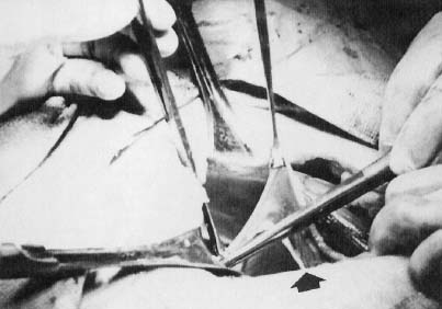
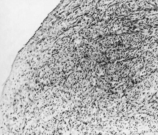
In monosomy X the gonad usually exists not as the typical ovoid structure but as a white fibrosis streak, 2 to 3 cm long and approximately 0.5 cm wide, located in the position ordinarily occupied by the ovary (Fig. 1). A streak gonad is characterized histologically by interfacing waves of dense fibrosis stroma (Fig. 2).

Fig. 1. Photograph taken at the time of celiotomy (Pfannenstiel’s incision) demonstrating the usual appearance of a streak gonad (arrow). The clamp is elevating a fallopian tube. This particular individual had XY gonadal dysgenesis, but the appearance of a streak gonad would have been almost identical in most 45,X individuals.(Simpson JL: Disorders of Sexual Differentiation: Etiology and Clinical Delineation. New York, Academic Press, 1976).

Fig. 2. Histologic appearance of a streak gonad from a 45,X individual.(Simpson JL: Disorders of Sexual Differentiation: Etiology and Clinical Delineation. New York, Academic Press, 1976).

That 45,X individuals show streak gonads as adults is not as obvious as might be expected, given that relatively normal ovarian development occurs in many other mammals (e.g., mice) with monosomy X. The presumptive explanation is that pivotal genes on the normal heterochromatic (inactive) X are being inactivated. Of the some 2000 genes on the X, only perhaps 5% escape inactivation.11 Most of these genes are on the X short arm (Xp), clustered in selected euchromatic regions. Candidate genes for ovarian maintenance genes will probably prove to lie in these euchromatic regions.

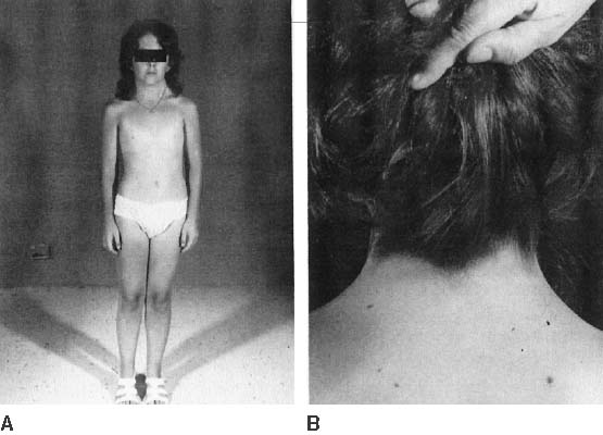
The endocrinologic correlates of ovarian failure are deficient secretion of sex steroids. Estrogen and androgen levels are thus decreased; follicle-stimulating hormone (FSH) and luteinizing hormone (LH) levels are compensatively increased. Deficiencies of estrogen-dependent processes leads to predictable effects of hormonal deficiency. Premenarchal uterine enlargement and growth spurt are not observed. Pubic and axillary hair fail to develop at puberty (Fig. 3). Breasts contain little parenchymal tissue, and areolar tissue is only slightly darker than the surrounding skin. External genitalia, vagina, and müllerian derivates are well differentiated, but remain small (unstimulated) in the absence of exogenous steroids.

Fig. 3. Photograph of a 45,X individual (A). This 140-cm tall patient had no secondary sexual development and several somatic anomalies comprising the Turner’s stigmata: webbing of the neck, low nuchal hair line, and pigmented nevi (B).(From Simpson JL: Disorders of Sexual Differentiation: Etiology and Clinical Delineation. New York, Academic Press, 1976).

Approximately 3% to 5% of adult 45,X patients menstruate spontaneously (at least twice) and show breast development. Many fertile patients have been reported, as recently reviewed by Abir and colleagues12 and Hovatta.13 An undetected 46,XX cell line (i.e., 45,X/46,XX mosaicism) should be suspected in menstruating 45,X patients. This is especially plausible in reports like that of Magee and colleagues14 who observed seven pregnancies in one ostensibly 45,X woman. However, it is not expected that some 45,X individuals could be fertile, inasmuch as germ cells are present in 45,X embryos. In addition, pregnancy can occasionally be achieved in hypergonadotropic women by sequential gonadotropin suppression followed by ovulation induction. Check and colleagues15 induced ovulation in 5% of 361 cycles in 100 hypergonadotropic women; the chromosomal complements of women were not stated.

Hormonal treatment of 45,X women usually involves hormone therapy (estrogen and cyclic progestogens). This can result in normal uterine size, which could be followed by assisted reproductive technology (ART) if pregnancy is desired. The process involves a partner’s sperm being mixed with ova donated from another woman, fertilization in vitro, and transfer of embryo to the uterus of the hormonally synchronized 45,X patient. The success rate (clinical pregnancies) is over 20% per cycle. Thus, Foudila and colleagues16 reported 20 clinical pregnancies among 18 women with Turner’s syndrome, the exact chromosomal complements not being stated. Although the clinical pregnancy rate per fresh embryo transferred was impressive at 46% (13/28), 7 of the 13 (54%) resulted in spontaneous abortion, for a take-home baby rate of 46% (6/13). Among transferred frozen embryos the rates were 28% (7/25) and 14% (1/7), respectively. Low pregnancy rates were also reported by Khastgir and colleagues17 and Tarani and colleagues.18 The ability of a 45,X woman to carry her pregnancy to term must be addressed before embarking on ART. Specifically, women with coaratation of the aorta may be unsuitable candidates. The increased prevalence of autoimmune disease (e.g., thyroiditis), carbohydrate dysfunction (diabetes mellitus), hypertension, and other adult-onset diseases further places many 45,X patients in high-risk situations.

In the opinion of the author, offspring of 45,X women show little, if any, increased risk for chromosomal abnormalities.19,20 Claims to the contrary12 need to be tempered by biases of biases of ascertainment not being taken into account. Outcomes are relatively normal in pregnancies observed after the index case was diagnosed (truncate analysis). Adverse outcomes prior to that time were probably the reason cytogenetic studies were initiated. Spontaneous abortions are increased donor oocyte ART. This probably reflects hormonal dysfunction or uterine factors (hypoplasia), rather than transmission of aneuploid (monosomic) gametes.

Somatic Anomalies

45,X individuals not only are short (less than 4 feet 10 inches) but often exhibit various somatic anomalies (Turner stigmata) (Table 1). No single feature(s) of Turner stigmata is pathognomonic, but in aggregate a characteristic spectrum exists that is more likely to exist in 45,X individuals than in individuals having most other sex chromosomal abnormalities. Systematic evaluation of renal, vertebral, cardiac, and auditory function is obligatory, irrespective of the patient’s age when diagnosed.

Growth

45,X neonates are usually low birth weight. Total body length at birth is less than normal, but often close to the 50th percentile. Before puberty, height velocity falls in the 10th to 15th percentile,21 and the mean height of untreated 45,X adults (younger than 16 years old) is between 141 and 146 cm,22,23 perhaps 20 cm less than normal. In normal females the predicted adult height can be estimated by summing the heights of both parents, dividing by 2, and subtracting by 13 cm.24 Taking into account decreased expected height for 45,X individuals, correlation of the height of a 45,X offspring with midparental height holds for Turner syndrome as it does for normal 46,XX females.25 That is, absolute height predicted in Turner syndrome is less but the midparental height correlation still holds.

Various treatments for short stature in 45,X patients have proposed, including growth hormone (GH), anabolic steroids and low-dose estrogen.26 Most treatment regimens show ostensible benefit, especially immediately after onset of therapy. Consensus is now that that ultimate height can be increased by 6 to 8 cm by GH treatment alone.27,28 The most popular form of treatment is human recombinant DNA-derived human GH. GH results in an 8.4 ± 4.5 cm increases in height over that predicted; final height was 150.4 ± 5.5 cm in one heterogeneous group.29 With growth hormone and oxandrolone, the increase was 10.3 ± 4.7 cm. Treatment regimens are discussed in standard pediatric endocrinologic treatsies (e.g., Grumbach and Conte 30). In general, treatment is begun at 2 to 5 years of age, and stopped at approximately 15 years of age.31 Low-dose estrogen is deferred until final height is near29; high-dose estrogen should then be given to stimulate secondary sexual development. The latter regime should begin at 14 to 15 years of age, starting with 0.3 to 0.625 mg conjugated equine estrogens daily for 6 to 12 months; dose is then increased to 1.25 mg.

A potential reason for limited efficacy of growth hormone treatment may be that epiphyses in 45,X individuals are structurally abnormal. Not only long bones, but teeth32 and skull33 are also abnormal. Thus, patients with a 45,X chromosomal complement could be said to have a skeletal dysplasia.

Some wonder whether presence of an unappreciated Y-bearing line (e.g., 45,X/46,XY) could lead to neoplastic consequences with treatment. However, few ostensibly nonmosaic 45,X cases will prove to have a 46,XY even after analysis of thousands of cells using FISH with a Y probe.

Intelligence

Most 45,X patients are of normal intelligence, but any given patient has a higher probability of being retarded than a 46,XX person.22 Performance IQ is lower than verbal IQ, the latter being similar to 46,XX matched controls. 45,X individuals may have a cognitive defect characterized by poor spatial processing skills (space-form blindness). Ross and colleagues34 opined that only loss of distal Xp (Xp 22.33) produced this phenotype; regions responsible for neurocognitive deficits were believed distant from statural or ovarian abnormalities.

Psychosocial deficits primarily reflect behavioral immaturity and difficulties in social relationships. These are probably secondary to delayed sexual development and statural growth.35,36 The possibility has been raised that parental origin influences phenotype Xm (X of maternal origin). Xm cases are said to show cognitive deficits more often than Xp.37 If true, this would indicate the X contains imprinted genes.

Adult-Onset Diseases

Many adult-onset disorders occur in 45,X cases with frequencies greater than expected in the general population. Hypertension deserves special comment, given its presence in about one third of adults 45,X individuals. Hypertension need not alter hormonal therapy; however, careful monitoring is required, and exogenous estrogen therapy may need to be reduced. Frequencies of diabetes mellitus and autoimmune thyroiditis are increased.

Is a Particular Region of the X Responsible for Somatic Anomalies in 45,X?

The region of the X responsible that if deleted results in somatic anomalies is unclear, but it is not necessarily the same as that for ovarian maintenance. The distal X short arm (Xp) has in particular been implicated in somatic development. A pseudoautosomal gene implicated in somatic development and short stature (Turner stigmata) is SHOX. This pseudoautosomal locus is not subject to inactivation. RPS4X is another candidate for the same reason. Zinn and colleagues38 and Zinn and Ross39 have attempted to correlate somatic anomalies with Xp perturbations, using molecular markers to define deletions and X/autosomal translocation. High-arched palate, short stature, and autoimmune thyroid disease were associated with terminal deletions of Xp11.2-22.1, the same region noted to contain ovarian determinants. Boucher and colleagues40 concluded that Xp11.4 is critical for lymphoedema. Bioné and Toniolo41 have also discussed candidate genes on Xp and Xq that could be important for somatic differentiation.

Back to Top
45,X/46,XX AND 45,X/47,XXX MOSAICISM

If nondisjunction or anaphase lag occurs in the zygote or embryo, two or more cell lines may result (mosaicism). The final chromosomal complement depends on the stage at which abnormal cell division occurs and the types of daughter cells that survive after the abnormal cell division (nondisjunction). Ability to detect mosaicism depends on the number of cells analyzed per tissue and the number of tissues analyzed. For example, counting 50 cells without detecting one nonmodal cell excludes (p < .005) mosaicism for a minority line of 10% or more.42 The common practice of a cytogenetic laboratory counting 20 cells carries a probability of 0.122 for failing to detect at least one cell representing a minority cell line of 10% frequency. With 50 cells, probability falls to 30%, the probability of failing to detect a 10% minority line. If the minority constituted 30%, the probability of failing to detect would be 0.001 if 20 cells were counted.42

The most common form of mosaicism associated with ovarian failure or gonadal dysgenesis is 45,X/46,XX. Population-based data do not exist, but it indeed seems that 45,X/46,XX individuals show fewer anomalies than 45,X individuals. One survey revealed 12% of 45,X/46,XX individuals menstruate, compared with only 3% of 45,X individuals.22 Among 45,X/46,XX individuals in that survey, 18% undergo breast development, compared with 5% of 45,X individuals. Mean adult height is greater in 45,X/46,XX than in 45,X; more mosaic (25%) than nonmosaic (5%) patients reach adult heights greater than 162 cm.22 Somatic anomalies are less likely to exist in 45,X/46,XX than in 45,X. 45,X/47,XXX is less common but phenotypically similar to 45,X/46,XX individuals.

Some 45,X/46,XY individuals may also show bilateral streak gonads; however, they often show a unilateral streak gonad and a contralateral dysgenetic testis (mixed gonadal dysgenesis) or bilateral testes. This phenotype is discussed in Chapter 5-83.

Back to Top
X SHORT ARM DELETIONS AND OVARIAN FAILURE: 45,X,del(Xp) OR 45,X/46,X, del (Xp) DELETIONS

Different terminal deletions of the short arm of the X chromosome exist, reflecting different amount of persisting Xp. Breakpoints presumably do not occur in just selected places, but rather throughout the chromosome. However, pooling structurally similar terminal deletions reveals the most common breakpoint for terminal deletions to be Xp11.2 → 11.4. In 46,X, del(X)(p11) only proximal Xp remains; the del(Xp) chromosome thus appears acrocentric or telocentric. More distal (telomeric) breakpoints also exist: Xp21, 22.1, 22.2, 22.3. Sequencing and analysis with polymorphic DNA markers are beginning to permit precise determinations of these breakpoints, but still relatively little molecular information exists.38,39,43,44

In 45,X,del(Xp)(p11) most of Xp is missing. Approximately half of these individuals show primary amenorrhea and gonadal dysgenesis. In a tabulation made 15 years ago, 12 of 27 reported del(X)(p11.2 → 11.4) individuals menstruated spontaneously; however, menstruation was rarely normal.4 More recent calculations1,2,45 have not materially altered the fundamental conclusions concerning location of key determinants. Figure 4 shows a compilation of cases reported through 1999. Similar conclusions were reached by Ogata and Matsuo,46 who estimate that 50% of del(Xp11) cases show primary amenorrhea and 45% show secondary amenorrhea. Several groups are approaching the topic molecularly, but to date their clinical conclusions generally seem similar.38,39,47,48 Both Wandstrat and colleagues47 and Thomas and Huson48 were impressed by single nonmosaic cases del(X) (p11.2) cases showing normal height and fertility. However, similar observations had been made earlier.

Fig. 4. Schematic diagram of the X chromosome showing ovarian function as a function of nonmosaic terminal deletions.(Simpson JL, Rajkovic A: Ovarian differentiation and gonadal failure. Am J Med Genet 89:186, 1999.)

Women with more distal deletions [del(X)(p21.1 to p22.122)] menstruate more often, but many are still infertile or show secondary amenorrhea. Ovarian failure (menopause) is more likely to be premature (younger than 35 or 40 years of age, depending on definition). Thus, Xp21, 22.1 or 22.2 is less important for ovarian development1,45 than Xp11. Deletion of only the most telomeric portion of Xp (Xp22.3 → Xpter) does not result in amenorrhea.49 Zinn and colleagues38 and Zinn and Ross39 concluded only that the region Xp11.3 → 22.1 was paramount, although this large region encompasses most of Xp. Cases with an interstitial deletion will be necessary to narrow the region of interest.38,39 Interstitial deletions have involved Xp11-22 and Xp11.4-22.3.50,51

Familial cases have been reportedly observed. These include both mother and daughter showing the same Xp-deletions43,52 as well as X/autosome translocations. Among 10 del(Xp) cases studied by James and colleagues53 were two mother-daughter pairs. A later analysis reported 25 females with de novo deletions of Xp and four familial Xp deletions.48 Familial cases have involved deletions at Xp11 as well as Xp22-12.38,54

Back to Top
X SHORT-ARM DEFICIENCIES CAUSED BY ISOCHROMOSOME FOR THE X LONG ARM [i(Xq)]

If division of the centromere occurs in the transverse rather than the longitudinal plane, an isochromosome results. The resulting metacentric chromosome consists of isologous arms. Both arms are structurally identical and contain the same genes. Isochromosome for the X long arm [i(Xq)] differs from terminal deletion of Xp in that not just the terminal portion but all of the Xp is deleted. An isochromosome for the X long arm is the most common X structural abnormality, but coexisting 45,X cells (mosaicism) are common.

Almost all reported 46,i(Xq) patients show streak gonads, and short stature, and Turner’s stigmata have long been accepted as almost universal.22 Only rarely do 46,X,i(Xq) patients seem to menstruate. The more often complete lack of gonadal development in 46,X,i(Xq) individuals contrasts with that in 46,X,del(Xp11) individuals, about half of whom menstruate or develop breasts (see above). This could indicate that gonadal determinants exist at several different locations on Xp. That is, a locus near the centromere could be deleted in i(Xq) yet retained in del(X)(p11).

Back to Top
X SHORT-ARM DELETIONS AND SOMATIC ANOMALIES

Most women with deletions of Xp are short in stature; thus, statural determinants responsible for the short stature in 45,X must exist on Xp. However, this is not invariate as witnessed by normal height in a 46,X, del(X)(p11.2) case of Thomas and Huson.48 Given that del(Xp) women who menstruate may still be short, genes on Xp responsible for statural determinants must be distinct from those responsible for ovarian development.45,54,55,56 Attempts have been made to localize our components of the Turner’s stigmata. Statural genes on Xp are more telomeric than the Xp ovarian determinants.

The short stature and skeletal features of Turner stigmata have been thought to be caused by haplo-insufficiency for SHOX. This gene is localized in the pseudoautosomal region (PAR), and is homologous to the Y-linked gene SHOY.57 Perturbation of SHOX causes Leri-Weill dyschondrosteosis, a dominantly inherited disorder characterized by short stature and skeletal abnormalities that include Madelung’s deformity. Short stature cubitus valgus, short metacarpals, short neck, scoliosis have also been observed with SHOX mutations.57,58,59,60

Zinn and colleagues38 concluded that high arched palate, short stature, and autoimmune thyroid disease were governed by a region on Xp11.2 → 22.1. Thomas and colleagues49 reported autism in three of eight cases involving perturbation of Xp22.31 → .33. Impaired visual spatial/perceptual abilities (space-form blindness) were thought to be localized to distal Xp 22.33 by Ross and colleagues.34

Interest has also been placed on localizing a putative locus protecting against hymphodema, given the potential of the latter as an anatomic abnormality underlying disparate pleiotropic features. Webbing of the neck, low posterior hairline, puffy hands/feet, and nail dysplasia are thought to all reflect superficial hypoplasia of lymphatic vessels. Boucher and colleagues40 studied seven Xp deletions and two Xp translocations, finding features suggesting lymphoedema in four or five. They observed considerable variability in phenotype despite ostensibly similar deletions. However, a putative locus was considered to exist in the proximal portion of Xp, between OTC and DJ109C12. If the locus suggested by Boucher and colleagues40 is confirmed, it would clearly be distinct from SHOX, which is pseudoautosomal. On the other hand, the two cases of James and colleagues61 who showed lymphedema had more distal deletions (del(X)(p22).

Back to Top
CANDIDATE GENES ON Xp

Several candidate genes on Xp have been proposed. Zinn and colleagues38 proposed ZFX, a DNA binding protein; a homologous gene exists on the Y (ZFY). Jones and colleagues62 proposed DFRX, located on Xp11.4 and homologous to a locus on Yq11.2. DFRX (or USP9X) targets proteins for degradation by the ubiquitan pathway. USP9X is homologous to the Drosphila gene fat facets (faf). Both DFRX (USP9X) and ZFX escaped inactivation in two de novo (X)(p11.2) deletions. James and colleagues61 concluded DFRX was an unlikely candidate after observing ovarian function in two cases despite haplo-insufficiency; however, neither of the cases reported by James and colleagues61 were completely normal, for which reason a role for DFRX in gonadal development is not completely excluded. After studying an interstitial deletion of Xp11.2-11.4, Zinn and colleagues38 hypothesized the following Xp candidate genes: DFRX (USP9X) (discussed above), UBE1 (another ubiquitan pathway enzyme) and BMP15 (a member of the transforming growth factor-β [TGF-β] family of signaling compounds). BMP15 is structurally similar to GDF-9, gene knockout of which causes germ cell failure in mice.63

Many other candidate X-ovarian genes are plausible, as reviewed by Bioné and Toniolo,41 Zinn,64 and Zinn and Ross.39 A host of other candidate genes can also be deduced from homologues in the mouse.2 However, most of these genes are autosomal in the mouse, thus, these could be better candidate genes for the disorders of XX gonadal dysgenesis to premature ovarian failure (POF) discussed in Chapter 5-86.

Back to Top
X LONG-ARM DELETIONS: 46,X,del(Xq) AND 45,X/46,X,del(Xq)

Like those involving the X short arm, deletions of the X long arm (Xq) vary in composition. The most extensive deletions originate at Xq13 and have long been shown to be associated with primary amenorrhea, lack of breast development, and complete ovarian failure. 1,2,45,46,61 Xq13 is thus the most pivotal region for ovarian maintenance. That this region contains the human X-inactivation center (XIC) is not obligatorily relevant, but probably is important. The abnormal phenotype associated with del(Xq13) could reflect perturbation of XIC rather than loss of a gene per se. Irrespective, key loci lie no more distal than proximal Xq21 given that menstruation occurs in deletions of breakpoints Xq21 or beyond (see Fig. 4). Menstruating del(X)(q21) women could have retained a region containing an ovarian maintenance gene, whereas del(X)(q13 or 21) women with primary amenorrhea could have lost such a locus. Figure 4 schematically shows phenotypes associated with various terminal deletions of Xq.

In more distal Xq deletions, the phenotype is usually POF, not primary amenorrhea.2,41,65,66 Distal Xq is thus less pivotal for ovarian maintenance than proximal Xq, although the former still contains regions important for ovarian maintenance. Although there is no clear step-wise demarcation into discrete breakpoints regions, it is heuristically useful to stratify terminal deletions in this fashion: Xq13 → 21,Xq22-25, and Xq26-28. In 1995, Ogata and Matsuo46 correlated ovarian function using such stratification, and our tabulations (see Fig. 4) are generally consistent.

Molecular mapping of the regions of Xq integral for ovarian development has begun by Bioné and colleagues.66 Sala and colleagues67 first studied seven X/autosome translocations involving Xq21-22; five of the seven had primary amenorrhea. A region of Xq spanning 15 mb encompassed breakpoints in all seven cases. Breakpoints in four other X-autosome translocations studied by Philippe and colleagues68 were localized to the same region. The YAC contig encompassing these breakpoints spanned most of Xq21, from DXS233 to DXS1171.6 If the breakpoints associated with ovarian failure truly spanned the entire Xq21 region, it would be unlikely that only a single gene in this region is responsible for ovarian failure. Alternatively, ovarian failure associated with these translocations could be the result not of perturbation of any specific gene but rather of generalized cytologic (meiotic) instability. Incidentally, the official nomenclature labels genes causing POF as POF1 and POF2. By this, the Xq25-26 region is POF1, whereas the more pivotal Xq13 regions is POF2.

Distal Xq deletions may be familial. Some familial aggregates are derivative of Xq/autosome translocations, but del(X)(q26) and other Xq deletions also may be familial.43,54,69,70,71,72 The amount of persistent ovarian function may vary among different family members having the same deletion.

Especially informative are the rare families ascertained for reasons other than POF. An example is an Xq interstitial deletion ascertained through amniotic fluid analysis by our group.43 Both fetus and mother had the same abnormality, suggesting that additional previously unrecognized families might be ascertained if prometaphase analysis or molecular studies of polymorphic loci were more often performed in POF. Brown and colleagues73 similarly ascertained a del(Xq) deletion through amniotic fluid analysis. Vegnetti and colleagues74 detected one del(Xq) case among 82 Italian women with POF. Susca and colleagues65 found 2 del(Xq) cases among 20 women with POF.

Distal Xq deletions seem to have a less deleterious effect on stature than proximal deletions, analogous to their effect on gonadal development. Somatic anomalies of Turner stigmata are uncommon, arguably no more common than in the general population.

One unusual case involved Xp21-22 duplication and Xp21→qter deletion [46,X,der(X):(pter→q21::p21→pter)].75 The affected 20-year-old woman had streak gonads, tall stature of 174 cm (more than 2 standard deviations [SD]) over age-matched Japanese women), multiple fractures, and endometriosis. It is tempting to postulate that the Xp duplication resulted in overexpression of SHOX, whereas Xq21→qter deletion was responsible for the gonadal failure.75

Back to Top
CANDIDATE GENES ON Xq

The most popular candidate gene for ovarian failure on the X long arm is the human homologue of the Drosophila melanogaster gene diaphanous (dia). Drosophila dia is a member of a family of proteins that establish cell polarity, govern cytokinesis, and organize the cytoskeleton mutation of the dia gene causes sterility in male and female flies.76 Sequence comparisons between dia and the human EST (expressed sequence tag) DRE25 revealed significant homology. In turn, DRE25 maps to human Xq21-22,76 a region already noted to be important for ovarian maintenance. Studying an Xq21/autosome translocation originally reported by Phillipe and colleagues68; Sala and colleagues67 found disruption of DRE2566 in association with ovarian failure.

Another candidate gene is XPNPEP2, which encodes a Xaa-Pro aminopeptidase (metalloprotease) that hydrolyzes proline bonds. XPNPEP2 is ubiquitously expressed in many tissues, and can influence a host of biologically active peptides that could include regulations ovarian function. XPNPEP2 was disrupted in an Xq translocation that involved Xq25.77 Zinn64 and Bioné and Toniolo41 considered several other Xq candidate genes and a host of mouse models point to other homologous genes on the X and autosomes (see Simpson and Rajkovic2). Edwards78 recently discussed still other genes in Drosophila melanogaster and Caenorhabditis elegans, in particular dealing with oocyte polarity, the homologs of which could be relevant to humans.

Other candidate POF genes were suggested by Davison and colleagues79 after analysis of a 46,X,del(X)(q26) case associated with the fragile X permutation phenotype. Based on the deleted chromosomal region, three candidate genes were identified by NIX and BLAST analysis: (1) heparan-sulfate-6-transferase (HS6ST), (2) an E2F-related transcription factor, and (3) some gene(s) in the region in which LINE 1 elements (long interspersed nuclear elements) were found. Davison and colleagues79 considered HS6ST, which showed 82% nucleotide and 65% amino acid homology, the most likely candidate gene sequence. Sulfated proteoglycans are involved in regulation of cell proliferation and migration, and in Drosophila are known to influence oocyte polarity. E2F, which showed 75% homology with the deleted Xq candidate sequence, is known to regulate somatic cell cycles and can imitate apoptosis. Other genes in the E2F family are active in mammalian embryos, including oocytes. The deleted candidate sequences also were homologous to LINE 1 elements (97% homology). LINE 1 are retrotransposons that are potentially mutagenic and capable of interfering with transcription or gene expression. They could be important per se or could indicate another nearby gene of importance.

Back to Top
OTHER X CHROMOSOME ABNORMALITIES

Other structural abnormalities of the X chromosome have been reported with the phenotype all ultimately involving deletions of specific regions. The most common group shows centric fragments and ring X[r(X)] chromosomes. These chromosomes are mitotically unstable, frequently associated with secondarily derived monosomic (45,X) lines. Fluorescent in situ hybridization (FISH) or molecular studies may be necessary to verify that the fragment or ring originates from an X and not a Y. Deserving of special mention are women once thought to have a 46,X,i(Xp) complement. They were of great interest because they showed primary amenorrhea and were of normal stature. However, it later developed that i(Xp) did not exist, the karyotype in these individuals instead being del(X)(q22 or 24).

A considerable body of literature exists on X/autosome translocations. Those involving Xq are more likely to cause gonadal failure than those involving Xp. Clinical management of gonadal failure due to X/autosomal translocation is comparable to any patient with an X chromosome and ovarian failure; however, genetic counseling can be complex because familial transmission is possible.

Sometimes duplication as well as deficiency exists. Excluding those exceptional cases6 in which Xp11 (DAX1) is duplicated in XY individuals to produce (XY gender reversal), the phenotype is generally predictable.

Back to Top
REFERENCES

1. Simpson JL: Genetic programming in ovarian development and oogenesis. In Lobo RA, Kelsey JMR (eds): Menopause Biology and Pathobiology. pp 77, 94. London, Academic Press, 2000

2. Simpson JL, Rajkovic A: Ovarian differentiation and gonadal failure. Am J Med Genet 89:186, 1999

3. Simpson JL, Elias S: Genetics in Obstetrics and Gynecology. 3rd ed.. Philadelphia, W.B. Saunders, 2002

4. Simpson JL: Phenotypic-karyotypic correlations of gonadal determinants:current status and relationship to molecular studies. In Sperling KVF (ed): Human Genetics. Proceedings of the Seventh International Congress, Berlin. pp 224, 232 Heidelberg, Springer-Verlag, 1987

5. German J: Gonadal dimorphism explained as a dosage effect of a locus on the sex chromosomes, the gonad-differentiation locus (GDL). Am J Hum Genet 42:414, 1988

6. Bardoni B, Zanaria E, Guioli S, et al: A dosage sensitive locus at chromosome Xp21 is involved in male to female sex reversal. Nat Genet 7:497, 1994

7. Yu RN, Ito M, Saunders TL, et al: Role of Ahch in gonadal development and gametogenesis. Nat Genet 20:353, 1998

8. Jirasek J: Principles of reproductive embryology. In Simpson JL (ed): Disorders of Sexual Differentiation: Etiology and Clinical Delineation. pp 51, 110 New York, Academic Press, 1976

9. Mathur A, Stekol L, Schatz D, et al: The parental origin of the single X chromosome in Turner syndrome: Lack of correlation with parental age or clinical phenotype. Am J Hum Genet 48:682, 1991

10. Cockwell A, MacKenzie M, Youings S, et al: A cytogenetic and molecular study of a series of 45,X fetuses and their parents. J Med Genet 28:152, 1991

11. Willard HF: The sex chromosomes and X chromosome inactivation. In Scriver CR, Beaudet AL, Sly WS, et al (eds): The Metabolic and Molecular Bases of Inherited Disease. pp 1191, 1211 Vol 8:New York, McGraw-Hill, 2001

12. Abir R, Fisch B, Nitke S, et al: Morphological study of fully and partially isolated early human follicles. Fertil Steril 75:141, 2001

13. Hovatta O: Pregnancies in women with Turner’s syndrome. Ann Med 31:106, 1999

14. Magee AC, Nevin NC, Armstrong MJ, et al: Ullrich-Turner syndrome: Seven pregnancies in an apparent 45,X woman. Am J Med Genet 75:1, 1998

15. Check JH, Nowroozi K, Chase JS, et al: Ovulation induction and pregnancies in 100 consecutive women with hypergonadotropic amenorrhea. Fertil Steril 53:811, 1990

16. Foudila T, Soderstrom-Anttila V, Hovatta O: Turner’s syndrome and pregnancies after oocyte donation. Hum Reprod 14:532, 1999

17. Khastgir G, Abdalla H, Thomas A, et al: Oocyte donation in Turner’s syndrome: An analysis of the factors affecting the outcome. Hum Reprod 12:279, 1997

18. Tarani L, Lampariello S, Raguso G, et al: Pregnancy in patients with Turner’s syndrome: Six new cases and review of literature. Gynecol Endocrinol 12:83, 1998

19. Dewhurst J: Fertility in 47,XXX and 45,X patients. J Med Genet 15:132, 1978

20. Simpson JL: Pregnancies in women with chromosomal abnormalities. In In Schulman JD, Simpson JL (eds): Genetic Diseases in Pregnancy. pp 439, 471 New York, Academic Press, 1981

21. Brook CG, Murset G, Zachmann M, et al: Growth in children with 45,XO Turner’s syndrome. Arch Dis Child 49:789, 1974

22. Simpson JL: Gonadal dysgenesis and abnormalities of the human sex chromosomes: Current status of phenotypic-karyotypic correlations. Birth Defects Orig Artic Ser 11(4):23, 1975

23. Ranke MB, Pfluger H, Rosendahl W, et al: Turner syndrome: Spontaneous growth in 150 cases and review of the literature. Eur J Pediatr 141:81, 1983

24. Tanner JM, Goldstein H, Whitehouse RH: Standards for children’s height at ages 2–9 years allowing for heights of parents. Arch Dis Child 45:755, 1970

25. Brook CG, Gasser T, Werder EA, et al: Height correlations between parents and mature offspring in normal subjects and in subjects with Turner’s and Klinefelter’s and other syndromes. Ann Hum Biol 4:17, 1977

26. Ranke MB, Blum WF, Haug F, et al: Growth hormone, somatomedin levels and growth regulation in Turner’s syndrome. Acta Endocrinol 116:305, 1987

27. Rosenfeld RG, Grumbach MM: Turner Syndrome. New York, Marcel Dekker, 1990

28. Rosenfeld RG, Frane J, Attie KM, et al: Six-year results of a randomized, prospective trial of human growth hormone and oxandrolone in Turner syndrome. J Pediatr 121:49, 1992

29. Rosenfeld RG, Attie KM, Frane J, et al: Growth hormone therapy of Turner’s syndrome: beneficial effect on adult height. J Pediatr 132:319, 1998

30. Grumbach MM, Conte FA: Disorders of sex differentiation. In Wilson JD, Foster DW, Kronenberg H (eds): Williams Textbook of Endocrinology. pp. 1303, Vol. 9:Philadelphia, WB Saunders, 1998

31. Saenger P: Turner’s syndrome. N Engl J Med 335:1749, 1996

32. Filippson R, Lindsten J, Almqvist S: Time of eruption of the permanent teeth, cephalometric and tooth measurement and sulphationfactor activity in 45 patients with Turner’s syndrome with different types of X chromosome aberration. Acta Endocrinol 48:91, 1965

33. Lindsten J, Fraccaro M: Turner’s syndrome. In Rashad MN, Mortion WRM (eds): Genital Anomalies,. pp 396, Springfield, Charles C. Thomas Publishers, 1969

34. Ross JL, Roeltgen D, Kushner H, et al: The Turner syndrome-associated neurocognitive phenotype maps todistal Xp. Am J Hum Genet 67:672, 2000

35. McCauley E, Sybert VP, Ehrhardt AA: Psychosocial adjustment of adult women with Turner syndrome. Clin Genet 29:284, 1986

36. McCauley E, Kay T, Ito J, et al: The Turner syndrome: Cognitive deficits, affective discrimination, and behavior problems. Child Dev 58:464, 1987

37. Skuse DH, James RS, Bishop DV, et al: Evidence from Turner’s syndrome of an imprinted X-linked locus affecting cognitive function. Nature 387:705, 1997

38. Zinn AR, Tonk VS, Chen Z, et al: Evidence for a Turner syndrome locus or loci at Xp11.2-p22.1 Am J Hum Genet 63:1757, 1998

39. Zinn AR, Ross JL: Molecular analysis of genes on Xp controlling Turner syndrome and premature ovarian failure (POF). Semin Reprod Med 19:141, 2001

40. Boucher CA, Sargent CA, Ogata T, et al: Breakpoint analysis of Turner patients with partial Xp deletions: Implications for the lymphoedema gene location. J Med Genet 38:591, 2001

41. Bione S, Toniolo D: X chromosome genes and premature ovarian failure. Semin Reprod Med 18:51, 2000

42. Ford CE: Mosaics and chimaeras. Br Med Bull 25:104, 1969

43. Tharapel AT, Anderson KP, Simpson JL, et al: Deletion (X)(q26.1→q28) in a proband and her mother: Molecular characterization and phenotypic-karyotypic deductions Am J Hum Genet 52:463, 1993

44. Davison RM, Fox M, Conway GS: Mapping of the POF1 locus and identification of putative genes for premature ovarian failure. Mol Hum Reprod 6:314, 2000

45. Simpson JL: Genetics of female infertility. In Filicori M, Flamigni C (eds): Proceedings of the Conference, Treatment of Infertility: The New Frontiers. pp 37, 52 Boca Raton, FL, Communications Media for Education, 1998

46. Ogata T, Matsuo N: Turner syndrome and female sex chromosome aberrations: Deduction of the principal factors involved in the development of clinical features. Hum Genet 95:607, 1995

47. Wandstrat AE, Conroy JM, Zurcher VL, et al: Molecular and cytogenetic analysis of familial Xp deletions. Am J Med Genet 94:163, 2000

48. Thomas NS, Huson SM: Atypical phenotype in a female with a large Xp deletion. Am J Med Genet 104:81, 2001

49. Thomas NS, Sharp AJ, Browne CE, et al: Xp deletions associated with autism in three females. Hum Genet 104:43, 1999

50. Wilson MG, Modebe O, Towner JW, et al: Ullrich-Turner syndrome associated with interstitial deletion of Xp11.4 leads to p22.31 Am J Med Genet 14:567, 1983

51. Herva R, Kaluzewski B, de la Chapelle A: Inherited interstitial del(Xp) with minimal clinical consequences: With a note on the location of genes controlling phenotypic features. Am J Med Genet 3:43, 1979

52. Fraccaro M, Maraschio P, Pasquali F, et al: Women heterozygous for deficiency of the (p21 leads to pter) region of the X chromosome are fertile. Hum Genet 39:283, 1977

53. James RS, Dalton P, Gustashaw K, et al: Molecular characterization of isochromosomes of Xq. Ann Hum Genet 61:485, 1997

54. Massa G, Vanderschueren-Lodeweyckx M, Fryns JP: Deletion of the short arm of the X chromosome: a hereditary form of Turner syndrome. Eur J Pediatr 151:893, 1992

55. Simpson JL: Genetics of oocyte depletion. In Lobo RA (ed): Perimenopause. pp 36, 45 New York, Springer, 1997

56. Simpson JL: Ovarian maintenance determinants on the X chromosome and on autosomes. In Coutifaris CML (ed): New Horizons in Reproductive Medicine. pp 439, 444 Proceedings of the IXth World Congress on Human Reproduction. New York and London, Parthenon, 1997

57. Clement-Jones M, Schiller S, Rao E, et al: The short stature homeobox gene SHOX is involved in skeletal abnormalities in Turner syndrome. Hum Mol Genet 9:695, 2000

58. Belin V, Cusin V, Viot G, et al: SHOX mutations in dyschondrosteosis (Leri-Weill syndrome). Nat Genet 19:67, 1998

59. Shears DJ, Vassal HJ, Goodman FR, et al: Mutation and deletion of the pseudoautosomal gene SHOX cause Leri-Weill dyschondrosteosis. Nat Genet 19:70, 1998

60. Kosho T, Muroya K, Nagai T, et al: Skeletal features and growth patterns in 14 patients with haploinsufficiency of SHOX: Implications for the development of Turner syndrome. J Clin Endocrinol Metab 84:4613, 1999

61. James RS, Coppin B, Dalton P, et al: A study of females with deletions of the short arm of the X chromosome. Hum Genet 102:507, 1998

62. Jones MH, Furlong RA, Burkin H, et al: The Drosophila developmental gene fat facets has a human homologue in Xp11.4 which escapes X-inactivation and has related sequences on Yq11.2 Hum Mol Genet 5:1695, 1996

63. Dong J, Albertini DF, Nishimori K, et al: Growth differentiation factor-9 is required during early ovarian folliculogenesis. Nature 383:531, 1996

64. Zinn AR: The X chromosome and the ovary. J Soc Gynecol Invest 8:S34, 2001

65. Susca F, Aoam R, Vucubim M, et al: Xq deletion and premature ovarian failure. Hum Reprod 14:236, 1999

66. Bione S, Sala C, Manzini C, et al: A human homologue of the Drosophila melanogaster diaphanous gene is disrupted in a patient with premature ovarian failure: Evidence for conserved function in oogenesis and implications for human sterility. Am J Hum Genet 62:533, 1998

67. Sala C, Arrigo G, Torri G, et al: Eleven X chromosome breakpoints associated with premature ovarian failure (POF) map to a 15-Mb YAC contig spanning Xq21. Genomics 40:123, 1997

68. Philippe C, Arnould C, Sloan F, et al: A high-resolution interval map of the q21 region of the human X chromosome. Genomics 27:539, 1995

69. Fitch N, de Saint VJ, V, Richer CL, et al: Premature menopause due to a small deletion in the long arm of the X chromosome: a report ofthree cases and a review. Am J Obstet Gynecol 142:968, 1982

70. Krauss CM, Tukray RN, Atkins L, et al: Familial premature ovarian failure due to interstitial deletion of the longarm of theX chromosome. N Engl J Med 317:125, 1987

71. Veneman TF, Beverstock GC, Exalto N, et al: Premature menopause because of an inherited deletion in the long arm of the X-chromosome. Fertil Steril 55:631, 1991

72. Brown S, Yu C, Lanzano P, et al: A de novo mutation (Gln2Stop) at the 5′ end of the SRY gene leads to sex reversal with partial ovarian function. Am J Hum Genet 62:189, 1998

73. Brown LY, Alonso ML, Yu J, et al: Prenatal diagnosis of a familial Xq deletion in a female fetus: a case report. Prenat Diagn 21:27, 2001

74. Vegetti W, Grazia TM, Testa G, et al: Inheritance in idiopathic premature ovarian failure: analysis of 71 cases. Hum Reprod 13:1796, 1998

75. Nakamura Y, Suehiro Y, Sugino N, et al: A case of 46,X,der(X)(pter→q21::p21→pter) with gonadal dysgenesis, tall stature, and endometriosis. Fertil Steril 75:1224, 2001

76. Castrillon DH, Wasserman SA: Diaphanous is required for cytokinesis in Drosophila and shares domains of similarity with the products of the limb deformity gene. Development 120:3367, 1994

77. Prueitt RL, Ross JL, Zinn AR: Physical mapping of nine Xq translocation breakpoints and identification of XPNPEP2 as a premature ovarian failure candidate gene. Cytogenet Cell Genet 89:44, 2000

78. Edwards RG: Ovarian differentiation and human embryo quality. Reproductive Biomedicine Online 3:138, 2001

79. Davison RM, Fox M, Conway GS: Mapping of the POF1 locus and identification of putative genes for premature ovarian failure. Mol Hum Reprod 6:314, 2000

Back to Top