Chapter 109
Preimplantation Diagnosis for Mendelian Disorders
Yury Verlinsky and Anver Kuliev
Main Menu   Table Of Contents

Search

Yury Verlinsky, MD
Reproductive Genetics Institute, Chicago, Illinois (Vol 5, Chap 109)

Anver Kuliev, MD
Reproductive Genetics Institute, Chicago, Illinois (Vol 5, Chap 109)

 
GENETIC ANALYSIS IN SINGLE CELLS AND AVOIDING MIDIAGNOSIS
EXPANDING PGD APPLICATION
CONCLUSION
REFERENCES

PGD has become an established procedure for avoiding the birth of affected children with single gene disorders and having a healthy unaffected offspring of their own. PGD is performed through polar body or blastomere biopsy, which have no deleterious effect on the preimplantation and postimplantation development. The review describes recent developments in improving the accuracy of PGD for Mendelian disorders and current changes in the spectrum of conditions for which PGD has been applied. Among the most recent applications of PGD were congenital malformations, blood group incompatibility, and increasing number of late-onset disorders with genetic predisposition that have not previously practiced in prenatal diagnosis. Despite ethical concerns, PGD has been further applied for preselection of unaffected and HLA-matched embryos and, recently, also for preimplantation HLA matching without testing for causative gene. This extends the practical value of PGD, with its usefulness being no longer limited to prevention of single gene disorders, but expanded to treatment of siblings requiring stem cell transplantation.

Preimplantation genetic diagnosis (PGD) is a novel approach to detect genetic disorders before pregnancy, avoiding the need for prenatal diagnosis and abortion. It is based on testing of oocytes or embryos to preselect and transfer only normal embryos back to the patient, achieving an unaffected pregnancy and birth of a healthy child. PGD is performed either by testing single blastomeres removed from the 8-cell preimplantation embryos or by testing female gametes (Fig. 1).1 The latter is based on the removal and genetic analysis of the first and second polar bodies (PB1 and PB2), which represent the byproducts of the first and second meiotic divisions (see Fig. 1A, 1B, 1C). Because neither PB1 nor PB2 have any biologic significance for embryo development, they may be removed and tested for the genetic contents to predict the resulting maternal contribution to the embryo. However, this approach cannot be applied for testing of paternally derived abnormalities and gender determination, which may be detected by genetic analysis of single blastomeres, removed from the cleavage stage embryos (see Fig. 1D). Both methods, therefore, are complementary and may be applied depending on the PGD objectives in each patient.

Fig. 1. A. First polar body removal. B. Second polar body removal. C. First and second polar body removal. D. Blastomere biopsy.

More that 5000 PGD cycles have been performed and resulted in the birth of more than 1000 children after the procedure.2 The overall experience of using PGD for single gene disorders is presently almost 1500 cases, showing that it is an established alternative to traditional prenatal diagnosis, which may be reliably applied as an integral part of genetic practices.3,4,5 As will be demonstrated, PGD has appeared to be applicable to a wider range of inherited conditions, which have never been indications for prenatal diagnosis, and also made possible to initiate nondisease testing to preselect HLA-compatible offspring in addition to PGD for causative gene, which makes possible to facilitate treatment of siblings requiring stem cell transplantation.

Back to Top
GENETIC ANALYSIS IN SINGLE CELLS AND AVOIDING MIDIAGNOSIS

Sources for Misdiagnosis

Because PGD for single gene disorders is based on single cell genetic analysis, its accuracy largely depends on the limitations of single cell DNA analysis, which may potentially cause misdiagnosis (Table 1). One of the key contributors to misdiagnosis is the phenomenon of preferential amplification, also known as allele-specific amplification failure (allele dropout [ADO]), requiring the application of special protocols to ensure the highest ADO detection rate (Fig. 2).1,6 A few previously reported misdiagnoses, involving PGD for beta-thalassemia, myotonic dystrophy (DM), fragile-X syndrome (XMR1), and cystic fibrosis (CF), might have been caused by this phenomenon, which has not initially been fully realized.7,5

Table 1. Problems of PGD for Single Gene Disorders


Prevention and elimination of DNA contamination
Optimization of reaction conditions
Detection of allele drop-out (ADO) and treferential amplification
Detection of aneuploidy


Fig. 2. Detection by linked markers for CFTR N1303 mutation.

It has been demonstrated that ADO rates in single cells might be different for different types of heterozygous cells.8 The ADO rate may exceed 20% in blastomeres compared with ADO rate in single fibroblasts and PB1, which was shown to be less than 10% (Fig. 3). A high rate of ADO in blastomeres may lead to an obvious misdiagnosis, especially in compound heterozygous embryos. As mentioned, most misdiagnoses, especially those at the initial stage of application of PGD for single gene disorders, were in the cases of blastomere biopsy from apparently compound heterozygous embryos.

Fig. 3. ADO rate in different types of single cells.

Use of Polymorphic Markers

To avoid a misdiagnosis caused by preferential amplification, a simultaneous detection of mutant gene together with up to three highly polymorphic markers closely linked to the gene tested was introduced.1,9 Each additional linked marker reduced misdiagnosis rate approximately by half, so with one linked marker amplified together with mutation, a misdiagnosis risk in blastomere analysis may be reduced from 20% to 10%, with two from 10% to 5% and with three from 5% to practically zero (Fig. 4). So, a multiplex nested polymerase chain reaction (PCR) analysis is performed, with the initial PCR containing all the pairs of outside primers, so that after the first-round PCR, separate aliquots of the resulting PCR product may be amplified using the inside primers specific for each site (Fig. 5). Only when the polymorphic sites and the mutation agree are embryos transferred. So, multiplex amplification allows detecting of ADO and prevents the transfer of misdiagnosed affected embryos.

Fig. 4. ADO detection with application of additional linked markers.

Fig. 5. Multiplex nested PCR for CFTR G551D mutation.

Sequential PB1 and PB2 DNA Analysis

Another efficient approach for avoiding misdiagnosis may be a sequential genetic analysis of the PB1 and PB2 in PGD for maternally derived mutations.1 Detection of both mutant and normal alleles in the heterozygous PB1, together with the mutant allele in the corresponding PB2, leaves no doubt that the resulting maternal contribution to the embryo is normal, even without testing for the linked markers as a control (Fig. 6). However, it will be ideal to test simultaneously at least for one linked marker to confirm the diagnosis.

Fig. 6. Principle of preselection of mutation-free oocytes using sequential force and second polar analysis and linked polymorphism in PGD for sickle cell disease.

Alternatively, the mutation-free oocytes may be also predicted when corresponding PB1 is homozygous mutant (Fig. 7). In that scenario, the corresponding PB2 should be hemizygous-normal, similar to the resulting maternal pronucleus. However, the genotype of the resulting maternal contribution may be quite opposite, in other words, mutant, if the corresponding PB1 is in fact heterozygous but erroneously diagnosed as homozygous-normal because of ADO of the normal allele. In the aforementioned scenario, the extrusion of the normal allele with PB2 would lead to the mutant allele left in the resulting oocyte. Therefore, the embryos resulting from the oocytes with homozygous mutant PB1 cannot be acceptable for transfer, unless the heterozygous status of PB1 is excluded by the use of linked markers as described. The example of misdiagnosis, caused by ADO of the normal allele in PB1 has been described earlier in a PGD cycle performed for FMR1 (Table 2).10 To completely avoid misdiagnosis, a sequential PB1 and PB2 may be required to combine with multiplex PCR to exclude the possibility of an undetected ADO in heterozygous PB1. The analysis of more than 1000 oocytes tested by sequential PB1 and PB2 analyses showed that more than half of ADOs were detected by sequential analysis of PB1 and PB2, with the remaining cases detected by multiplex PCR.9 The accuracy of this approach may be demonstrated by the reports of PGD for thalassemia and familial dysautonomia (FD), resulting in the transfer of three unaffected embryos in each case, which were confirmed by the birth of the two sets of triplets free from thalassemia and FD (Fig. 8).11,12

Fig. 7. Possible distribution of thalassemia mutation after types I and II meiotic divisions.


Table 2. PGD for FMRI Gene by Linkage Analysis in Case of Misdiagnosis


Oocyte # Cell Type DXs1193 (in bp) DXs548 (in bp) Predicted Oocyte Genotype
1 PB1 154*/156 245/247 Affected
  PB2 156 247  
3 PB1 154/156 245/247 Affected
  PB2 156 247  
5 PB1 154/156 245/247 Normal
  PB2 154 245  
6 PB1 154/156 245/247 Normal
  PB2 154 245  
9 PB1 154 245 Normal
  PB2 156 247  
10 PB1 154/156 245/247 Affected
  PB2 156 247  
Cord blood   154 245 Affected

PB 1 first polar body; PB 2 second polar body; Bp, size of PCR product in base pairs.
*Bold figures show markers linked to the expanded allele.
†Potential 5% risk for ADO (allele drop-out)


Fig. 8. Family pedigree. PGD for familial dysautonomia resulting in birth of triplets free from disease.

Fluorescent and Real Time PCR

The other method with the proved potential for detecting and avoiding misdiagnosis caused by preferential amplification is fluorescence PCR (F-PCR), which may allow detection of some of the heterozygous PB1 or blastomeres misdiagnosed as homozygous in conventional PCR, therefore having potential of reducing the ADO rates at least to some extent (Fig. 9).9 In addition, the method also allows a simultaneous gender determination, DNA fingerprinting, and detection of common aneuploidies. With further improvement and simplification, F-PCR combined with a multiplex system and sequential PB1 and PB2 analysis in cases of maternally derived mutations may allow excluding almost completely the risk for misdiagnosis caused by preferential amplification.

Fig. 9. Comparison of ADO rates after fluorescent or conventional PCR.

The accuracy of PGD has been further improved with the application of fluorescent PCR with the expand long template (ELT) kit, which enabled reduction of the ADO rate from as high as 30% to 35% in both conventional and fluorescent PCR to as low as 5% in testing for DM.13 Another development in improving the accuracy of single cell PCR analysis involves the application of real-time PCR, which was found to reduce the ADO rate almost by half, compared with conventional or fluorescent PCR (unpublished data). The application of these approaches together with simultaneous testing for the causative mutation along with at least one or two linked markers may allow avoiding reliably he risk for misdiagnosis.

Simultaneous Testing for Aneuploidy

Finally, because of high rate of mosaicism at the cleavage stage, testing for the chromosome, in which the gene in question is mapped, is of an obvious value to exclude the lack of mutant allele caused by monosomy of this chromosome in the biopsied blastomere. As mentioned, aneuploidy testing is technically feasible and may be performed by adding primers for chromosome-specific microsatellite markers to the multiplex PCR protocols worked out for specific genetic disorder.14 The development of multiplex nested PCR systems will also allow performing PGD for different conditions simultaneously, as attempted for PGD for CFTR mutation together with XMR1 or gender determination.15,16

Custom-Made PGD Design

Because of the need for the development of a custom-made PGD design for each mutation and each couple, a preparatory work has become an integral part of PGD for single gene disorders to ensure avoiding the potential misdiagnosis. For example, in some cases a particular set of outside primers has to be designed to eliminate false priming to the pseudogene, as described in PGD for long-chain 3-hydroxyacyl-Coa dehydrogenase deficiency (Fig. 10).17 Also, the preparatory work may frequently involve a single sperm-typing needed for establishing paternal haplotypes so that linked marker analysis could be performed in addition to mutation analysis, especially in cases of paternally derived dominant conditions or PGD combined with preimplantation HLA-matching (see later). The availability of the parental haplotypes allows not only the confirmation of the absence of the mutant gene but also the presence of both maternal and paternal wild alleles in PGD by blastomere analysis, especially when only one informative marker is available, as demonstrated in PGD for sonic hedgehog mutation (Table 3).18

Fig. 10. PGD for long-chain 3-Hydoxyacyl-CoA dehydrogenase deficiency.


Table 3. Results of Multiplex PCR Analysis With Simultaneous Testing for SHH Mutation and Linked Marker (D7S550) and the Follow Up Embryo Analysis for Confirmation of Single Blastomere Diagnosis


Embryo # Mutation Analysis (SHH gene) Linked Marker Analysis (D7S550)* Predicted Genotype Transfer/Follow-Up Analysis
2 N/ADO 2/4 Carrier (affected) Confirmed
4 N/N 1/3 Normal Transferred
5 N/N 1/3 Normal Transferred
8 N/N 2/3 Normal Frozen
9 N/ADO 2/4 Carrier (affected) Confirmed
10 ADO/M 1/4 Carrier (affected) Confirmed
16 N/N 2/3 Normal Frozen
17 N/ADO 1/4 Carrier (affected) Confirmed
19 N/ADO 1/4 Carrier (affected) Confirmed
Mother N/N 1/2 Normal  
Father N/M 3/4 Carrier (mosaic)  

N, normal allele; M, mutant allele; ADO, allele dropout.
*Linked markers:
Maternal PCR product 138 base pairs = 1
Maternal PCR product 158 base pairs = 2
Paternal PCR product 152 base pairs = 3 (linked to normal gene)
Paternal PCR product 156 base pairs = 4 (linked to mutant gene)


Back to Top
EXPANDING PGD APPLICATION

The list of disorders, presently comprising more than 50 different conditions, for which PGD was applied is being extended beyond the indications for prenatal diagnosis, although the most frequent ones are still CF and hemoglobin disorders (Table 4).3,5,7 According to our experience of approximately 400 PGD cycles for single gene disorders, almost half of these cycles were performed for CF and hemoglobin disorders, followed by DM and XMR1 (unpublished data), similar to the experiences in other active centers.5 There have been many previous reports on PGD for different single gene disorders as well extensive reviews on the subject.9,19–32 We present only the most recent developments and the expanding application of PGD, which has been shown to be useful for much wider indications than those used in traditional prenatal diagnosis.


Table 4. List of Conditions Tested by PGD


Cystic fibrosis
Tay-Sachs disease
Hemophilia A and B
Retinitis pigmentosa
Sickle cell disease
Thalassemia
Alport disease
Alpha-1-antitrypsin deficiency
Fragile-X syndrome
Duchenne muscular dystrophy
Myotonic dystrophy
Becker muscular dystrophy
Marfan's syndrome
Familial adenamatous polyposis coli
Fancony anemia A and C
Huntington's disease
Phenylketonuria
X-linked hydrocephalus
Ornithine transcarbamylase deficiency
Long-chain 3-hydroxyacyl-CoA dehydrogenase deficiency
Medium-chain acyl-CoA dehydrogenase deficiency
Myotubular myotonic dystrophy
p53 oncogene mutations
Neurofibromatosis I and II
Niemann-Pick disease
ADA deficiency
Wiscott-Aldrich syndrome
Lowe syndrome
Early-onset Alzheimer disease
Mochado-Joseph disease
Von Hippel Lindau syndrome
Hypophosphatasia
Familial dysautonomia
Polycystic kidney disease 1 and 2
Citrullinemia
Kell I group genotyping
Holoprosencephaly (SSH)
Gaucher's disease
Cerebellar ataxia
Epidermolysis bullosa
Ectodermal displasia (PKP1)
Multiple epiphyseal displasia
Charcot-Marie-tooth disease 1A and 1B
Hurler syndrome
Crouson
Currarino
Triade
Brain tumor
MLD
Norrie disease
Osteogenesis imperfecta
X-linked Adrenoleukodistrophy
Immunodeficiency with hyper-IgM
Type 1 (HIGM1)
Tuberous sclerosis
SMA
ARPKD


Specific Diagnosis for X-linked Diseases

More than one half of PGD cases for single gene disorders were performed by gender determination for X-linked conditions, which were the most straightforward application from the very beginning, either using PCR or FISH technique.3,5 This was not only because the sequence information was not always available but also because it was technically easier to identify female embryos by DNA analysis or FISH technique, despite the obvious cost of discarding 50% healthy male embryos. However, testing for X-linked genetic disorders may be entirely limited to oocytes, because the mutations involved are fully maternally derived. Therefore, testing of oocytes for maternally derived specific mutations makes it possible to avoid further testing of the resulting embryos, which may be transferred irrespective of gender or any contribution from the father. Initially, the approach was applied for ornithine transcarbamylase deficiency (OTC)33 and then was extended to specific diagnosis of other X-linked disorders.10 It presently comprises the experience of specific diagnosis in 35 cycles performed for OTC, XMR1, myotubular myotonic dystrophy, and X-linked hydrocephalus. This resulted in transfer of 68 mutation-free embryos in 33 cycles and yielded a dozen unaffected clinical pregnancies. Specific diagnosis for X-linked mutations has also been 
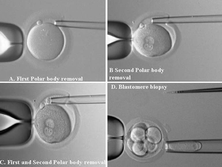
performed at the cleavage stage.34,35 The data demonstrate the clinical usefulness of the specific polar body or blastomere testing for X-linked disorders as an alternative to PGD by gender determination.

Couples with Homozygous Affected Partners

PGD was also provided for couples with one homozygous or double heterozygous-affected partner who were affected patients with thalassemia or phenylketonuria (PKU), resulting in an unaffected pregnancy and birth of healthy children.1,36 Although the risk for producing an affected child in such couples is as high as 50%, irrespective of maternal or paternal affected status, the strategy of PGD in such cases will depend on whether father or mother is affected. In couples with the affected fathers, PGD may concentrated on the preselection of mutation-free oocytes, while with the affected mothers, a cleavage stage PGD is required to identify those few embryos containing the normal gene.

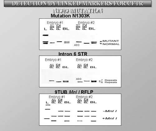
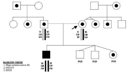
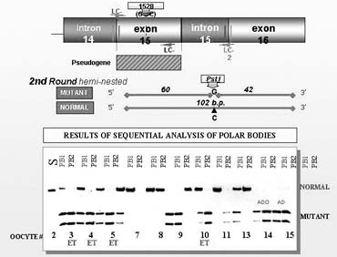
The testing is particularly complicated if the parents are carrying different mutations. In one such case performed for thalassemia, the affected mother was double heterozygous (IVS I-110; IVS I-1-6) while the male partner was heterozygous carrier of the third mutation (IVS II-745).1 This required a complex PGD design to exclude preferential amplification of each of the three alleles tested in blastomeres that underwent biopsy (Fig. 11), as also described in PGD for PKU.36 In this case, the affected father was compound heterozygous for R408 and Y414C mutations in exon 12 of phenylalanine hydroxylase (PAH) gene, and the carrier mother was heterozygous for R408W mutation in same exon. PGD strategy was based on the preselection of the mutation-free oocytes using a sequential PB1 and PB2 DNA analysis. Based on the multiplex heminested PCR analysis, four embryos resulting from the zygotes predicted to contain no mutant allele of PHA gene were transferred, yielding an unaffected twin pregnancy and birth of the healthy twins (Fig. 12).

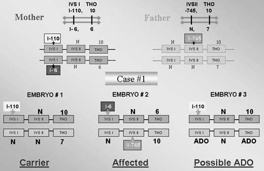
Fig. 11. PGD analysis for three different mutations in beta-globin gene in a couple with homozygous-affected female partner.

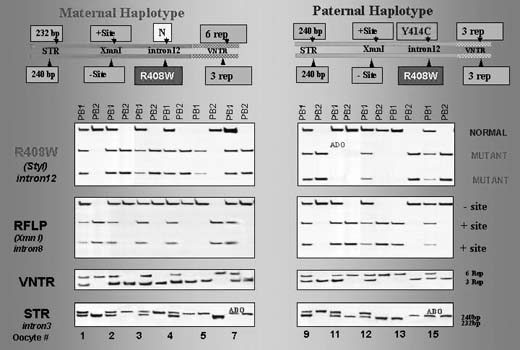
Fig. 12. PGD for R408W in phenylalanine hydroxylase gene.

With the improvement of treatment and improved life expectancy for increasing number of genetic disorders, an increasing number of couples with affected maternal or paternal partners may require PGD as the only means for having unaffected children of their own.

Cancer Predisposition

Cancer predisposition has not traditionally been considered as an indication for prenatal diagnosis, because this would lead to pregnancy termination, which is not justified on the basis of genetic predisposition alone. However, the possibility of choosing embryos free of genetic predisposition for transfer would obviate the need for considering pregnancy termination, because only potentially normal pregnancies are established. PGD for such conditions appears acceptable on ethical grounds because only a limited number of the embryos available from hyperstimulation are selected for transfer.

The first PGD for inherited cancer predisposition have been performed for couples carrying p53 tumor-suppressor gene mutations known to determine a strong predisposition to the majority of cancers.37 The couple was with the paternally derived missense mutation caused by a transversion of a G-to-A in exon 5 of the p53 tumor-suppressor gene. The carrier was a 38-year-old proband with Li-Fraumeni syndrome (LFS), diagnosed with rhabdomyosarcoma of the right shoulder at the age of 2 followed by right upper extremity amputation. At the age of 31, he was also diagnosed with a high-grade leiomyosarcoma of the bladder and underwent a radical cystoprostatectomy. His mother was diagnosed with leiomyosarcoma at age 37.

PGD was performed by blastomere biopsy and multiplex nested PCR analysis with simultaneous testing for p53 tumor-suppressor gene mutation and linked polymorphic markers, allowing preselecting and transferring back to the patient only mutation-free embryos (Fig. 13). A singleton pregnancy and birth of a mutation-free child resulted, and the child is currently healthy and free from the mutation predisposing to LFS.

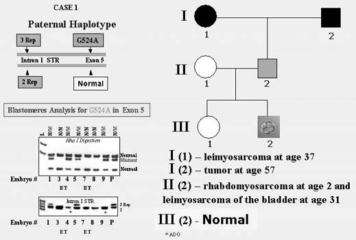
Fig. 13. PGD for p53 tumor-supressor gene mutations.

At present, PGD is also applied for other cancers, including familial adenomatous polyposis coli, Von Hippel Lindau syndrome, retinoblastoma, neurofibromatosis types I and II, and familial posterior fossa brain tumor.38 Overall, 20 PGD cycles were performed for 10 couples, resulting in preselection and transfer of 40 mutation-free embryos, which yielded five unaffected clinical pregnancies and four healthy children born by the present time. Despite the controversy of PGD use for late-onset disorders, the data demonstrate the usefulness of this approach as the only acceptable option for at-risk couples to avoid the birth of children with inherited predisposition to cancer and to have a healthy child.

Other Late-Onset Disorders With Genetic Predisposition

One of the first experiences of PGD for late-onset disorders was PGD for genetic predisposition to one of the forms of Alzheimer disease (AD)39 caused by an autosomal-dominant familial predisposition to a presenile form of dementia, determined by nearly completely penetrant autosomal-dominant mutation in the amyloid precursor protein (APP) gene, for which no treatment is available despite a possible predictive diagnosis. A 30-year-old women had no signs of AD but was a carrier of V717L mutation, resulting from G-to-C substitution in exon 17 of the APP gene. The predictive testing in the patient was performed because of the early-onset AD in her sister carrying this mutation in whom symptoms of AD developed at age 38. This sister is still alive, but her cognitive problems progressed to the point that she was placed in an assisted living facility. Her father had died at age 42 and also had a history of psychological difficulties and marked memory problems. V717L mutation was also detected in one of her brothers who experienced mild short-term memory problems as early as age 35, with a moderate decrease in memory, new learning, and sequential tracking over the next 2 to 3 years. The other family members, including one brother and two sisters, were asymptomatic, although predictive testing was performed only in sisters, who appeared to be free from mutation in APP gene (Fig. 14).

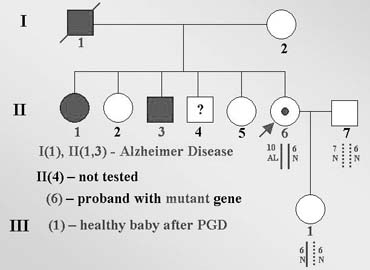
Fig. 14. Pedigree of family with early-onset Alzheimer disease determined by mutation V717L.

PGD was performed by DNA analysis of PB1 and PB2 to preselect and transfer back to the patient only the embryos resulting from mutation-free oocytes. Based on both mutation and the linked marker analysis, unaffected embryos resulting from mutation-free oocytes were preselected for transfer back to the patients, resulting in a singleton clinical pregnancy and birth of an unaffected mutation-free child.

PGD, therefore, provides a nontraditional option for patients who may wish to avoid the transmission of the mutant gene predisposing to the late-onset disorders in their potential children. Because such diseases present beyond early childhood or later and may not be expressed in 100% of the cases, the application of PGD for this group of disorders is still controversial. However, for diseases with no current prospect for treatment arising despite presymptomatic diagnosis and follow-up, PGD may be offered as the only relief for the at-risk couples.

Blood Group Incompatibility

The first PGD for maternal–fetal incompatibility resulting in the healthy pregnancy outcome was performed for Kell (K1) genotype, which is one of the major antigenic systems in human red blood cells, comparable in importance with RhD, causing maternofetal incompatibility leading to severe hemolytic disease of the newborn (HDN) in sensitized mothers.40 K1 allele is present in 9% of the populations, in contrast to its highly prevalent allelic variant K2. The gene is located on chromosome 7 (7q33), consisting of 19 exons with the only C-to-T base substitution in exon 6 in K1 compared with K2 antigen.

In cases of pregnancy by the K1 fetus in the K2 mother, antibodies to K1 may be developed, leading to maternofetal incompatibility causing severe HDN. Although prenatal diagnosis is available for identification of pregnancies at risk for HDN, this may not always prevent the potential complications for the fetus, such as stillbirth or neonatal death, making PGD a possible option for preventing both Kell and Rhesus hemolytic diseases.

PGD for Kell disease was performed for two at-risk couples with a history of neonatal death in previous pregnancies caused by HDN. The preselection and transfer of the embryos free from K1 allele of KEL gene was possible in each case, yielding a clinical pregnancy and the birth of healthy twins, confirmed to be free of K1 allele (Fig. 15).

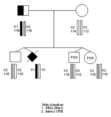
Fig. 15. PGD for Kell genotype (pedigree).

A number of attempts have also been undertaken to perform PGD for Rhesus disease, which, however, has not yet resulted in a clinical pregnancy.41 Both of these conditions are quite prevalent, taking into consideration the approximately 15% frequency for RhD and 9% for KEL antigen, presenting the risk for alloimmunization that may lead to HDN in some of the at-risk couples. Therefore, PGD may be a useful option for these couples to avoid the establishment of the RhD or K1 pregnancy in the sensitized mothers.

Although the at-risk pregnancies detected by prenatal diagnosis may be treated by an intrauterine transfusion, the potential complication for the fetus cannot be completely excluded even after this procedure. Pregnancy termination in such cases will also be unacceptable, because the antibodies to K1, for example, are developed only in 5% of persons obtaining incompatible blood. However, some of the at-risk couples have had such unfortunate experience with HDN, resulting in neonatal death as in both of our couples, that they regard PGD as their only option to plan another pregnancy. This makes PGD attractive for patients at risk for alloimmunization, although such conditions have rarely been an indication for prenatal diagnosis.

Congenital Malformations

Congenital malformations are highly prevalent (29.3/1000 live births) and are usually sporadic. However, with progress of the human genome project, an increasing number of inherited forms are being described, which therefore may be avoided through PGD. For example, sonic hedgehog (SHH) gene mutation, for which the first PGD has recently been performed,18 causes the failure of cerebral hemispheres to separate into distinct left and right halves and leads to holoprosencephaly (HPE), which is one of the most common developmental anomalies of forebrain and midface. Although most HPE are sporadic, familial cases are not rare, with clear autosomal-dominant inheritance.

A great intrafamilial clinical variability of HPE from alobar HPE and cyclopia, to cleft lip and palate, microcephaly, ocular hypertelorism, and even normal phenotype, suggests the interaction of SHH gene with other genes expressed during craniofacial development and the possible involvement of environmental factors. This may explain the fact that almost one third of carriers of SHH mutations may be clinically unaffected. Therefore, even in familial cases, the detection of SHH mutations in prenatal diagnosis might not justify pregnancy termination, making PGD a more attractive option for couples at risk for producing a progeny with HPE, as demonstrated in the first PGD for this mutation.18

This couple presented for PGD because of the previous two children with the clinical signs of HPE. One of them, a female with severe HPE and cleft lip and palate, died soon after birth. Both the child and the parents were chromosomally normal, but DNA analysis in the child's autopsy material demonstrated the presence of SHH nonsense mutation caused by GAG-to-TAG sequence change, leading to premature termination of the protein at position 256 (Glu256 → stop). The same mutation was found in their 5-year-old son who was born after a full-term normal pregnancy. This child has less severe facial dysmorphisms, which included microcephaly, Rathke's pouch cyst, single central incisor, and choanal stenosis. There was also clinodactyly of the fifth fingers and curved-in fourth toes bilaterally. The child's growth was slow in the first 2 years, but thereafter he has been maintaining a reasonably good growth (Fig. 16).

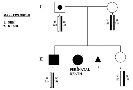
Fig. 16. PGD for sonic hedgehog (SHH) mutation: family pedigree.

PGD was performed by blastomere biopsy and multiplex nested PCR analysis involving specific mutation testing simultaneously with linked marker analysis (Table 3). Of nine tested embryos, four embryos were free of mutant gene, from which two were transferred back to patient, resulting in a singleton pregnancy and birth of a healthy child after confirmation of the mutation-free status by amniocentesis (see Fig. 16). Similar approach was used for PGD of Crouson syndrome.42

The data suggest the clinical usefulness of PGD for familial cases of congenital malformations. Because of high prevalence of congenital anomalies, the approach may have practical implications for the at-risk couples as a preventive measure to be used in genetic practices.

Preimplantation HLA Matching Combined With PGD

Preimplantation HLA-matching was first introduced in combination with mutation analysis for Fanconi anemia with the objective of establishing an unaffected pregnancy yielding a potential donor progeny for transplantation in an affected sibling.43 This resulted in a clinical pregnancy and birth of an unaffected child whose cord blood was transplanted to the affected sibling, thus saving her life (Fig. 17).

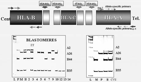
Fig. 17. HLA detection system by allele-specific primers in case of PGD for Fanconi anemia.

The strategy would not likely be clinically acceptable through traditional prenatal genetic diagnosis because of a possible clinical pregnancy termination after HLA-matching. However, PGD for such purpose should be acceptable, because only a limited number of the embryos are usually preselected for transfer, which in this case will represent unaffected embryos with a perfect match for affected siblings in need of transplantation. Because the multiplex single cell PCR used in PGD presents the opportunity for combined PGD and HLA testing, it has become a useful way to preselect an embryo that may be an HLA-match to the affected sibling requiring stem cell transplantation.

The method has currently been applied for the HLA-genotyping in two dozen cycles in combination with PGD for thalassemia, Fanconi anemia, hyper immunoglobulin M syndrome, X-linked adrenoleukodystrophy, and Wiskott-Aldrich syndrome, confirming the usefulness of preimplantation HLA-matching as part of PGD, with prospect of application of this approach to other inherited conditions also requiring an HLA-compatible donor for bone marrow transplantation (Verlinsky et al, unpublished data). This provides a realistic option for the couples desiring to avoid the birth of an affected child, together with the establishment of a healthy pregnancy, potentially providing an HLA-matched progeny for treatment of an affected sibling.

Back to Top
CONCLUSION

Important ethical issues have recently been raised with an increasing use of PGD for late-onset disorders with genetic predisposition and preimplantation HLA-typing to produce an HLA-compatible donor for treating a family member with fatal bone marrow disease or cancer requiring stem cell transplantation.44,45,46,47 Although there is no actual difference in the application of PGD for the latter conditions, the controversy can be explained by the fact that in traditional prenatal diagnosis, if the fetus would be found to carry the gene predisposing to late-onset disorder or to be HLA-unmatched, then the couple would have to make an extremely difficult decision of pregnancy termination, which could not be justified by such findings. Alternatively, PGD technology allows genetic testing of human eggs and embryos before pregnancy is established, making it realistic to establish only HLA-matched or potentially normal pregnancies without genetic predisposition to late-onset disorders.

In any case, as seen from this review, PGD is now becoming an established clinical option in reproductive medicine and is applied using separate consent forms and the research protocols approved by Institutional Ethics Committees. The number of apparently healthy children born after PGD has passed its first 1000, showing that there is no evidence of any incurred adverse effect. However, these protocols would still require confirmatory CVS or amniocentesis and a follow-up monitoring of its safety and accuracy. Although PGD will help solve some of the long-standing ethical problems, such as the abortion issue (which will be avoided as a result of this new approach), others could become a serious obstacle, particularly those related to designer babies. These considerations are highly relevant to the subject of PGD, as well as to any other new methods as we proceed with further development of appropriate technology for controlling genetic disability.

Back to Top
REFERENCES

1. Verlinsky Y, Kuliev A: Atlas of Preimplantation Genetic Diagnosis. London, NY, Parthenon Publishing Group, 2000

2. Verlinsky Y, Cohen J, Munne S et al: The first thousand babies born after preimplantation genetic diagnosis. Fertil Steril (in press)

3. International Working Group on Preimplantation Genetics (IWGPG): Preimplantation Genetic Diagnosis--Experience of Three Thousand Clinical Cycles. Report of the 11th Annual Meeting International Working Group on Preimplantation Genetics, in conjunction with 10th International Congress of Human Genetics, Vienna, May 15, 2001 Reprod BioMed Online 3:49-53, 2001

4. Kuliev A, Verlinsky Y: Current features of preimplantation genetic diagnosis. Reprod BioMed Online 5:296-301, 2002

5. ESHRE Preimplantation Genetic Diagnosis Consortium: Data Collection III, May 2002. Hum Reprod 17:233-246, 2002

6. Rechitsky S, Verlinsky O, Amet T et al: Reliability of preimplantation diagnosis for single gene disorders. Mol Cell Endocrinol 183:S65-S68, 2001

7. International Working Group on Preimplantation Genetics (IWGPG): Tenth Anniversary of Preimplantation Genetic Diagnosis: Report of the 10th Annual Meeting International Working Group on Preimlantation Genetics, in association with 3rd International Symposium on Preimplantation Genetics, Bologna, Italy, June 23, 2000. J Assist Reprod Genet 18:66-72, 2001

8. Rechitsky S, Strom C, Verlinsky O et al: Allele drop out in polar bodies and blastomeres. J Assist Reprod Genet 15:253-257, 1998

9. Rechitsky S, Verlinsky O, Amet T et al: Reliability of preimplantation diagnosis for single gene disorders. Mol Cell Endocrinol 183:S65-S68, 2001

10. Verlinsky Y, Rechitsky S, Verlinsky O et al: Polar body based preimplantation diagnosis for X-linked genetic disorders. Reprod BioMed Online 4:38-42, 2002

11. Kuliev A, Rechitsky S, Verlinsky O et al: Birth of healthy children after reimplantation diagnosis of thalassemias. J Assist Reprod Gent 16:207-211, 1999

12. Rechitsky S, Verlinsky O, Kuliev A et al: Preimplantation genetic diagnosis for familial dysautonomia. Reprod BioMed Online 6:488-496, 2003

13. Sermon K, Seneca S, De Rycke M et al: PGD in the lab for triplet diseases-myotonic dystrophy, Huntington's disease and Fragile-X syndrome. Mol Cell Endocrinol 183:S77-S85, 2001

14. Katz MG, Mansfield J, Gras L et al: Diagnosis of trisomy 21 in preimplantation embryos by single-cell DNA fingerprinting. Reprod BioMed Online 4:43-50, 2002

15. Harper J, Wells D, Piyamongkol W et al: Preimplantation genetic diagnosis for single gene disorders: experience with five single gene disorders. Prenat Diagn 22:525-533, 2002

16. Ray PF, Frydman N, Attie T et al: Birth of healthy twins after preimplantation diagnosis of Cystic fibrosis combined with gender determination. Mol Hum Reprod 8:688-694, 2002

17. Verlinsky Y, Rechitsky S, Verlinsky O et al: Preimplantation diagnosis for long-chain 3-hydroxyacyl-coa dehydrogenase deficiency. Reprod BioMed Online 2:17-19, 2001

18. Verlinsky Y, Rechitsky S, Verlinsky O et al: Preimplantation Diagnosis for Sonic Hedgehog Mutation Causing Familial Holoprosencephaly. N Engl J Med 348:1449-1454, 2003

19. Handyside AH, Lesko JG, Tarin JJ et al: Birth of a normal girl after in vitro fertilization and preimplantation diagnosis testing for cystic fibrosis. N Engl J Med 327:905-909, 1992

20. Sermon K, Lissens W, Joris et al: Clinical application of preimplantation diagnosis for miotonic dystrophy. Prenat Diagn 17:925-932, 1997

21. Sermon K, Lissens W, Messiaen L et al: Preimplantation genetic diagnosis of Marfan syndrome with the use of fluorescent polymerase chain reaction and automated lazer fluorescence DNA sequencer. Fertil Steril 71:163-166, 1999

22. Sermon K, Henderix P, Lissens W et al: Preimplantation genetic diagnosis for medium-chain acyl-CoA dehydrogenase (MCAD) deficiency. Mol Hum Reprod 16:1165-1168, 2000

23. Kuliev A, Rechitsky S, Verlinsky O et al: Preimplantation diagnosis of thalassemias. J Assist Reprod Genet 15:219-225, 1998

24. Xu K, Shi ZM, Veeck LL et al: First unaffected pregnancy using preimplantation genetic diagnosis for sickle cell disease. JAMA 281:1701-176, 1999

25. Kanavakis E, Vrettou C, Palmer G et al: Preimplantation genetic diagnosis in 10 couples at risk for transmitting beta-thalassemia major: Clinical experience including the initiation of six singleton pregnancies. Prenat Diagn 19:1217-1222, 1999

26. De Vos A, Sermon K, Van de Velde H et al: Two pregnancies after preimplantation genetic diagnosis of osteogenesis imperfecta type I and type IV. Hum Genet 106:605-613, 2000

27. Goossens V, Sermon K, Lissens W et al: Clinical application of preimplantation genetic diagnosis for cystic fibrosis. Prenat Diagn 20:571-581, 2000

28. Daniels G, Pettigrew R, Thornhill A et al: Six unaffected livebirths following preimplantation diagnosis for spinal muscular athrophy. Molec Hum Reprod 7:995-1000, 2001

29. Georgiou I, Sermon K, Lissens W et al: Preimplantation genetic diagnsis for spinal and bulbar muscular atrophy (SBMA). Hum Genet 108:494-498, 2001

30. Carvalho F, Sousa M, Fernandes S et al: Preimplantation genetic diagnosis for familial amyloidotic polyneuropathy (FAP). Pren Diagn 21:1093-1099

31. Stern HJ, Harton GL, Sisson ME et al: Non-disclosing preimplantation genetic diagnosis for Huntington disease. Prenat Diagn 22:503-507, 2002

32. Hellani A, Lauge A, Ozand P et al: Pregancy after preimplantatio genetic diagnosis for Ataxia Telangeectasia. Mol Hum Reprod 8:785-788, 2002

33. Verlinsky Y, Rechitsky S, Verlinsky O et al: Preimplantation diagnosis for ornithine transcarbamilase deficiency. Reprod BioMed Online 1:45-47, 2000

34. Ray P, Harper J, Ao A et al: Successful preimplantation genetic diagnosis for sex linked Lesch-Nyhan syndrome using specific diagnosis. Prenat Diagn 19:1237-1241, 1999

35. Ray PF, Gigarel N, Bonnefont JP et al: First specific preimplantation genetic diagnosis for ornithine transcarbamylase deficiency. Prenat Diagn 20:1048-1054, 2000

36. Verlinsky Y, Rechitsky S, Verlinsky O et al: Preimplantation diagnosis for PKU. Fertil Steril 76:346-349, 2001

37. Verlinsky Y, Rechitsky S, Verlinsky O et al: Preimplantation diagnosis for p53 tumor suppressor gene mutations. Reprod BioMed Online 2:102-105, 2001

38. Rechitsky S, Verlinsky O, Chistokina A et al: Preimplantation genetic diagnosis for cancer predisposition. Reprod BioMed Online 5:148-155, 2002

39. Verlinsky Y, Rechitsky S, Verlinsky O et al: Preimplantation diagnosis for early onset Alzheimer disease Caused by V717L Mutation. J Am Med Assoc 287:1018-1021, 2002

40. Verlinsky Y, Rechitsky S, Verlinsky O et al: Preimplantation diagnosis for Kell genotype. Fertil Steril 80:1047-1051, 2003

41. Van Den Veyver IB: Prenatal and Preimplantation Genetic Diagnosis for RhD Alloimmunization, pp 181–185. 10th International Conference on Prenatal Diagnosis and Therapy. Barcelona (Spain), June 19–21, 2000. Barcelona, University of Barcelona, 2000

42. Abou-Sleiman PM, Apessos A, Harper JC et al: Pregnancy following preimplantation genetic diagnosis for Crouson syndrome. Mol Hum Reprod 8:304-309, 2002

43. Verlinsky Y, Rechitsky S, Verlinsky O et al: Preimplantation diagnosis for Fanconi anemia combined with HLA matching. JAMA 285:3130-3133, 2001

44. Damewood MD: Ethical implications of a new application of preimplantation diagnosis. JAMA 285:3143-3144, 2001

45. Towner D, Loewy RS: Ethics of preimplantation diagnosis for a woman destined to develop early-onset Alzheimer disease. JAMA 287:1038-1040, 2002

46. Edwards RG: Social and ethical issues of PGD, cloning and gene therapy. Reprod BioMed Online 6:170-180, 2003

47. Robertson JA: Extending preimplantation genetic diagnosis: the ethical debate. Hum Reprod 18:465-471, 2003

Back to Top