Chapter 59 Infertility Aspects of Endometriosis Robert S. Schenken Table Of Contents |
|
Robert S. Schenken, MD |
INTRODUCTION |
Endometriosis is the presence of tissue that structurally and functionally resembles endometrial glands or stroma outside the uterine cavity. The structures most commonly affected by endometriosis include the ovaries, uterosacral ligaments, pelvic peritoneum, rectovaginal septum, and lower genital tract. Several theories have been proposed to explain the histogenesis of endometriosis.1 The implantation theory proposes that endometrial tissue from the uterus is shed during menstruation and transported through the fallopian tubes, where it gains access to and implants upon pelvic structures. The direct transplantation theory is the probable explanation for endometriosis that develops in episiotomy, cesarean section, and other scars following surgery. Endometriosis in locations outside the pelvis is explained by dissemination of endometrial cells or tissue through lymphatics and blood vessels. The coelomic metaplasia theory proposes that the coelomic (peritoneal) cavity contains undifferentiated cells or cells capable of dedifferentiating into endometrial tissue. This theory is based on embryologic studies demonstrating that all pelvic organs, including the endometrium, are derived from the cells lining the coelomic cavity. Retrograde menstruation, altered immunity, and genetics may play causative roles in the development of endometriosis. Anatomic alternations of the pelvis that increase tubal reflux of menstrual endometrium may increase a woman's chance of developing endometriosis. An increased incidence of endometriosis is found in girls with genital tract obstructions that prevent expulsion of menses into the vagina and increase the likelihood of tubal reflux.2 The incidence of retrograde menstruation is similar in women with and without endometriosis. Consequently, the development of endometriosis could depend on the quantity of endometrial tissue reaching the peritoneal cavity, the capacity of a woman's immune system to eliminate the refluxed menstrual debris, and/or the inherent ability of the endometrium cell to adhere to the peritoneum or ovary. Women with endometriosis typically present with one of three problems: an adnexal mass, pelvic pain, or infertility. Surgery for adnexal masses is generally indicated to remove the endometrioma and/or other pathologic pelvic conditions. Pelvic pain from endometriosis often can be treated successfully with surgery or medical therapy, but treatment of endometriosis in the female partner of an infertile couple raises a large number of complex clinical questions that do not have simple answers. There are few infertility problems requiring greater clinical skills than those required to plan therapy for an infertile woman with endometriosis. |
FECUNDITY IN INFERTILE WOMEN WITH ENDOMETRIOSIS | ||||||||||||||||||
In normal couples, the monthly fecundity (f) is in the range of 0.15 to 0.20.3 In most studies of women with endometriosis and infertility who have not received treatment, the monthly fecundity is in the range of 0.02 to 0.1 (Table 1).4,5,6,7,8 Some women with endometriosis and infertility have a markedly reduced monthly fecundity. Early studies suggested that 25% to 50% of infertile women have endometriosis and that 30% to 50% of women with endometriosis are infertile.9,10 These initial estimates were based on the incidence of histologically documented endometriosis in women undergoing laparotomy for gynecologic procedures in referral medical centers. Recent studies in women undergoing laparoscopy have strengthened the epidemiologic association with infertility. A prospective study of infertile women and women undergoing tubal ligation demonstrated a higher incidence of endometriosis in infertile women (48%) compared with fertile women (5%).11 Other studies have confirmed that infertile women are 10 to 20 times more likely to have endometriosis than fertile women.12,13 The limitations of the above retrospective studies should be noted. These observations raise many questions. For example, in women with endometriosis and infertility, the low fecundity might be from factors other than endometriosis.
|
ENDOMETRIOSIS AND INFERTILITY: CAUSE AND EFFECT |
The hypothesis that endometriosis causes infertility or a decrease in fecundity in humans remains controversial. To address a question of proof of causation, Jansen14 reviewed the pregnancy rate in patients undergoing artificial insemination by donor. Seven patients had untreated minimal endometriosis, and the remaining women had normal pelvic findings at laparoscopy. A fecundity rate of 12.0% was reported in patients with a normal pelvis compared with 3.6% in patients with endometriosis. The results of this prospective study were inconsistent, with two retrospective studies demonstrating expected fecundity rates in women with minimal endometriosis undergoing donor insemination.15,16 If endometriosis does cause infertility, then eradication of the disease process should improve pregnancy rates. One retrospective study and one quasi-randomized study suggest that surgery improves fecundity.17,18 Two randomized studies have assessed pregnancy rates following surgical treatment or expectant management. The Canadian Collaborative Group of Endometriosis reported a randomized trial of laparoscopy with and without treatment in 341 women with minimal or mild disease. The fecundity rate in treated patients was 4.7 versus 2.4 per 100 person-months in controls (95% CI 1.2 to 3.1).19 However, the Gruppo Italiano per lo Studio dell' Endometriosi conducted a similar study in 111 patients with stage I or II endometriosis. One year after surgery, the pregnancy rate was 29% in the no treatment group and 24% in the ablation/resection group. These conflicting studies may be attributed to different patient population or responses of individual patients to therapy. They do indicate that the pathophysiology of infertility in patients with endometriosis remains enigmatic.20 Experiments on rabbits, rats, and monkeys, however, strongly suggest that surgically induced endometriosis causes a decrease in fecundity in all three of these species.21,22,23,24 In most of these animal studies, endometriosis is surgically induced by suturing small squares of endometrium on pelvic peritoneal surfaces or bowel mesentery. For example, Schenken and Asch removed the right uterine horn from New Zealand white rabbits, minced the uterine horn into squares measuring 2 × 5 × 5 mm, and sutured the squares onto pelvic peritoneal surfaces.21 Control animals had the right uterine horn removed but had 2 × 5 × 5-mm square of adipose tissue sutured onto their peritoneal surfaces. A second laparotomy was performed 25 days after the initial surgery. All the uterine horn grafts survived transplantation and developed into vascularized hemorrhagic solid masses or multioculated cystic masses. Five of eight animals in the experimental group developed adhesions from bowel to the implants. The adipose tissue transplants were well vascularized and viable. There was no cyst formation in these implants. One of eight animals in the control group developed adhesions. Fourteen days later, the animals received human chorionic gonadotropin and were inseminated with rabbit semen from bucks with proven fertility. Six of eight control animals became pregnant. Only two of eight animals with endometriosis became pregnant. Hahn and co-workers observed similar results.22 They also noted an inverse relationship between the severity of the endometriosis and the fecundity: The number of implanted embryos decreased as the total weight of the endometriotic implants increased. Vernon and Wilson transplanted uterine squares or adipose tissue to the bowel mesentery of Sprague-Dawley rats.23 The transplanted uterine squares developed into vascular cysts that measured 5 mm in diameter and contained clear fluid. Moderate to severe adhesions were present in all animals with surgically induced endometriosis. Approximately 20% of the sham-operated animals (controls) developed adhesions. Twenty-five rats with autotransplanted uterine square and 15 controls were mated. In the controls, the average number of day 14 embryos was 11.3; in the animals with endometriosis, the average number of day 14 embryos was 8. In another group of experimental animals, the average number of live-born pups was 9 for the controls versus 4.5 for the rats with endometriosis. Interestingly, pregnancy appeared to induce regression of the endometriotic lesions. Schenken and colleagues examined the effects of surgically induced endometriosis on fertility in monkeys.24 Animals with surgically induced endometriosis and control animals with adipose tissue autografts were mated. Pregnancy rates were similar between control animals (42%) and animals with mild endometriosis (35%). Animals with moderate and severe endometriosis, however, had reduced pregnancy rates (12%). Pelvic adhesions appeared to account, in part, for the reduction in fertility. Animals that had endometriosis and ovulated from an ovary with adhesions had a 0% per cycle pregnancy rate. Animals that had endometriosis and ovulated from an adnexa without adhesions had a 33% per cycle pregnancy rate. In addition, some of the animals with endometriosis appeared to have luteinized unruptured follicles, which accounted for a part of the reduction in fertility. These studies strongly suggest that the experimental induction of endometriosis reduces fecundity. The experimental observations in monkeys may be especially relevant to improving our understanding of endometriosis in humans. D'Hooghe and colleagues assessed cycle pregnancy rates in baboons with spontaneous (n = 16) or induced (n = 21) endometriosis.25 There were 9 with minimal, 14 with mild, and 7 each with moderate and severe disease. All had histologically proven disease, whereas all control animals (n = 34) had a normal pelvis at laparoscopy. The cycle pregnancy rates were 24%, 10%, 8%, and 13% in women with minimal, mild, moderate, and severe disease, respectively. The cycle pregnancy rates were significantly lower in baboons with stage III–IV endometriosis (9%, 6/64) and with stage II disease (10%, 7/73) than in animals with stage I endometriosis (24%, 11/45) or in controls (19%). The normal pregnancy rate in baboons with stage I disease and decreased rate in advanced disease suggests that fertility declines with increasing stages of endometriosis. Although the animal data support the hypothesis that endometriosis can cause infertility, the situation in humans is more complex and controversial. A major problem is that infertility is a clinical problem of couples, and many couples have multiple factors that decrease fecundity. The complexity of infertility in the clinical situation often makes it difficult to identify a single causative factor. In addition, our understanding of the causes of infertility is incomplete. It is possible that in infertile couples who appear to have a decrease in fecundity from endometriosis, a defect in endometrial or oocyte function may actually be present. |
BIOLOGIC MECHANISMS THAT MAY LINK ENDOMETRIOSIS AND INFERTILITY |
Several mechanisms have been proposed to clarify the association between endometriosis and infertility: Distorted pelvic anatomy None of these mechanisms has been proved to cause decreased fecundity in women. These mechanisms are briefly discussed below. Distorted Pelvic Anatomy Major pelvic adhesions resulting from any disease can impair egg release from the ovary, block sperm entry into the distal fallopian tube, and inhibit ovum pickup. In animal models of endometriosis, pelvic adhesions appear to contribute to the observed decreased fecundity noted in animals with advanced endometriosis.24 Altered Peritoneal Function Many studies demonstrate that women with endometriosis have an increased volume of peritoneal fluid, increased macrophage concentration and function, and increased peritoneal fluid concentrations of prostaglandin, interleukin-l, tumor necrosis factor, and proteases. These alterations may impair oocyte, sperm, embryo, and fallopian tube function.26,27,28,29 Altered Systemic Immune Function IgG and IgA antibodies and lymphocytes may be increased in the endometrium of women with endometriosis. These abnormalities may alter endometrial receptivity to embryo implantation.30,31 Endocrine and Ovulatory Abnormalities Numerous endocrine and ovulatory disorders may be present in women with endometriosis, including the luteinized unruptured follicle syndrome, luteal phase dysfunction, abnormal follicular growth, and premature and multiple luteinizing hormone (LH) surges.32,33,34 In a monkey model, advanced endometriosis appeared to cause a decrease in fecundity, which was attributed partly to luteinized unruptured follicle syndrome and partly to luteal phase defects.24 Abnormal Tubal Function Peritoneal fluid from women with endometriosis reportedly contains an ovum capture inhibitor that prevents normal cumulus-fimbria interaction.35 Ovum capture inhibitor, a protein with a molecular mass of greater than 100,000 daltons, remains to be purified. Abnormal Fertilization and Implantation In women with endometriosis, it is not clear whether abnormal fertilization and implantation contribute to decreased fecundity. In a rabbit model of endometriosis, Hahn and colleagues reported that blastocysts failed to implant normally in rabbits with endometriosis.36 Lessey reported that some women with endometriosis lack endometrial αvβ3 integrin expression.37 They hypothesized that decreased endometrial integrin production in the mid-luteal phase causes infertility by impairing implantation. These observations support the concept that endometriosis may represent one component of a disease that is characterized by dysfunction in multiple components of the müllerian tract, including the cervix, endometrium, fallopian tubes, and peritoneum. |
DIAGNOSIS AND STAGING |
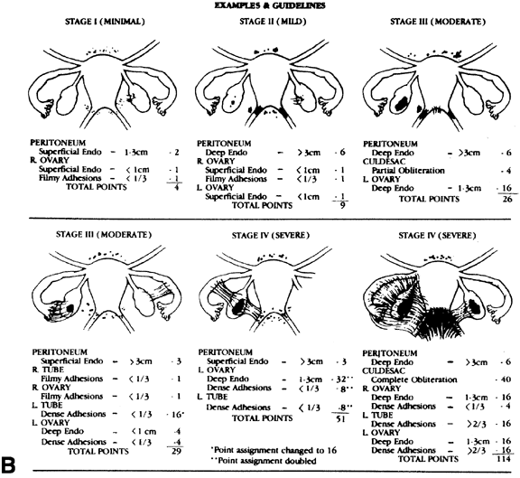
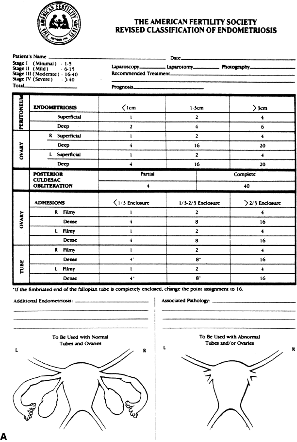
The current clinical opinion is that a surgical procedure such as laparoscopy or laparotomy is required for definitive diagnosis of endometriosis. Given this state of clinical practice, an important question is when to laparoscope a patient to determine whether endometriosis is present. A history and physical examination can yield a number of significant historical findings: Pertinent Historical Finding Secondary dysmenorrhea Pertinent Physical Findings Nodular uterosacral ligaments Endometriosis is a heterogenous disease spanning a spectrum from a single 1-mm peritoneal implant to 10-cm endometriomas with cul-de-sac obliteration. Consequently, a clinical staging system is absolutely necessary to allow clinicians to communicate effectively with regard to prognosis and treatment. The revised American Fertility Society (rAFS) system currently is the most widely accepted staging system (Fig. 1).38 In this system, points are assigned for severity of endometriosis based on the size and depth of the implant and the severity of the adhesions. Unfortunately, the staging system does not correlate well with a woman's chance for conception following therapy. It is unlikely that any accurate staging system will be introduced until we have a better understanding of the pathophysiology of endometriosis-associated infertility. |
PHARMACEUTICAL THERAPY FOR ENDOMETRIOSIS | |||||||||||||||||||||||||||||||||||||||||||||||||||||||||||||||||||||||||||||||||
Pharmaceutical therapeutic options for endometriosis continue to expand rapidly. Six major options are currently available: danazol, nafarelin, leuprolide, goserelin, progestins, and combined estrogen-progestin therapy. The efficacy of these agents in the treatment of endometriosis is attributed to the steroid responsiveness of endometriosis implants. The majority of human endometriosis implants contain estrogen receptors, androgen receptors, and progesterone receptors.39,40 Clinical and laboratory observations demonstrate that estrogen is critical to the growth and function of endometriotic implants.41 Clinical observations suggesting that estrogen is critical to the growth of endometriosis include the findings that endometriosis rarely occurs before menarche; menopause, either surgical or natural, usually produces an improvement in preexisting endometriosis; and new cases of endometriosis occur very rarely after menopause unless exogenous estrogen is administered. As discussed below, there is no evidence that medical treatment of endometriosis in infertile women improves fecundity. A meta-analysis by Hughes and colleagues included 7 studies comparing medical therapy to no treatment or placebo.42 The common odds ratio for pregnancy following medical therapy was 0.85 (95% CI, 0.95 to 1.22). However, hormonal treatment is extremely effective in reducing pelvic pain associated with endometriosis. For infertile women with endometriosis accompanied by dyspareunia, dyschezia, dysmenorrhea, or nonspecific pelvic pain, hormonal treatment may improve sexual function as well as the frequency of sexual relations, thereby increasing fecundity. In addition, hormone therapy may be of value in the postoperative treatment of infertile women with endometriosis considering in vitro fertilization/embryo transfer(IVF/ET).43,44 Danazol is an isoxazole derivative of ethinyl testosterone (Fig. 2). The pharmacology of danazol is best understood by examining its interaction with intracellular steroid receptors, circulating steroid-binding globulins, and enzymes of ovarian steroidogenesis.
Danazol binds to three classes of intracellular steroid receptors: the androgen receptor, the progestin receptor, and the estrogen receptor45 (Table 2). Danazol has high affinity for the androgen receptor, moderate affinity for the progestin receptor, and poor affinity for the estrogen receptor.46 Danazol is both an androgen and estrogen agonist. Although danazol is an estrogen agonist, the affinity of danazol for the estrogen receptor is so poor that for clinical purposes danazol has no major effect on the estrogen receptor system. In contrast, because danazol is an androgen agonist with a very high affinity for the androgen receptor, danazol has potent androgenic effects in vivo. Many of danazol's side effects can be attributed to its androgenic properties. These side effects include the following: (1) a decrease in high-density lipoprotein cholesterol; (2) an increase in low-density lipoprotein cholesterol; (3) a decrease in hepatic production of sex hormone-binding globulin (SHBG) and thyroxine-binding globulin; and (4) an increase in bone density. The effects of danazol on the intracellular progestin receptor are complex. In some model systems, danazol can produce atypical secretory changes in estrogen-primed endometrium, suggesting that danazol is a progestin agonist.47 In other model systems, danazol blocks the effects of progesterone, suggesting that danazol is a progestin antagonist. Danazol is best regarded as a mixed progestin agonist-antagonist. Table 2. Danazol Interaction with Intracellular Androgen, Progestin, and
Estrogen Receptors and Circulating Binding Globulin
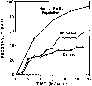
CBG, corticosteroid-binding globulin; SHBG, sex hormone-binding globulin. Danazol has high affinity for SHBG and can displace testosterone and dihydrotestosterone from SHBG.48 The observation that danazol increases free and bioavailable testosterone suggests that part of its androgenic effects may be from the displacement of testosterone and dihydrotestosterone from SHBG. Danazol inhibits multiple enzymes of ovarian steroidogenesis, including cholesterol side-chain cleavage enzyme, 3β-hydroxysteroid dehydrogenase-isomerase, 17-ketosteroid reductase, 17α-hydroxylase, 17,20-1yase, and aromatase.49 Experiments using monkey models and human subjects suggest that danazol inhibits ovarian estrogen production by a direct effect on ovarian steroidogenesis.50,51 The pharmacologic properties of danazol create an acyclic, high-androgen, low-estrogen environment that is hostile to the growth of endometriotic tissue. Danazol produces a high-androgen environment because it is inherently androgenic and because it decreases the hepatic production of SHBG and displaces testosterone from SHBG, resulting in an increase in free testosterone. By direct actions on the ovaries and the hypothalamic-pituitary axis, danazol produces a low-estrogen environment by inhibiting follicular growth. Acting through hypothalamic and pituitary androgen and progestin receptors, danazol decreases LH and FSH secretion. By inhibiting multiple enzymes of ovarian steroidogenesis, danazol decreases ovarian estrogen production. The low-estrogen, high-androgen environment created by danazol is hostile to the growth of endometriotic tissue. In addition, the acyclic endocrine environment produced by danazol results in amenorrhea and prevents the reseeding of the peritoneum with new implants of endometrium. The major side effects of danazol are weight gain, bloating, decreased breast size, ache, oily skin, hirsutism, headache, deepening of the voice, hot flashes, and muscle cramps.52 In women receiving 800 mg/day of danazol, the weight gain averages 4 kg. Approximately 80% of women receiving danazol will report one or more of the above side effects. Danazol can produce a large number of biochemical abnormalities including mild increases in serum levels of creatine phosphokinase, lactate dehydrogenase, glutamic-oxaloacetic transaminase, and glutamic-pyruvic transaminase. These typically are minor, clinically insignificant alterations. Danazol therapy produces marked alterations, however, in the circulating lipid profile. At doses of 600 mg/day, danazol produces a 50% decrease in high-density lipoprotein cholesterol and a 40% increase in low-density lipoprotein cholesterol. These lipid alterations resolve within 3 months of discontinuation of danazol therapy. The clinical impact of this atherogenic lipid profile in women of reproductive age is unclear. Seibel and co-workers and Bayer and colleagues conducted prospective, controlled, randomized studies of the effectiveness of danazol in the treatment of infertility associated with minimal endometriosis.8,53 Couples who had 1 year of infertility were eligible for entry into the study after they completed a basic infertility evaluation, which included semen analysis, basal body temperature chart, postcoital test, hysterosalpingogram, and endometrial biopsy. Patients (n = 73) then underwent a diagnostic laparoscopy and tubal lavage. If Kistner stage I endometriosis was observed, the female patients were randomized to receive danazol (n = 37) or no treatment (n = 36).54 Women who were randomized to the danazol treatment group received an initial danazol dosage that tapered off incrementally after the first 2 months, as follows:danazol 800 mg/day for 2 months, 600 mg/day for 2 months, and 400 mg/day for 2 months. Patients were then observed for 12 months, beginning either immediately after laparoscopy in the untreated group or after completion of danazol therapy in the treated group. During the 12 months of follow-up, 35% of the danazol-treated women and 47% of the untreated group became pregnant; this difference was not statistically significant (Fig. 3). In conclusion, treatment of women with mild endometriosis with danazol did not improve fecundity. In a randomized study comparing gestrinone with placebo, Thomas and Cooke observed similar results.55
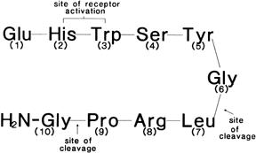
Gonadotropin-Releasing Hormone Analogs: Nafarelin, Leuprolide, Goserelin Gonadotropin-releasing hormone (GnRH) is a hypothalamic decapeptide (Fig. 4) that controls the pituitary secretion of LH and FSH. In the follicular phase of the menstrual cycle, the hypothalamus releases one pulse of GnRH per hour into the portal circulation. The ability of GnRH to stimulate LH and FSH secretion is critically dependent on the frequency and amplitude of the GnRH pulse. If the GnRH pulse frequency is chronically slow or too fast, the pituitary will not secrete LH and FSH in response to the GnRH pulse. A continuous infusion of GnRH, through molecular mechanisms that are poorly understood, markedly decreases pituitary production of LH and FSH. Because much of the information in the GnRH signal is contained in the pulse frequency, GnRH has a very short half-life (4 minutes). GnRH is metabolized by endopeptidases present in the pituitary circulation and pituitary gonadotropes, which cleave the 5–6 and 6–7 peptide bonds. By chemically altering the sixth amino acid, GnRH analogs that are resistant to cleavage by endopeptidases can be produced. These GnRH analogs have a long half-life (2 to 4 hours) and are perceived by the pituitary as a continuous infusion of GnRH. Consequently, chronic administration of these GnRH analogs decreases LH and FSH secretion and causes a cessation of ovarian estrogen production. GnRH analogs currently available include: leuprolide, nafarelin, and goserelin. Leuprolide is administered as a monthly depot injection. Nafarelin is administered in the form of a nasal spray and goserelin as a monthly subcutaneous implant. The doses of nafarelin, leuprolide, and goserelin necessary to reduce circulating estradiol to 15 pg/mL or 30 pg/mL are listed in Table 3. Table 3. Dose Effects of Leuprolide, Nafarelin, and Goserelin
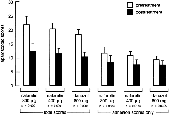
In a large randomized clinical trial comparing the efficacy of danazol and nafarelin in the treatment of endometriosis, Henzl and co-workers observed that both drugs produced symptomatic improvement in 80% of subjects and noted a 43% improvement in the rAFS endometriosis score56 (Fig. 5). Most of the side effects associated with nafarelin therapy (e.g., hot flashes, headaches, mood changes, decreased libido, vaginal dryness) were attributed to hypoestrogenism. Recent observations indicate that GnRH analogs can produce a decrease in vertebral bone density. For example, Dawood and colleagues reported that 6 months of GnRH analog therapy with buserelin (1200 μg/day intranasally) resulted in a 7% decrease in trabecular bone density.57 Six months after completing GnRH agonist therapy, the trabecular bone density remained 4% below pretreatment values. Similar losses in trabecular bone density during GnRH agonist therapy have been reported with the use of nafarelin and buserelin.58,59 Interestingly, Matta and associates reported that the bone loss was completely reversible once the GnRH analog was discontinued.59
In a recently reported controlled trial, Fedele and associates randomized 71 women with rAFS stage I or II endometriosis either to receive hormonal treatment with the GnRH agonist analog buserelin (400 μg 3 times a day for 6 months) or to receive no treatment.60 Median follow-up was 17 months in the buserelin group and 18 months in the control group, The 1-year actuarial pregnancy rate was similar in both groups: buserelin, 30%; control, 37%. The 2-year actuarial pregnancy rate was also similar in both groups: buserelin, 61%; control, 61%. This study suggests that hormonal treatment of rAFS stage I or II endometriosis with buserelin does not improve fecundity. Progestin-Only Regimens Numerous progestins (e.g., medroxyprogesterone acetate, norethindrone acetate, norgestrel acetate, lynestranol) have been used as single agents for the treatment of endometriosis. These agents produce a hypoestrogenic hormonal environment by suppressing ovarian estrogen production by inhibition of LH and FSH secretion. In addition, these agents have a direct antiestrogenic effect on endometriotic tissue by binding to intracellular progesterone and androgen receptors. Oral administration of medroxyprogesterone acetate (30 to 50 mg/day) is effective in the treatment of endometriotic lesions.61 Side effects associated with this therapy include bloating, weight gain, depression, and irregular uterine bleeding. Parenteral medroxyprogesterone acetate should not be used for the treatment of endometriosis in infertile women because it can produce prolonged amenorrhea after termination of therapy. Despite the longstanding use of progestational agents in the treatment of endometriosis, only three studies have reported the effects of these medications on subsequent fertility. In one study, 9 months of treatment with dydrogesterone resulted in a 53% cumulative pregnancy rate in 19 women.62 Another study utilizing 3 months of oral medroxyprogesterone acetate reported 12 pregnancies in 26 patients (46%).63 In women with mild and moderate disease, the cumulative pregnancy rate was 55%; however, neither of the above studies was controlled. Hull and associates reported a controlled comparative trial with oral medroxyprogesterone, danazol, and expectant management in women with stage I and II endometriosis.64 On the basis of life table analysis, pregnancy rates in the three groups, respectively, were 71%, 46%, and 55%, and there were no significant differences among the treatment groups. This study suggests that progestational therapy is no more effective than expectant management in the infertile patient with endometriosis. Combined Estrogen-Progestin Regimens In 1959, Kistner reported that combined estrogen-progestogen contraceptives were effective in the treatment of endometriosis when used in a continuous “pseudopregnancy” regimen.65 Long-term administration of a combination of estrogen and a progestin results in suppression of LH and FSH, resulting in the absence of ovarian follicular development and decidualization of the endometrium. Pseudopregnancy regimens produce an acyclic hormone environment but do expose the endometriotic lesions to a significant amount of estrogen. Although most synthetic progestins have sufficient androgenic and progestational activity to block the effects of co-administered estrogen, in some patients the administered estrogen actually stimulates metabolic activity in endometriotic lesions. Therefore, the use of pseudopregnancy for women with severe endometriosis is not recommended. One group of patients may be especially suitable candidates for pseudopregnancy treatment. Young women with severe, incapacitating dysmenorrhea and minimal or mild endometriosis usually report marked improvement in their pain during “mini-pseudopregnancy” therapy. A typical mini-pseudopregnancy regimen consists of 15 weeks of continuous estrogen-progestin therapy (5 packages of birth control pills), followed by a 1-week withdrawal of hormone therapy, again followed by 15 weeks of continuous estrogen-progestin therapy, and so on. This is continued as long as pain relief is maintained.66 In a recent randomized, controlled trial, an estrogen-progestin regimen was compared with a GnRH analog regimen for the treatment of pelvic pain caused by endometriosis.67 The GnRH analog was more efficacious than the estrogen-progestin regimen in reducing dyspareunia. The estrogen-progestin and GnRH analog regimens were equally efficacious in reducing dysmenorrhea. Because the estrogen-progestin regimen is much less expensive than any GnRH analog regimen, it may offer an attractive cost-benefit ratio for the treatment of dysmenorrhea caused by endometriosis. Unfortunately, there were no randomized-controlled trials assessing the efficacy of oral contraceptive therapy in women with endometriosis-associated infertility. |
SURGERY FOR ENDOMETRIOSIS |
In general, principles of surgical management of endometriosis are based on clinical opinion, not on data from controlled trials. Commonly recommended guidelines include:
Several studies have reported acceptable pregnancy rates after laparoscopic surgery for endometriosis.70 Unfortunately, the two randomized, controlled studies on the effectiveness of laparoscopic conservative surgery in enhancing fertility in women with endometriosis report conflicting results.19,20 Overall, cumulative pregnancy rates have ranged from 34% to 75% when electrical energy or the argon laser was used for coagulation of endometriotic implants. In one study, a monthly fecundity rate of 2.5% after this treatment compared favorably with results after other forms of therapy.71 Pregnancy rates after CO2 laser vaporization of endometriotic implants have ranged from 25% to 100% for mild, 19% to 66% for moderate, and 25% to 50% for severe disease.72 Pregnancy rates after laser vaporization do not appear to be related to the stage of disease, an observation also found with conservative laparotomy. There are two large studies using the CO2 laser in the treatment of endometriosis-associated infertility.73,74 One study used life table analysis and stratified outcome by stage of disease and other infertility factors.73 The life tables failed to reveal any significant difference in pregnancy rates when patients were stratified by stage of endometriosis. Monthly fecundity rates in patients treated exclusively with CO2 laser were 3.5%, 2.5%, and 5.6% with stage I, II, and III endometriosis, respectively. |
COMBINATION MEDICAL-SURGICAL THERAPY |
Combination medical-surgical therapy for endometriosis consists of either preoperative or postoperative medical therapy. Preoperative therapy is reported to reduce pelvic vascularity and the size of endometriotic implants, thus reducing intraoperative blood loss and decreasing the amount of surgical resection needed. Postoperative medical therapy is advocated to eradicate residual endometriotic implants. This is applicable to patients with extensive disease in whom resection of all implants is impossible or inadvisable. Postoperative hormonal therapy has also been recommended to treat “microscopic disease.” Although theoretically advantageous, combination medical-surgical treatment has not consistently been shown to enhance fertility. The efficacy of preoperative danazol, lynestrenol, gestrinone, and buserelin in the treatment of endometriosis-associate infertility has been reported.75 None of the studies involved randomized control groups. The overall pregnancy rates are consistent with those after medical or surgical therapy alone. One nonrandomized study did conclude that preoperative danazol resulted in a higher pregnancy rate than postoperative danazol.76 The difference, however, was not statistically significant. There is one study comparing different preoperative medical regimens after staging laparoscopy. Patients with moderate and severe endometriosis received 6 months of medical therapy with danazol, lynestrenol, gestrinone, or buserelin.77 Laparotomy with microsurgical resection of the implants was performed, and patients were followed for 18 months. Twenty patients treated with preoperative buserelin had a significant reduction in disease severity. The investigators concluded that buserelin afforded significantly higher pregnancy rates than danazol or progestins, but no significant differences among treatment groups were demonstrated when correcting for stage of disease. Studies of the efficacy of postoperative medical therapy have involved conservative surgery using operative laparoscopy followed by testosterone, estrogen and progestin combinations, danazol, gestrinone, or lynestrenol.75 Pregnancy rates do not appear to be enhanced with postoperative pseudopregnancy or testosterone therapy. In contrast, one study reported significantly higher pregnancy rates in patients treated with conservative surgery and postoperative danazol compared with surgery alone. Rönnberg and Järvinen suggested that medical therapy alone was superior for patients with mild or moderate endometriosis.78 Buttram and associates also were unable to demonstrate improved pregnancy rates with postoperative danazol compared with surgery alone.76 Telimaa and colleagues evaluated postoperative danazol, medroxyprogesterone, and placebo.79 No significant differences in pregnancy rates were observed among treatment groups. Chong and colleagues compared danazol alone, CO2 laser laparoscopy alone, and CO2 laser laparoscopy with postoperative danazol in infertile patients with minimal endometriosis.80 Pregnancy rates were not statistically different among the three groups (49%, 45%, and 51%, respectively). In conclusion, there is only one nonrandomized study in the literature demonstrating that postoperative danazol improves fertility.81 The preponderance of data suggests no efficacy of preoperative or postoperative medical therapy.75,82 |
ASSISTED REPRODUCTIVE TECHNOLOGY AND ENDOMETRIOSIS | ||||||||||||||||||
Dodson and Haney reported success with controlled ovarian hyperstimulation/intrauterine insemination (COH/IUI) in the treatment of endometriosis-associated infertility.83 They observed monthly fecundity rates of 0.10% for stage I (n = 263), 0.09% for stage II (n = 146), 0.18% for stage III (n = 51), and 0% for stage IV (n = 14) endometriosis. Deaton and colleagues, in a randomized controlled trial, compared clomiphene citrate and IUI with preovulatory intercourse in patients with unexplained infertility and surgically corrected endometriosis.84 A statistically significant increase in cycle fecundity was seen with four cycles of clomiphene citrate/IUI compared with controls (0.095 vs 0.033, respectively). No difference as seen between patents with explained infertility and those with treated endometriosis. Chaffkin and colleagues randomized patients to receive either human menopausal gonadotropin (HMG) with intercourse or HMG with IUI.85 All patients had endometriosis previously treated with laser laparoscopy. The monthly fecundity rate (0.13%) was greater in the insemination group (n = 109) than in the intercourse group (mean, 0.07%; n = 76). Fedele and colleagues randomized 40 women with stage I or II endometriosis and infertility to receive either three cycles of HMG-IUI or no treatment (expectant management).5 The cycle fecundity was 0.15 in the HMG-IUI group and 0.045 in the untreated group (p < 0.05). Kemmann and associates reported on the effects of expectant management, clomiphene citrate, HMG, or IVF/ET on cycle fecundity in women with infertility and minimal or mild endometriosis.6 The cycle fecundity rates were 0.028, 0.066, 0.114, and 0.22, respectively. The improved fecundity with HMG and IVF/ET was statically significant. Both of these studies suggest that treatment for women with infertility and minimal or mild endometriosis should involve a gradual increase in the intensity of empiric therapy (Table 4).5,6
HMG, human menopausal gonadotropins; IVF, in vitro fertilization. There are no large randomized, prospective studies that definitively demonstrate that in IVF/ET or gamete intrafallopian transfer (GIFT) is more effective than expectant management in the treatment of infertility associated with endometriosis. In one small randomized trial, 21 women with endometriosis and infertility were randomized to receive either IVF (n = 15) or expectant (n = 6) management. None of the women in the expectant management group became pregnant. Five of the 15 women who received IVF/ET became pregnant (33%, p = NS).86 In women with endometriosis, the stage of the disease and a history of previous bilateral ovarian surgery may influence the response to IVF/ET. Matson and Yovich analyzed the relationship between the rate of oocyte recovery, fertilization, and pregnancy during IVF/ET and stage of endometriosis in 154 women with endometriosis.87 Forty women with tubal factor infertility served as a control group. In this study, the number of oocytes recovered was similar in all the groups: tubal factor, 4.3 oocytes; stage I endometriosis, 4.2 oocytes; stage II endometriosis, 3.8 oocytes; stage III endometriosis, 3.2 oocytes; and stage IV endometriosis, 3.1 oocytes. The fertilization rate was similar in all groups. The clinical pregnancy rate was much lower in the women with advanced endometriosis. The per cycle clinical pregnancy rates were as follows: tubal factor, 18%; stage I endometriosis, 13%; stage II endometriosis, 8%; stage III endometriosis, 6%; and stage IV endometriosis, 2%. Similar findings have been observed by other investigators who have made the additional observations that advanced endometriosis is associated with a high cycle cancellation rate, a poor estradiol response to gonadotropin stimulation, a decreased number of oocytes per retrieval, and an implantation inhibitory factor.88,89,90 Women with advanced endometriosis and a history of a previous oophorectomy and a contralateral ovarian cystectomy appear to do very poorly in IVF/ET programs.88 It is possible that the previous ovarian surgery has depleted the available oocyte pool, making the women perimenopausal. In addition, women with large endometriomas at the start of an IVF/ET cycle may also be at risk for a poor cycle outcome.91 It is our recommendation that women with an endometrioma have the ovarian mass resected before initiation of an IVF/ET cycle. Several studies suggest that in women with advanced endometriosis, long-term treatment with a GnRH analog prior to initiation of a cycle may improve fecundity.43,92,93 Among patients with severe endometriosis, 6 months of hormonal suppression with GnRH-a resulted in an increased number of oocytes retrieved, embryos transferred, and pregnancies. The investigators concluded that long-term GnRH-a therapy may reduce preclinical abortions in patients with severe endometriosis who are undergoing IVF/ET.43 Remorgida and colleagues studied the effect of 6 months of GnRH agonist therapy prior to GIFT in patients with minimal and mild endometriosis.44 No difference in oocyte recovery rates or quality was noted compared with patients receiving a “flare-up” stimulation protocol or HMG with GnRH-a. Pregnancy rates were 55% among 18 women treated with 6 months of GnRH-a before beginning HMG therapy, 31% among 19 patients receiving the flare-up stimulation protocol, and 33% among 18 women receiving HMG alone. The difference among groups was not statistically significant. Two recent studies demonstrated the benefits of prolonged down-regulation with GnRH-a before initiation of IVF/ET in patients with endometriosis.94,95 In their prospective, randomized study, Kim and associates achieved significantly better results in patients with advanced-stage endometriosis (stage III and IV but not stage I and II) with longer GnRH-a treatment regimen (4 weeks).95 In accumulated experience with 1047 patients undergoing IVF/ET, pregnancy rates improved significantly with increased time receiving GnRH-a treatment before IVF/ET.95 These and other studies have suggested that longer periods of pretreatment with GnRH-a will improve implantation rates in patients with endometriosis who undertake IVF, although not all studies agree.96,97,98 |
CLINICAL APPROACH TO INFERTILE WOMEN WITH ENDOMETRIOSIS | ||||||||||
Planning therapy for an infertile woman with endometriosis requires considerable clinical skill. Contributing to the difficulty of clinical decision-making is the fact that few randomized prospective clinical trials of treatment for infertility associated with endometriosis have been reported. A few clinical rules help guide clinicians in planning therapy for these women. The most important is that the entire infertility evaluation should be complete, including a semen analysis, postcoital test, tests of ovulatory function, and documentation of tubal patency. The value of a careful and complete infertility evaluation cannot be overemphasized. For infertile women with suspected stage I or stage II endometriosis, a decision must be made whether to perform laparoscopy before empirical treatment with clomiphene, HMG, or IVF/ET. Expectant management after laparoscopy is an option for younger women. As discussed previously, the largest randomized trial suggests that laparoscopic treatment of mild endometriosis improves fecundity, whereas a similar study with fewer patients shows no benefit of surgery. This should be openly discussed with the patient when planning a treatment paradigm. Of course, if pain is also a concern, laparoscopy and surgical treatment would seem prudent. The age of the female partner of the infertile couple is an important factor in designing therapy. After age 35, there is a significant decrease in fecundity, and the spontaneous abortion rate significantly increases as women age. The decrease in fecundity due to the two variables of endometriosis and age may be additive. Consequently, the older the female partner of an infertile couple, a more aggressive therapeutic plan with COH-IUI or IVF/ET in lieu of expectant management seem reasonable (Table 5). Table 5. Treatment of Women with Infertility and Stage I and II Endometriosis
IUI, intrauterine insemination; IVF, in vitro fertilization; GIFT, gamete intrafallopian tube transfer. For infertile women with rAFS stage III or stage IV endometriosis and no other identifiable infertility factor, conservative surgery, laparoscopy and possible laparotomy should be recommended. Three studies suggest that surgical therapy may increase fertility in women with advanced endometriosis. Olive and Lee retrospectively analyzed 130 infertile women treated with expectant management or surgery, or both.99 In women with mild (Acosta classification) or moderate endometriosis, expectant management and surgical treatment resulted in similar fecundity.100 In contrast, in women with severe endometriosis, surgical treatment resulted in statistically significant enhanced fecundity as compared with expectant management. Of 32 women with severe endometriosis treated with expectant management, none became pregnant within the 231 cumulative months of follow-up. Of 34 women with severe endometriosis, 10 became pregnant after surgical management within 702 cumulative months of follow-up. Garcia and David reported similar findings.101 In another important finding, Telimaa reported that the surgical removal of an endometrioma may improve fecundity in women with infertility and endometriosis.102 These studies suggest that surgery plays a role in the treatment of infertility associated with advanced endometriosis. These studies also suggest that no treatment (expectant management) has no role to play in the treatment of infertility associated with advanced endometriosis. Table 5 outlines a treatment plan for women with infertility and advanced endometriosis. Expectant management is not a good option for women with infertility and advanced endometriosis. For infertile women who have stage III or stage IV endometriosis and have previously had one or more infertility operations, IVF/ET is often a better therapeutic options that another infertility operation. There is no prospective randomized trial comparing the effect of surgical treatment followed by ART and ART alone on pregnancy outcome. Pagidas and colleagues evaluated the efficacy of repeat operation of stage III/IV endometriosis versus IVF/ET.103 In a retrospective study, 23 women with severe endometriosis underwent IVF/ET and 18 women underwent repeat surgery. They reported a pregnancy rate after two cycles of IVF/ET of 70%, whereas the cumulative pregnancy rate after repeat operation during a 9-month period was 24%. They concluded that if initial surgery failed to restore fertility in patients with moderate to severe endometriosis, IVF/ET is an effective alternative. In summary, there are limited data available to enable us to estimate the effect of surgical treatment in addition to ART on the outcome of pregnancy in endometriosis-associated infertility. |