Circulating Cell-free DNA in Women's Medicine
Authors
INTRODUCTION
ISOLATION AND IDENTIFICATION OF CCF DNA IN SERUM AND PLASMA
Extraction of ccf DNA
Peripheral blood samples for coagulant serum and/or EDTA plasma should be taken from individuals prior to any invasive procedures. The blood samples should be processed immediately by centrifugation at 1600 g for 10 minutes. The plasma and serum layers should then be transferred to new Eppendorf tubes and centrifuged again at maximum speed (16,000 g) for 10 minutes to remove cellular DNA completely from the plasma and/or serum fractions. Because of the relatively low concentration of ccf DNA in blood, various methods have been developed and tested to optimize the extraction of the molecules from plasma/serum.19 The ccf DNA can be extracted from each 400–1000 ml plasma and serum sample according to study aims using commercial kits for DNA extraction. In our group, we compared three different methods including a manual method using the High Pure PCR Template Preparation Kit from Roche Diagnostics, a manual method using the QIAamp DNA mini kit from QIAGEN, and an automated system using the MagNA Pure LC Instrument from Roche Applied Science, for the extraction (Fig. 1). The automated method with the MagNA Pure LC DNA isolation kit seemed visually to give higher amounts of ccf DNA, however, no significant differences in quantities were observed using the different commercial kits in our laboratory.20 The DNA preparations can be eluted in 100 ml elution buffer according to MagNA Pure LC software.
Fig 1. DNA extraction using the automated system – MagNA Pure LC Instrument (A) and a manual system – QIAamp DNA mini kit (B).
A B
Quantitative analysis of ccf DNA in plasma and serum samples
Eukaryotic cells have nuclear DNA (nDNA) and additional cytoplasmic mitochondrial DNA (mtDNA). It has been demonstrated that cell-free nucleic acids, i.e., cell-free (cf) nuclear DNA (nDNA) and cf mtDNA exist in the circulation.20 Real-time quantitative polymerase chain reaction (PCR) can be applied to measure the levels of ccf DNA in blood (Fig. 2). For analyzing ccf nDNA, the GAPDH housekeeping gene has been used in our group with forward 5’-CCCCACACACATGCACTTACC-3’ and reverse 5’-CCTAGTCCCAGGGCTTTGATT-3’ primers, and 5’-MGB-TAGGAAGGACAGGCAAC–VIC-3’ as the probe. For determining mtDNA, a sequence of the MTATP 8 gene starting at locus 8446 with forward primer 5’-AATATTAAACACAAACTACCACCTACC-3’, reverse primer 5’-TGGTTCTCAGGGTTTGTTATAA-3’, and a probe 5’-6-FAM-CCTCACCAAAGCCCATA-MGB-3’21 has been applied. Five ml of DNA elution is used as a template for the real-time PCR analysis. The PCR is performed using the ABI PRISM 7000 Sequence Detection System (Applied Biosystems, ABI) in our laboratory. The real-time PCR is carried out in 25 µl of total reaction volume containing 5 µl of DNA, 12.5 μl of TaqMan® Universal PCR Master Mix, four primers, and two probes using a 2 minute incubation at 50°C. The reaction is processed by an initial denaturation step at 95°C for 10 minutes and 40 cycles of 1 minute at 60°C and 15 seconds at 95°C. For the multiplex TaqMan amplification of the two species (mtDNA and nDNA) simultaneously, the concentration of primers and probes was optimized. The optimal concentration of the primers and probes for duplex real-time PCR is 0.6 μM for each primer and 0.4 μM for each probe.20, 22 The positive reaction is detected by accumulation of a fluorescent signal. The cycles required for the fluorescent signal to cross the threshold are defined as cycle threshold (CT).
Fig. 2. Standard curves for detection of nDNA and mtDNA by real-time PCR. (A) The ABI Prism® 7000 Sequence Detection System. (B) The correlation of amplification efficiency for nDNA and mtDNA on a standard curve using multiplex real-time PCR.
CT values of GAPDH assay and MTATP 8 assay can be simply used for the quantitative evaluation. The CT values can also be converted into quantities according to standard curves generated by dilution of either HPLC-purified single-stranded synthetic DNA oligonucleotides (Microsynth) specifying a designed PCR amplicon, or human genomic DNA with a known concentration. In our group, we use a serial 5-fold dilution with six concentration points of a genomic DNA sample with a known concentration measured by NanoDrop® ND-1000 Spectrophotometer for generating the standard curves20, 23 (Fig. 2B). The quantities of ccf DNA in plasma or serum are expressed as genome equivalent (GE)/ml after conversion.
In our group, we analyzed the ccf DNA levels in paired plasma and serum samples. Our data showed that the concentration of ccf DNA in serum is about 8-fold higher than that in plasma. In healthy individuals, ccf DNA in plasma and serum samples is not correlated with human gender and human age. Frequent blood donation does not affect the quantity of ccf DNA.23
Qualitative analysis of ccf DNA in plasma and serum samples
Ccf tumor DNA in the plasma/serum of cancer patients and fetal mutant DNA in maternal wild-type background can be identified by sequencing, allele-specific PCR, methylation analysis, digital PCR, BEAMing approach (beads, emulsion, amplification and magnetics), and MALDI-TOF MS (matrix-assisted laser desorption/ionization-time of flight mass spectrometry), etc.24, 25, 26, 27, 28 Figure 3 shows a sensitive detection of minority mutant DNA in majority wild-type background DNA by MALDI-TOF MS.
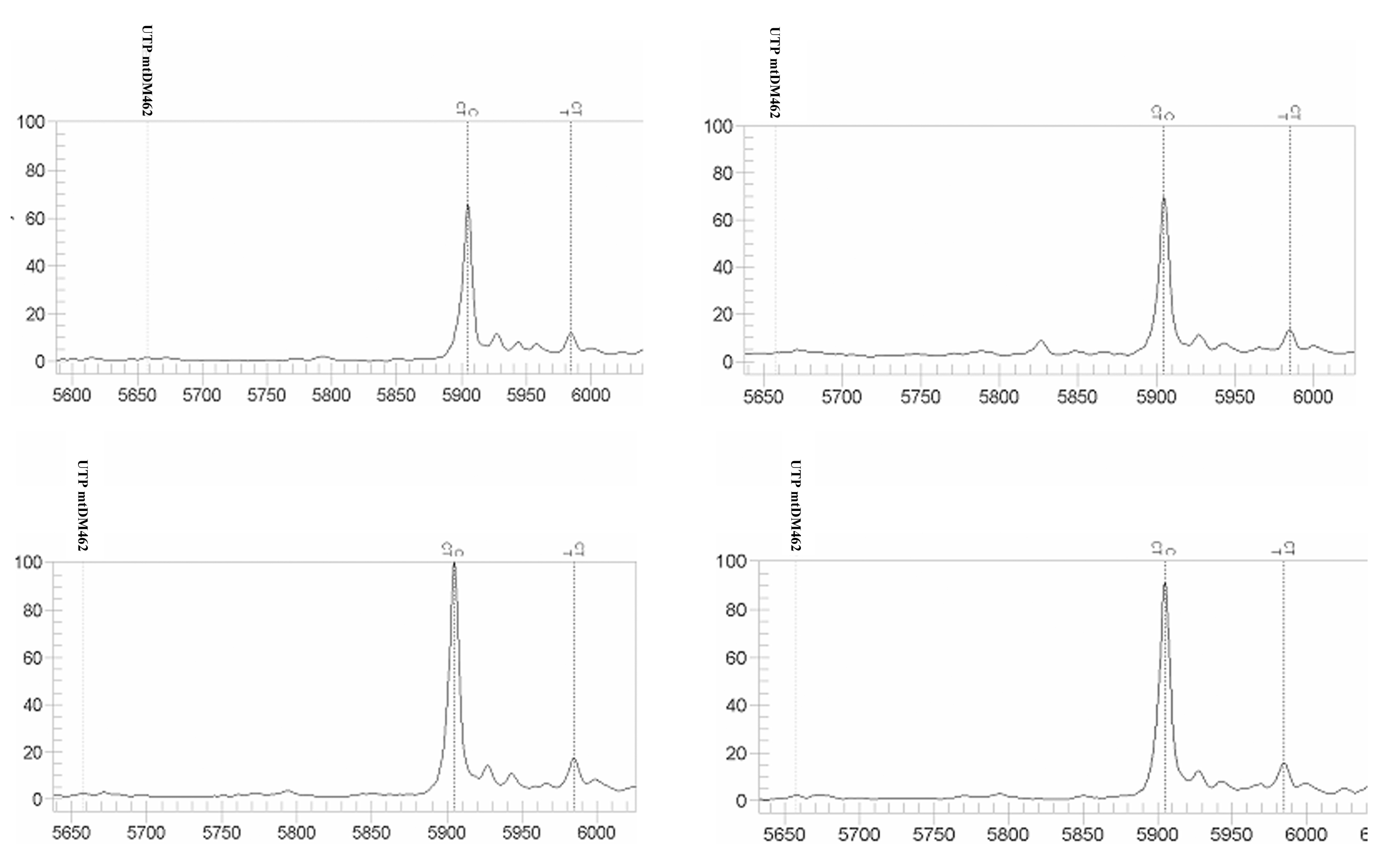 Fig. 3. Reproducible sensitive detection of less than 1.5% mutant DNA (T) as a minor component in more than 98.5% wild-type DNA (C) background using MALDI-TOF MS.
The group of Stroun and Anker was the first to identify cancer derived K-ras in the circulation of patients with colorectal cancer using sequence-specific primers to amplify mutant DNA by allele-specific PCR.3 A colorectal cancer tissue derived specific codon 12 K-ras mutation was found in the circulation of 86% of the patients, whereas the mutant DNA was not detected in the plasma specimens of patients without the K-ras alterations in their tumor tissues, or in healthy control subjects.3 N-ras mutations have also been found in the plasma of patients with myelodysplastic syndrome or acute myelogenous leukemia29 using the conventional method. Recently, Diehl et al.25 explored the possibility of using ccf tumor derived somatic mutant DNA on TP53, KRAS, APC, and PIK3CA genes for the management of colorectal cancer. They applied the BEAMing technique to measure the amount of ccf tumor mutant DNA and to monitor the dynamic changing of the molecules in the circulation during the management of the patients. Patients with detectable ccf tumor DNA suffered a relapse, whereas subjects without circulating tumor DNA did not experience tumor recurrence. The ccf tumor DNA detection seems to be more reliable for predicting a relapse than the standard biomarker, carcinoembryonic antigen (CEA), used for the management of colorectal cancer. Other than using cancer tissue mutations to identify the ccf tumor DNA in circulation, other genetic events such as microsatellite alterations, inversions, deletions, and aberrant methylationhave also been suggested to detect cancer derived ccf DNA by molecular biological approaches.30, 31, 32, 33, 34, 35 Recently, in the field of women’s medicine, quantitative and qualitative analysis of ccf DNA has been widely used for prenatal diagnosis and cancer research.
CCF FETAL DNA IN MATERNAL CIRCULATION FOR NONINVASIVE PRENATAL DIAGNOSIS
Prenatal diagnosis of inherited disorders is mainly based on genetic testing of fetal genetic materials obtained from amniocentesis or chorionic villus sampling. These invasive procedures carry a significant risk for both fetus and mother,36, 37 which burdens not only the affected pregnant women, but also their family members and friends.38, 39, 40, 41, 42 The discovery of ccff DNA in maternal plasma opens a great hope for risk-free noninvasive prenatal diagnosis (NIPD).43, 44, 45, 46, 47
Noninvasiveprenatal diagnosis of paternally inherited genetic traits
Autosomal dominant,paternally inherited genetic traits absent in the maternal genome can be examined for risk-free prenatal diagnosis using ccff DNA in the maternal circulation (Fig. 4). In our lab, we established a multiplex real-time PCR assay for the routine determination of fetal sex and RhD status using ccff DNA in the maternal plasma. We showed that ccff DNA could be used for the reliable and rapid examination of several fetal genetic traits, which was a significant achievement in the transition of a research technique to clinical diagnostic applications.9, 47 Recently, ccff DNA has been successfully applied to determine fetal sex for sex-linked disorders and RhD status for the management of RhD blood group incompatibility on a routine basis by several laboratories, such as the group of Jean-Marc Costa in Paris, the group of Martin Peter in Bristol, Sanquin in Amsterdam, and Sequenom in San Diego. Alloimmunization against the fetal Kell (KEL1) blood group antigen is the second most important cause of hemolytic disease of the fetus and newborn. In our group, we developed MALDI-TOF MS-based single allele-based extension reaction (SABER) (Fig. 4) to examine the fetal KEL1 gene from KEL1-negative pregnant women using ccff DNA in maternal plasma. An accuracy of 94% could be achieved by the risk-free noninvasive procedure for prenatal diagnosis.27 The strategy also permits determinationof autosomal dominant,paternally inherited mutations absent in maternal genome. Risk-free NIPD of fetal myotonic dystrophy, and achondroplasia using ccff DNA in the maternal circulation has been reported by Amicucci et al. and our group.48, 49
 Fig. 4. Strategies of NIPD for monogenic disorders using MALDI-TOF MS. (Zhong XY, Holzgreve W. MALDI-TOF MS in prenatal genomics. Transfusion Med Hemother 2009, submitted.)
Noninvasiveprenatal exclusion of autosomal recessive inherited genetic traits
For the risk-free NIPD of autosomal recessive inherited genetic traits, a strategy has been developed to exclude the compound heterozygous conditions throughthe detection of fetal DNA in maternal plasma (Fig. 4). An affected fetus with a compound heterozygous condition harbors different mutations, one from the father and another one from the mother. The presence of a wild-type paternal allele or absence of a mutant paternal allele in maternal plasma indicates that thefetus has not inherited the mutated paternal allele and thuscannot manifest such a disorder (Fig. 4).50 Thalassemias are hereditary anemias due to defects in hemoglobin production caused mostly by mutations. For the autosomal recessive forms of the disease, if both parents are carriers of a different mutation, there is a 25% chance with each pregnancy for an affected child with compound heterozygosity.51, 52, 53 It has been recently shown that the detection of paternally inherited ß-thalassemia mutations in maternal plasma could noninvasively exclude compound heterozygous pregnancies using the simple real-time PCR approach26 and the high throughput MALDI-TOF mass spectrometry-based assay.54 This strategy and approachcan be also applicable to otherautosomal recessive conditions, such as cystic fibrosis,55 congenital adrenal hyperplasia,56 Alpers syndrome, Tay-Sachs syndrome, Gaucher syndrome, connexin 26 disease, etc. Recently, detection of maternally inherited mutations, which are identical to the "maternal background noise", has also become possible. Lo et al. developed a digital relative mutation dosage (RMD) approach that determines whether the dosages of the mutant and wild-type alleles of a disease-causing gene are balanced or unbalanced in maternal plasma. The dosages of ccf mutant DNA in maternal circulation from a pregnant woman carrying a fetus having two mutant alleles is very slightly higher. By using digital technology to count this, an accurate assessment can be made.57, 58, 59 The digital PCR procedures are shown in Figure 5.
Fig. 5. The equipment and procedures for performing digital PCR.
Noninvasiveprenatal diagnosis of aneuploidies
Noninvasive prenatal detection of fetal chromosomal aneuploidies using ccf maternal plasma DNA is a considerable challenge, but exploration has become possible recently based on precise molecular biological technical developments. One approach is based on the measurement of the allelic ratio for multiple single nucleotide polymorphism (SNP) sites on chromosomes 13 and 21 after fetal DNA enrichment in maternal plasma by using formaldehyde treatments. In the study, only three cases in which a fetus affected by trisomy 21 was carried were analyzed.60, 61 Recently, Fan et al. developed a polymorphism-independent high-throughput shotgun sequencing technology to measure the over- and under-representation of chromosomes in maternal plasma from an aneuploid fetus. This method enabled successful identification of nine cases of trisomy 21 (Down syndrome), two cases of trisomy 18 (Edward syndrome), and one case of trisomy 13 (Patau syndrome) in a cohort of 18 normal and aneuploid pregnancies.62 Meanwhile, Chiu et al. also used massively parallel genomic sequencing to quantify overrepresented DNA sequences from fetal trisomy 21 in maternal plasma for the noninvasive prenatal detection. All 14 trisomy 21 fetuses and 14 euploid fetuses in the study were correctly identified.57 Figure 6 shows the approach for the identification of fetal aneuploidy using ccff DNA in the maternal circulation. Based on the small sample size in the studies, further testing with more cases is needed for the techniques to work. On the other hand, the techniques are cost- and labor-consuming to perform, thus they are not applicable for routine screening.
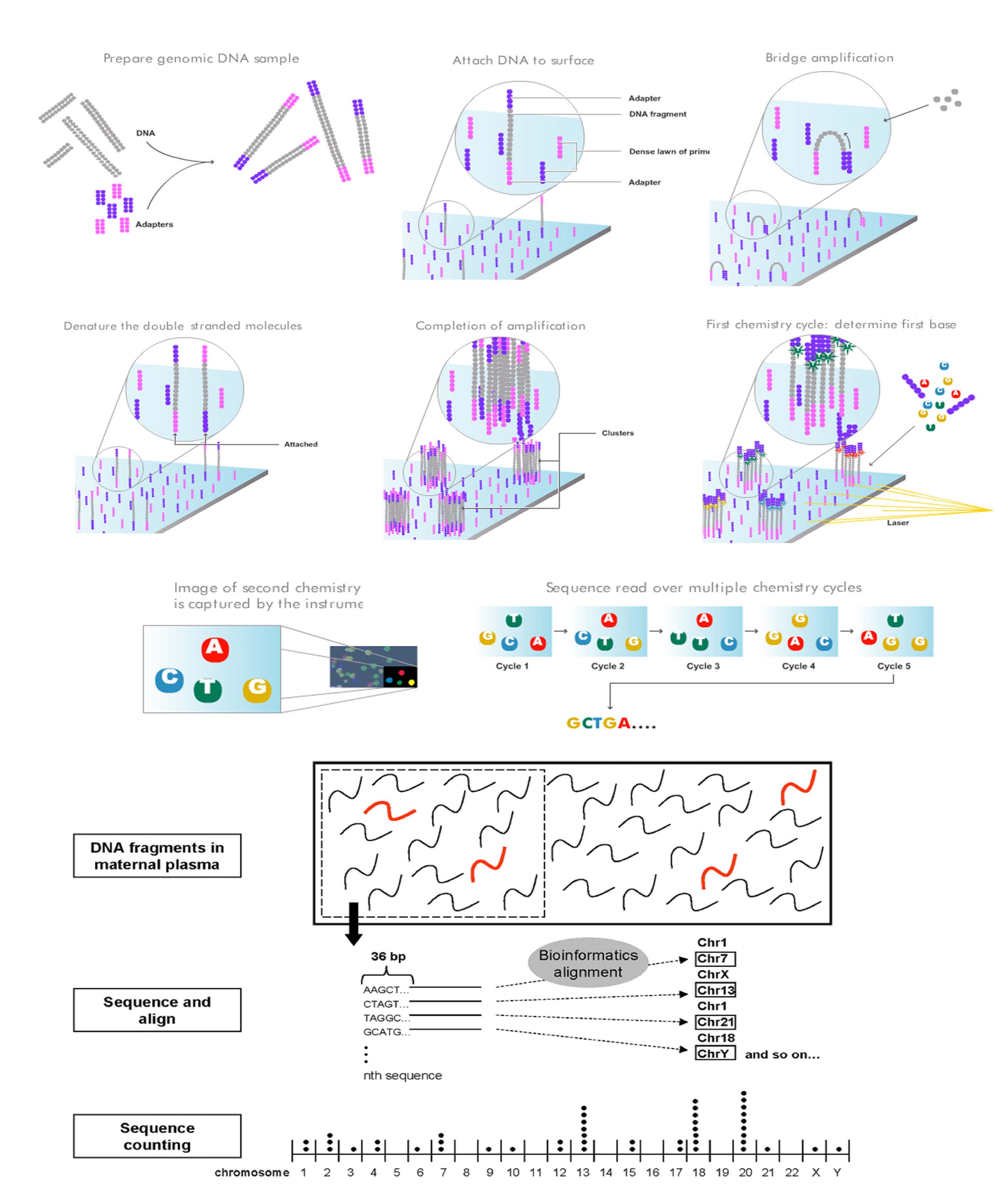 Figure 6. Massively parallel genomic sequencing for sequence counting.
Elevated levels of ccff DNA in maternal circulation in pathological pregnancies
It was also reported by many groups that pregnancies affected by certain pathological processes, such as preeclampsia,13, 63, 64 preterm labor,65, 66 polyhydramnios,67 hyperemesis,68, 69 placental insufficiency, and placental abruption70 are associated with quantitative abnormalities of ccff DNA in the maternal blood. The increased levels of ccff DNA in pathological pregnancies may stem from the shedding or trafficking of these materials from the placental damage. For the first time, our group observed that ccff DNA concentrations are elevated early in pregnancy before disease onset, in pregnancies which later develop preeclampsia, suggesting that the species can serve as a sensitive marker for earlier diagnosis of the disorder71 (Fig. 7). We also found that the increases in both ccff DNA and maternal ccf DNA levels in maternal blood corresponded to the severity of the disorder indicating that the concentration of the ccf DNA may be applicable to the monitoring of preeclampsia.13 Elevated ccff DNA has been observed in patients with true preterm labor, while lower concentrations of fetal DNA were associated with successful tocolytic therapy, suggesting that this technique may help to differentiate true and false preterm labor.65
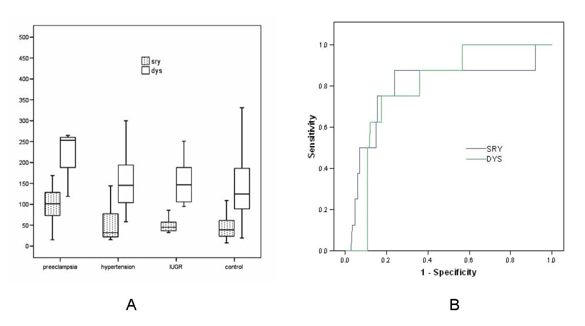 Fig. 7. Elevations of ccff DYS14 and SYR DNA in maternal plasma occur before onset of preeclampsia. Box plots showing concentrations of ccff DYS14 and SRY DNA in plasma samples obtained from pregnant women with preeclampsia, chronic hypertension, intrauterine growth restriction (IUGR), and normotensive controls (A). The medians are indicated by a line inside each box, the 75th and 25th percentiles by the box limits; the upper and lower error bars represent the 10th and 90th percentiles, respectively. (B) Two receiver operating characteristic (ROC) curves, for DYS14 and SRY plotted on the same graph. The area under the ROC curve is 0.8 for both the DYS14 assay and SRY assay. An optimal cut-off value chosen by ROC curve analysis could provide a sensitivity of 87.5% and a specificity of 76.4% for the SRY assay, and a sensitivity of 75% and a specificity of 82.4% for the DYS14 assay to discriminate between the cases at risk for preeclampsia and normal controls.
CCF DNA IN PATIENTS WITH ENDOMETRIOSIS
Elevated levels of ccf DNA have been found in inflammatory conditions, such as inflammatory second hit, sepsis, chronic illnesses, systemic lupus erythematosus, and rheumatoid arthritis.14, 72, 73, 74 Endometriosis, defined as the presence of endometrial tissue outside the uterus, is one of the most common benign gynecological inflammatory conditions in premenopausal women. Immunologic factors may play a role in its pathophysiology. Recently, anti-endometrial antibodies have been found in the sera of women with endometriosis in common with human autoimmunity, suggesting that endometriosis may correlate with autoimmune inflammation.75 Apoptosis has also been observed in eutopic and ectopic endometrium of patients with endometriosis.76 Based on the observations of autoimmune reaction and apoptotic event in endometriosis, we measured the levels of ccf DNA in patients with endometriosis (Fig. 8). The level of ccf nDNA in plasma was significantly higher in the endometriosis group than in the control group. A cut-off value selected by ROC could provide a sensitivity of 70% and a specificity of 87% to discriminate between the minimal/mild cases and healthy controls. The finding suggests that ccf nDNA might be a potential biomarker for developing noninvasive diagnostic test in endometriosis for tailoring treatment.
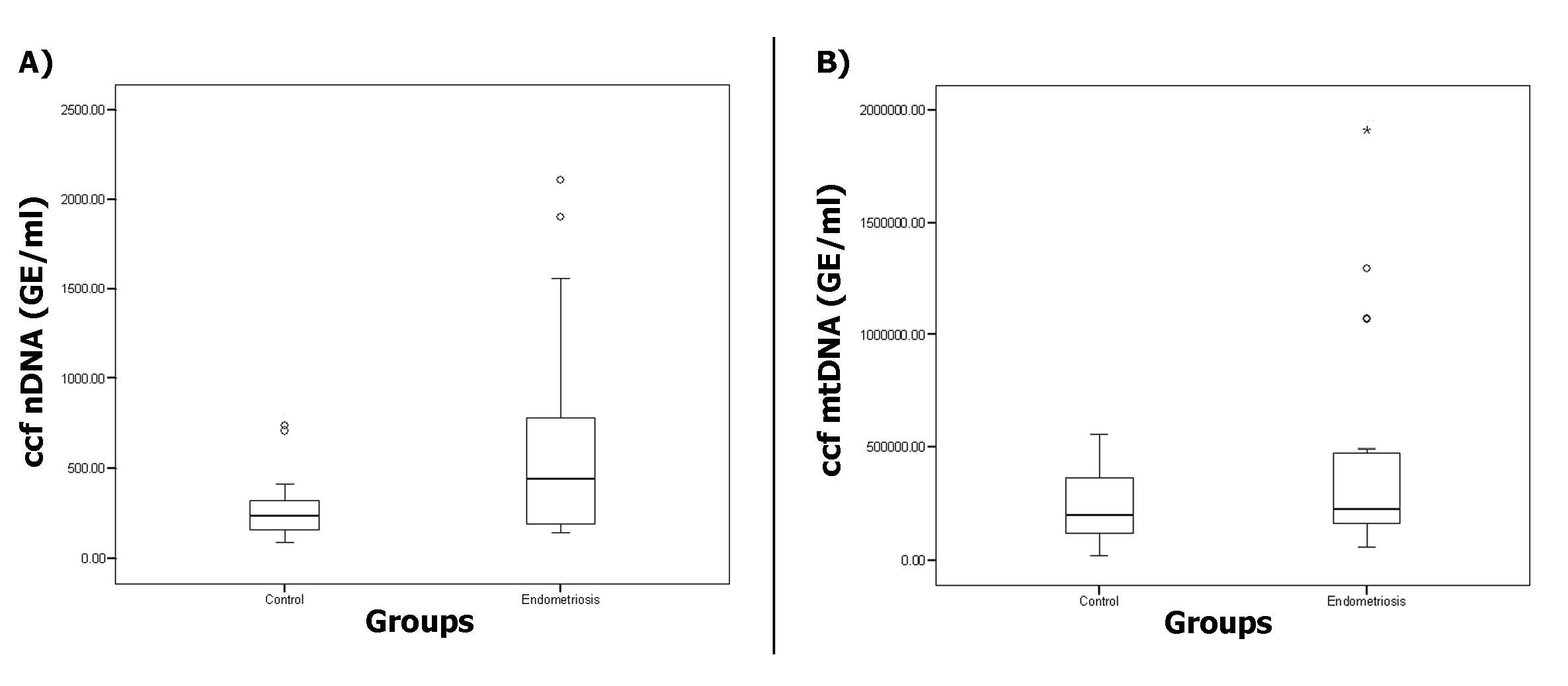 Fig. 8. Levels of plasma ccf nDNA (A) and ccf mtDNA (GE/ml) (B) in the endometriosis group and control group.
CCF DNA IN PATIENTS WITH BREAST TUMORS
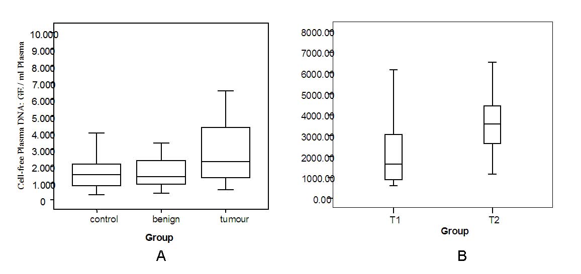
Several groups reported the findings of high levels of ccf DNA in the circulation of patients with breast cancer. In our group, whereas the high level of ccf plasma DNA was correlated with breast cancer, the high level of ccf serum DNA was associated with both malignant and benign breast tumors. In plasma ccf DNA is regarded mostly as a natural release of the species, and the main part of ccf DNA in serum is considered as an unnatural release of the species during blood clotting procedures. Our data suggest that ccf plasma DNA is more specific than ccf serum DNA in breast cancer. Combining the plasma and serum concentration of ccf DNA may have the potential to distinguish between malignant and benign conditions77, 78, 79 (Fig. 9). It was also observed that high levels of ccf plasma DNA related to tumor size and that levels decrease after breast surgery.80
Fig. 9. Ccf DNA in breast tumors. Box plot indicating ccf plasma DNA levels in normal individuals and patients with benign lesion and breast cancer (A). (B) Plasma ccf DNA levels in breast cancer, in which the tumor size is less than 2 cm (T1) or more than 2 cm (T2).
In patients with breast cancer ccf DNA contains cancer tissue-specific methylation changes, heterozygosity (LOH), and microsatellite instability (MI).
DNA methylation is related to carcinogenesis. Hypermethylation of human tumor suppressor genes (TSGs) leads to the silencing of the genes responsible for tumor suppression, thus causing cancers. Using a methylation-specific PCR technique, aberrant promoter methylation of TSGs, such as APC, RASSF1A RARbeta, cyclin D2, P16, ATM, CDH1, CDH13, and HIC-1 genes was found in plasma or serum samples of patients with breast cancer.81, 82, 83, 84, 85 It was shown that analysis of ccf RARbeta2 and RASSF1A methylated DNA in plasma samples could provide 95% diagnostic coverage in breast cancer patients, 60% in patients with benign lesions, and was without false-positive results in the healthy women observed.86 Hu et al. reported that cancer derived p16 methylation was significantly associated with nodal metastasis of patients with breast cancer. The ccf methylated DNA detection corresponded to the cancer tissue specific methylation changes, suggesting that ccf methylated DNA in breast cancer could be used to monitor the progress of the condition. However, the accurate determination of ccf methylated DNA in cancer is still a considerable challenge, so the results are contradictory.81, 82, 83, 84, 85 Recently, in our group, we applied the SEQUENOM’s EpiTYPER™ assay for high-throughput quantitative analysis of DNA methylation status using MALDI-TOF MS with MassCLEAVETM reagent, which is based on base-specific (T) cleavage reactions.87 We mixed full methylated DNA into the pure unmethylated DNA in different ratios of 100:0, 50:50, 25:75, 5:95, and 0:100. The assay was able to discriminate the 5% methylated in 95% unmethylated components according to the ratios by the quantitative assay.87 As little as 5 ng of DNA per PCR reaction allowed methylation analysis using this system. The approach may be applicable to detect tumor derived ccf methylated DNA for developing blood-based test in the field.
Microsatellites are repetitive DNA sequences comprising short tandem repeatsof 2–6 bp in size. The repeating units form variable lengths of DNA between individuals and/or species.Microsatellite lengths are highly polymorphic in humanpopulations, but appear stable during the life span of the individual. Instability of microsatellite sequences has been found in various human malignancies. The detection of microsatellite instability is an important step in the field of oncology. Using appropriate primers, DNA fragmentscan be amplified and analyzed as microsatellite markers. With a panelof such markers, the tumor-specific microsatellite profile can be identified. Cancer-specific microsatellite changes have been used to analyze cancer derived genetic materials in the circulation. The first report of this approach applied to breast cancer was from the group of Anker and Stroun.88 Using different markers, they analyzed the presence of microsatellite instability and loss of heterozygosity (LOH) in plasma or serum DNA from 61 patients. Microsatellite alterations werepresent in 35–81% of the breast cancer tissue samples. Identical microsatellite alterations were also found in 15–48% of the corresponding plasma samples. The cancer derived DNA changes in the plasma/serum could be detectable at an early stage of the condition, suggesting that microsatellite instability in plasma or serum might become a useful diagnostic tool for early and potentially curable breast cancer. A similar observation of a LOH in the serum of 21% of patients with early-stagebreast cancer based on eight markers has been shown by Taback et al.89 Using tumor-specific LOH as a marker, Pantel’s group analyzed cancer derived cellular and cell-free DNA in different sample types, such as serum, tissue, and bone marrow, from patients with breast cancer. The genomic aberrations on chromosomes 10, 16, and 17 were frequent in the circulating DNA of breast cancer patients. Circulating tumor DNA did not reflect the presence of tumor cells in blood or the level of tumor-associated protein markers such as CA 15-3. The significant association of LOH with some risk factors was found; however, the low rate of LOH changes in breast cancer may limit their use in clinical application.90, 91
Silva et al. combined several genetic changes, such as microsatellite instability, aberrant methylation, and point mutations serving as multiple markers to identify and characterize ccf tumor DNA in breast cancer. More than 90% of the cases had at least one molecular event in tumor tissues, and 66% of the cases showed a similar alteration in plasma DNA. In comparison with clinicopathological parameters, patients with and without ccf tumor DNA revealed significant differences in the axillary involvement, rate of invasive ductal carcinoma, high proliferative index, and the parameter comprised of lymph node metastases, histological grade II, and peritumoral vessel involvement.92 Use of multiple tumor markers may increase the accuracy of the approach.
The origin of ccf DNA is not completely understood, but apoptotic and necrotic cell death, especially blood cell death, contribute to ccf DNA.93 It was hypothesized that cancer derived DNA may be released mostly from cancer cell necrosis, and thus varies in size. Umetani et al. examined the integrity of ccf DNA in patients with breast cancer. The correlation between the ratio of longer fragments to total ccf DNA in serum and clinical risk factors was analyzed. Higher serum DNA integrity was associated with advanced stages, tumor size, lymphovascular invasion, and lymph node metastasis. Serum DNA integrity combined with lymphovascular invasion had value for predicting lymph node metastasis in a multivariate analysis.94 Adjuvant chemotherapy could change the integrity of ccf serum DNA in patients with breast cancer, suggesting that this information might be helpful in evaluating the response of patients to adjuvant systemic therapy.95, 96
Ccf DNA includes the proportion of histone-protein bound molecules, possibly as nucleosomes, and unbound part molecules. Because of supportive proteins, nucleosomes are more persistent than their unbound counterparts.97 Kuroi et al.98, 99 examined circulating nucleosomes in patients with breast cancer by enzyme-linked immunosorbent assay (ELISA). The high levels of circulating nucleosomes in patients were significantly elevated with breast cancer. No correlation between high levels of circulating nucleosomes and clinicopathological factors, such as tumor size, menopausal status, estrogen receptor status, histological type and lymphatic or venous spread in node-negative breast cancer, was found in a study cohort of more than 90 cases. Nevertheless, levels of circulating nucleosomes were decreased after therapy. In patients with recurrent breast cancer, circulating nucleosomes showed a transient increase, thus circulating nucleosomes seem to be a sensitive marker for monitoring of patients. However, our group showed a contradictory result. In our group, we examined the quantities of serum GAPDH, which represent the levels of cell-free total (bound and unbound) DNA, by quantitative PCR, and nucleosomes, which represent the levels of cell-free bound DNA, were examined by ELISA. Our data suggested that using quantitative real-time PCR to analyze the total circulating cell-free DNA is more efficient and sensitive than using the ELISA to analyze only histone-protein bound cell-free DNA. The ccf GAPDH DNA by quantitative PCR can be considered as a better marker than nucleosomes by ELISA in the patient with breast lesions.100
Since elevated levels of vascular endothelial growth factor (VEGF) and its soluble receptor (sVEGFR1) have been observed in the serum of patients with various cancers,101, 102 we compared the levels of ccf DNA and the levels of VEGF/sVEGFR1 in serum from patients with breast cancer. While higher levels of ccf serum DNA were observed in the patients with breast tumors, levels of VEGF and VEGFR1 in serum samples were not elevated with the conditions, implying that ccf serum DNA can be considered as a more sensitive marker than VEGF and VEGFR1 in patients with benign and malignant breast tumors. However, based on recent experimental findings, the application of cancer ccf DNA in malignancy has been not applicable for routine diagnosis and management. The value of longitudinal study using the approach for monitoring cancer derived ccf DNA remains to be determined.
CCF DNA IN PATIENTS WITH OVARIAN TUMORS
In 2006, Kamat et al.103 for the first time found higher levels of ccf GAPDH, beta-actin, and beta-globin DNA in patients with high-grade, advanced stage (III or IV) serous ovarian carcinomas, using real-time PCR. The study was performed on a small sample of 19 patients and 12 age-matched controls. No tumor-specific DNA markers were used to distinguish between cancer and noncancer origin of ccf DNA. However, these preliminary results could show that total ccf DNA in plasma of patients with ovarian cancer may be useful for noninvasive screening and disease surveillance in ovarian cancer. Subsequently, through animal experiments, the group reported that tumor-specific ccf DNA levels in ovarian cancer correlated with increasing tumor burden and declined following therapy, suggesting that tumor-specific ccf DNA may serve as a biomarker for predicting therapeutic response.104
In our group, we found elevated levels of ccf nuclear DNA (nDNA) and ccf mitochondrial DNA (mtDNA) in patients with ovarian tumors using a gold-standard multiplex real-time PCR on a sample size of more than 100 cases. Our data showed that the patients with epithelial ovarian cancer (EOC) have significantly higher amounts of ccf nDNA and ccf mtDNA in plasma compared to the healthy control group and to the other group with benign ovarian diseases. The possible diagnostic value of using the ccf DNA as markers was evaluated by ROC analysis. A sensitivity of 63–79% and a specificity of 62–69% can be achieved to discriminate between the cancer cases and normal controls, as well as between the malignant and benign cases105 (Fig. 10). However, the main sources of ccf DNA are hematopoietic cells, therefore tumor-specific genetic markers are needed to distinguish between tumor ccf DNA and nontumor ccf DNA.
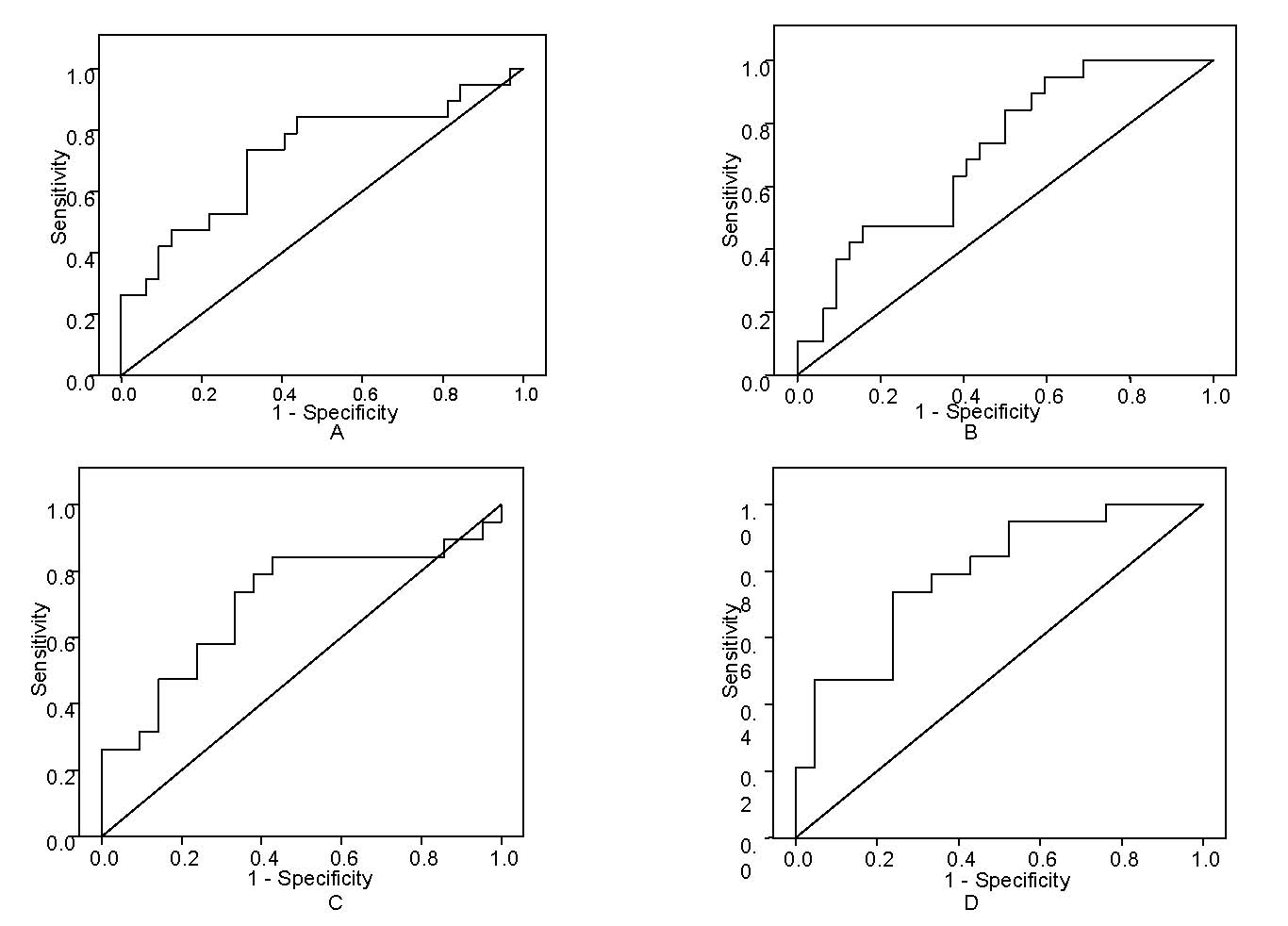 Fig. 10. Ccf nDNA and mtDNA in ovarian tumors. ROC curve of ccf nDNA shows a sensitivity of 74% and a specificity of 69% to discriminate malignant and healthy individuals (A). ROC curve of ccf mtDNA shows a sensitivity of 63% and a specificity of 62% to discriminate malignant and healthy individuals (B). ROC curve of ccf nDNA shows a sensitivity of 74% and a specificity of 67% to discriminate malignant and benign cases (C). ROC curve of ccf mtDNA shows a sensitivity of 79% and a specificity of 67% to discriminate malignant and benign cases (D).
Using conventional methylation specific PCR (MSP), Ibanez de Caceres et al.106 reported that tumor cell-specific BRCA1 and RASSF1A hypermethylation could be detectable in serum, plasma, and peritoneal fluid from ovarian cancer patients. Using the same method, Gifford et al.107 demonstrated that the acquired hMLH1 methylation in plasma DNA after chemotherapy predicted poor survival for ovarian cancer patients. Recently, using a conventional microarray-basedtechnique, Melnikov et al.108 analyzed differences in DNA methylation profilesin a panel of genes in serous papillary adenocarcinomas. The group identified ten genes as informative with 69% sensitivity and 70% specificity for cancer detectionin tissue samples, and five genes as informative with 85% sensitivity and 61% specificity for cancerdetection in plasma samples. However, the clinical relevance and usefulness of the approach for the management of ovarian cancer should be clarified by developing a robust approach and/or by increasing solid study sample size.
CONCLUSIONS
In the field of prenatal medicine, ccff DNA can be detected in maternal circulation during pregnancy, offering an excellent method for noninvasive prenatal diagnosis of the genetic status of fetus. Identification of fetal Rhesus D status using ccff DNA in maternal circulation of Rhesus D negative pregnant women has been successfully applied for the management of neonatal hemolytic disease (NHD) routinely. NIPD of fetal Kell status using ccff DNA has recently also become applicable for the management of NHD. Detection of the fetal Y chromosome and paternal inherited mutations in maternal circulation has been used for the prenatal diagnosis of X-linked diseases and autosomal inherited diseases by risk-free procedures. High levels of ccf DNA were found in several pregnancy associated complications, implying a diagnostic value for evaluating placental damage. However, NIPD of maternal inherited mutations and inherited diseases with homozygous, as well as fetal aneuploidy, remains a considerable challenge.
In the field of oncology, the blood based approach offers an alternative opportunity for noninvasive measurement of tumor derived genetic materials in the circulation in the management for patients with cancer. Elevated levels of ccf DNA have been observed in various of cancers. Ccf DNA in patients with cancers harbors tumor-specific genetic and epigenetic alterations, which can be used as cancer specific markers for monitoring. Ccf tumor DNA seemed to be more reliable for predicting a relapse than a standard biomarker, carcinoembryonic antigen (CEA), used for the management of colorectal cancer. Ccf tumor DNA in plasma can be detected earlier and more frequently than circulating tumor cells in blood of metastatic animal models.109, 110, 111, 112 The results suggest that the circulating molecules might be a more sensitive and dynamic biomarker for developing blood based test in clinical applications.
ACKNOWLEDGMENTS
This work was supported in part by Swiss National Science Foundation (320000-119722/1), Swiss Cancer League, Krebsliga Beider Basel, and Dr Hans Altschueler Stiftung. We thank Mr Ramin Radpour, Mr Alex Xiu-Cheng Fan, and Mrs Corina Kohler for their excellent assistance and help.
REFERENCES
Mandel, P, Metais, P. (1948). Les acides nucleiques du plasma sanguin chez I'Homme. CR Acad Sci Paris, 241-243. |
|
Tan, E.M., Schur, P.H., Carr, R.I. & Kunkel, H.G. (1966). Deoxybonucleic acid (DNA) and antibodies to DNA in the serum of patients with systemic lupus erythematosus. J Clin Invest, 45, 1732-40. |
|
Anker, P., Lefort, F., Vasioukhin, V., Lyautey, J., Lederrey, C., Chen, X.Q., Stroun, M., Mulcahy, H.E. & Farthing, M.J. (1997). K-ras mutations are found in DNA extracted from the plasma of patients with colorectal cancer. Gastroenterology, 112, 1114-20. |
|
Anker, P., Lyautey, J., Lefort, F., Lederrey, C. & Stroun, M. (1994). [Transformation of NIH/3T3 cells and SW 480 cells displaying K-ras mutation]. C R Acad Sci III, 317, 869-74. |
|
Anker, P., Mulcahy, H., Chen, X.Q. & Stroun, M. (1999). Detection of circulating tumour DNA in the blood (plasma/serum) of cancer patients. Cancer Metastasis Rev, 18, 65-73. |
|
Stroun, M., Anker, P., Lyautey, J., Lederrey, C. & Maurice, P.A. (1987). Isolation and characterization of DNA from the plasma of cancer patients. Eur J Cancer Clin Oncol, 23, 707-12. |
|
Stroun, M., Anker, P., Maurice, P., Lyautey, J., Lederrey, C. & Beljanski, M. (1989). Neoplastic characteristics of the DNA found in the plasma of cancer patients. Oncology, 46, 318-22. |
|
Lo, Y.M., Corbetta, N., Chamberlain, P.F., Rai, V., Sargent, I.L., Redman, C.W. & Wainscoat, J.S. (1997). Presence of fetal DNA in maternal plasma and serum. Lancet, 350, 485-7. |
|
Zhong, X.Y., Hahn, S. & Holzgreve, W. (2001a). Prenatal identification of fetal genetic traits. Lancet, 357, 310-1. |
|
Anker, P., Lyautey, J., Lederrey, C. & Stroun, M. (2001). Circulating nucleic acids in plasma or serum. Clin Chim Acta, 313, 143-6. |
|
Lo, Y.M. (2001). Circulating nucleic acids in plasma and serum: an overview. Ann N Y Acad Sci, 945, 1-7. |
|
Lo, Y.M. (2006). Recent developments in fetal nucleic acids in maternal plasma: implications to noninvasive prenatal fetal blood group genotyping. Transfus Clin Biol, 13, 50-2. |
|
Zhong, X.Y., Laivuori, H., Livingston, J.C., Ylikorkala, O., Sibai, B.M., Holzgreve, W. & Hahn, S. (2001c). Elevation of both maternal and fetal extracellular circulating deoxyribonucleic acid concentrations in the plasma of pregnant women with preeclampsia. Am J Obstet Gynecol, 184, 414-9. |
|
Zhong, X.Y., von Muhlenen, I., Li, Y., Kang, A., Gupta, A.K., Tyndall, A., Holzgreve, W., Hahn, S. & Hasler, P. (2007d). Increased concentrations of antibody-bound circulatory cell-free DNA in rheumatoid arthritis. Clin Chem, 53, 1609-14. |
|
Lam, N.Y., Rainer, T.H., Chan, L.Y., Joynt, G.M. & Lo, Y.M. (2003). Time course of early and late changes in plasma DNA in trauma patients. Clin Chem, 49, 1286-91. |
|
Anker, P. & Stroun, M. (2001). Tumor-related alterations in circulating DNA, potential for diagnosis, prognosis and detection of minimal residual disease. Leukemia, 15, 289-91. |
|
Lo, Y.M. (2008). Fetal nucleic acids in maternal plasma. Ann N Y Acad Sci, 1137, 140-3. |
|
Stroun, M. & Anker, P. (2005). Circulating DNA in higher organisms cancer detection brings back to life an ignored phenomenon. Cell Mol Biol (Noisy-le-grand), 51, 767-74. |
|
Huang, D.J., Mergenthaler-Gatfield, S., Hahn, S., Holzgreve, W. & Zhong, X.Y. (2008). Isolation of cell-free DNA from maternal plasma using manual and automated systems. Methods Mol Biol, 444, 203-8. |
|
Xia, P., Radpour, R., Zachariah, R., Fan, A.X., Kohler, C., Hahn, S., Holzgreve, W. & Zhong, X.Y. (2009). Simultaneous quantitative assessment of circulating cell-free mitochondrial and nuclear DNA by multiplex real-time PCR. Genetics and Molecular Biology, 32, 20-24. |
|
Walker, J.A., Hedges, D.J., Perodeau, B.P., Landry, K.E., Stoilova, N., Laborde, M.E., Shewale, J., Sinha, S.K. & Batzer, M.A. (2005). Multiplex polymerase chain reaction for simultaneous quantitation of human nuclear, mitochondrial, and male Y-chromosome DNA: application in human identification. Anal Biochem, 337, 89-97. |
|
Fan, A.X., Radpour, R., Haghighi, M.M., Kohler, C., Xia, P., Hahn, S., Holzgreve, W. & Zhong, X.Y. (2009). Mitochondrial DNA content in paired normal and cancerous breast tissue samples from patients with breast cancer. J Cancer Res Clin Oncol. |
|
Zhong, X.Y., Hahn, S., Kiefer, V. & Holzgreve, W. (2007a). Is the quantity of circulatory cell-free DNA in human plasma and serum samples associated with gender, age and frequency of blood donations? Ann Hematol, 86, 139-43. |
|
Chan, K.C., Ding, C., Gerovassili, A., Yeung, S.W., Chiu, R.W., Leung, T.N., Lau, T.K., Chim, S.S., Chung, G.T., Nicolaides, K.H. & Lo, Y.M. (2006). Hypermethylated RASSF1A in maternal plasma: A universal fetal DNA marker that improves the reliability of noninvasive prenatal diagnosis. Clin Chem, 52, 2211-8. |
|
Diehl, F., Schmidt, K., Choti, M.A., Romans, K., Goodman, S., Li, M., Thornton, K., Agrawal, N., Sokoll, L., Szabo, S.A., Kinzler, K.W., Vogelstein, B. & Diaz, L.A., Jr. (2008). Circulating mutant DNA to assess tumor dynamics. Nat Med, 14, 985-90. |
|
Li, Y., Di Naro, E., Vitucci, A., Zimmermann, B., Holzgreve, W. & Hahn, S. (2005). Detection of paternally inherited fetal point mutations for beta-thalassemia using size-fractionated cell-free DNA in maternal plasma. Jama, 293, 843-9. |
|
Li, Y., Finning, K., Daniels, G., Hahn, S., Zhong, X. & Holzgreve, W. (2008b). Noninvasive genotyping fetal Kell blood group (KEL1) using cell-free fetal DNA in maternal plasma by MALDI-TOF mass spectrometry. Prenat Diagn, 28, 203-8. |
|
Zimmermann, B.G., Grill, S., Holzgreve, W., Zhong, X.Y., Jackson, L.G. & Hahn, S. (2008). Digital PCR: a powerful new tool for noninvasive prenatal diagnosis? Prenat Diagn, 28, 1087-93. |
|
Vasioukhin, V., Anker, P., Maurice, P., Lyautey, J., Lederrey, C. & Stroun, M. (1994). Point mutations of the N-ras gene in the blood plasma DNA of patients with myelodysplastic syndrome or acute myelogenous leukaemia. Br J Haematol, 86, 774-9. |
|
Hsu, H.S., Chen, T.P., Hung, C.H., Wen, C.K., Lin, R.K., Lee, H.C. & Wang, Y.C. (2007). Characterization of a multiple epigenetic marker panel for lung cancer detection and risk assessment in plasma. Cancer, 110, 2019-26. |
|
Kolesnikova, E.V., Tamkovich, S.N., Bryzgunova, O.E., Shelestyuk, P.I., Permyakova, V.I., Vlassov, V.V., Tuzikov, A.S., Laktionov, P.P. & Rykova, E.Y. (2008). Circulating DNA in the blood of gastric cancer patients. Ann N Y Acad Sci, 1137, 226-31. |
|
Ludovini, V., Pistola, L., Gregorc, V., Floriani, I., Rulli, E., Piattoni, S., Di Carlo, L., Semeraro, A., Darwish, S., Tofanetti, F.R., Stocchi, L., Mihaylova, Z., Bellezza, G., Del Sordo, R., Daddi, G., Crino, L. & Tonato, M. (2008). Plasma DNA, microsatellite alterations, and p53 tumor mutations are associated with disease-free survival in radically resected non-small cell lung cancer patients: a study of the perugia multidisciplinary team for thoracic oncology. J Thorac Oncol, 3, 365-73. |
|
Muller, I., Beeger, C., Alix-Panabieres, C., Rebillard, X., Pantel, K. & Schwarzenbach, H. (2008). Identification of loss of heterozygosity on circulating free DNA in peripheral blood of prostate cancer patients: potential and technical improvements. Clin Chem, 54, 688-96. |
|
Nakamoto, D., Yamamoto, N., Takagi, R., Katakura, A., Mizoe, J.E. & Shibahara, T. (2008). Detection of microsatellite alterations in plasma DNA of malignant mucosal melanoma using whole genome amplification. Bull Tokyo Dent Coll, 49, 77-87. |
|
Schwarzenbach, H., Chun, F.K., Lange, I., Carpenter, S., Gottberg, M., Erbersdobler, A., Friedrich, M.G., Huland, H. & Pantel, K. (2007a). Detection of tumor-specific DNA in blood and bone marrow plasma from patients with prostate cancer. Int J Cancer, 120, 1465-71. |
|
Evans, M.I. & Andriole, S. (2008). Chorionic villus sampling and amniocentesis in 2008. Curr Opin Obstet Gynecol, 20, 164-8. |
|
Forabosco, A., Percesepe, A. & Santucci, S. (2009). Incidence of non-age-dependent chromosomal abnormalities: a population-based study on 88965 amniocenteses. Eur J Hum Genet. |
|
Cohen, S.M. & Yagel, S. (2009). Evaluating the rate and risk factors for fetal loss after chorionic villus sampling. Obstet Gynecol, 113, 437; author reply 437. |
|
Li, D.K., Karlberg, K., Wi, S. & Norem, C. (2008a). Factors influencing women's acceptance of prenatal screening tests. Prenat Diagn, 28, 1136-43. |
|
Odibo, A.O., Dicke, J.M., Gray, D.L., Oberle, B., Stamilio, D.M., Macones, G.A. & Crane, J.P. (2008a). Evaluating the rate and risk factors for fetal loss after chorionic villus sampling. Obstet Gynecol, 112, 813-9. |
|
Odibo, A.O., Gray, D.L., Dicke, J.M., Stamilio, D.M., Macones, G.A. & Crane, J.P. (2008b). Revisiting the fetal loss rate after second-trimester genetic amniocentesis: a single center's 16-year experience. Obstet Gynecol, 111, 589-95. |
|
Yukobowich, E., Anteby, E.Y., Cohen, S.M., Lavy, Y., Granat, M. & Yagel, S. (2001). Risk of fetal loss in twin pregnancies undergoing second trimester amniocentesis(1). Obstet Gynecol, 98, 231-4. |
|
Hahn, S., Zhong, X. & Holzgreve, W. (2007). Non-invasive prenatal diagnosis of Down's syndrome. Lancet, 369, 1997-8; author reply 1998-9. |
|
Hahn, S., Zhong, X.Y. & Holzgreve, W. (2008). Recent progress in non-invasive prenatal diagnosis. Semin Fetal Neonatal Med, 13, 57-62. |
|
Holzgreve, W., Hahn, S., Zhong, X.Y., Lapaire, O., Hosli, I., Tercanli, S. & Mindy, P. (2007). Genetic communication between fetus and mother: short- and long-term consequences. Am J Obstet Gynecol, 196, 372-81. |
|
Lo, Y.M. (2009). Noninvasive prenatal detection of fetal chromosomal aneuploidies by maternal plasma nucleic acid analysis: a review of the current state of the art. Bjog, 116, 152-7. |
|
Zhong, X.Y., Holzgreve, W. & Hahn, S. (2001b). Risk free simultaneous prenatal identification of fetal Rhesus D status and sex by multiplex real-time PCR using cell free fetal DNA in maternal plasma. Swiss Med Wkly, 131, 70-4. |
|
Amicucci, P., Gennarelli, M., Novelli, G. & Dallapiccola, B. (2000). Prenatal diagnosis of myotonic dystrophy using fetal DNA obtained from maternal plasma. Clin Chem, 46, 301-2. |
|
Li, Y., Holzgreve, W., Page-Christiaens, G.C., Gille, J.J. & Hahn, S. (2004). Improved prenatal detection of a fetal point mutation for achondroplasia by the use of size-fractionated circulatory DNA in maternal plasma--case report. Prenat Diagn, 24, 896-8. |
|
Chiu, R.W. & Lo, Y.M. (2004). Recent developments in fetal DNA in maternal plasma. Ann N Y Acad Sci, 1022, 100-4. |
|
Nikuei, P., Hadavi, V., Rajaei, M., Saberi, M., Hajizade, F. & Najmabadi, H. (2008). Prenatal diagnosis for beta-thalassemia major in the Iranian Province of Hormozgan. Hemoglobin, 32, 539-45. |
|
Steinberg, M.H. (2008). Sickle cell anemia, the first molecular disease: overview of molecular etiology, pathophysiology, and therapeutic approaches. ScientificWorldJournal, 8, 1295-324. |
|
Steinberg, M.H. (2009). Genetic etiologies for phenotypic diversity in sickle cell anemia. ScientificWorldJournal, 9, 46-67. |
|
Ding, C., Chiu, R.W., Lau, T.K., Leung, T.N., Chan, L.C., Chan, A.Y., Charoenkwan, P., Ng, I.S., Law, H.Y., Ma, E.S., Xu, X., Wanapirak, C., Sanguansermsri, T., Liao, C., Ai, M.A., Chui, D.H., Cantor, C.R. & Lo, Y.M. (2004). MS analysis of single-nucleotide differences in circulating nucleic acids: Application to noninvasive prenatal diagnosis. Proc Natl Acad Sci U S A, 101, 10762-7. |
|
Bustamante-Aragones, A., Gallego-Merlo, J., Trujillo-Tiebas, M.J., de Alba, M.R., Gonzalez-Gonzalez, C., Glover, G., Diego-Alvarez, D., Ayuso, C. & Ramos, C. (2008). New strategy for the prenatal detection/exclusion of paternal cystic fibrosis mutations in maternal plasma. J Cyst Fibros, 7, 505-10. |
|
Chiu, R.W., Lau, T.K., Cheung, P.T., Gong, Z.Q., Leung, T.N. & Lo, Y.M. (2002). Noninvasive prenatal exclusion of congenital adrenal hyperplasia by maternal plasma analysis: a feasibility study. Clin Chem, 48, 778-80. |
|
Chiu, R.W., Chan, K.C., Gao, Y., Lau, V.Y., Zheng, W., Leung, T.Y., Foo, C.H., Xie, B., Tsui, N.B., Lun, F.M., Zee, B.C., Lau, T.K., Cantor, C.R. & Lo, Y.M. (2008). Noninvasive prenatal diagnosis of fetal chromosomal aneuploidy by massively parallel genomic sequencing of DNA in maternal plasma. Proc Natl Acad Sci U S A, 105, 20458-63. |
|
Lo, Y.M., Lun, F.M., Chan, K.C., Tsui, N.B., Chong, K.C., Lau, T.K., Leung, T.Y., Zee, B.C., Cantor, C.R. & Chiu, R.W. (2007). Digital PCR for the molecular detection of fetal chromosomal aneuploidy. Proc Natl Acad Sci U S A, 104, 13116-21. |
|
Lun, F.M., Tsui, N.B., Chan, K.C., Leung, T.Y., Lau, T.K., Charoenkwan, P., Chow, K.C., Lo, W.Y., Wanapirak, C., Sanguansermsri, T., Cantor, C.R., Chiu, R.W. & Lo, Y.M. (2008). Noninvasive prenatal diagnosis of monogenic diseases by digital size selection and relative mutation dosage on DNA in maternal plasma. Proc Natl Acad Sci U S A, 105, 19920-5. |
|
Dhallan, R., Au, W.C., Mattagajasingh, S., Emche, S., Bayliss, P., Damewood, M., Cronin, M., Chou, V. & Mohr, M. (2004). Methods to increase the percentage of free fetal DNA recovered from the maternal circulation. Jama, 291, 1114-9. |
|
Dhallan, R., Guo, X., Emche, S., Damewood, M., Bayliss, P., Cronin, M., Barry, J., Betz, J., Franz, K., Gold, K., Vallecillo, B. & Varney, J. (2007). A non-invasive test for prenatal diagnosis based on fetal DNA present in maternal blood: a preliminary study. Lancet, 369, 474-81. |
|
Fan, H.C., Blumenfeld, Y.J., Chitkara, U., Hudgins, L. & Quake, S.R. (2008). Noninvasive diagnosis of fetal aneuploidy by shotgun sequencing DNA from maternal blood. Proc Natl Acad Sci U S A, 105, 16266-71. |
|
Lo, Y.M., Leung, T.N., Tein, M.S., Sargent, I.L., Zhang, J., Lau, T.K., Haines, C.J. & Redman, C.W. (1999). Quantitative abnormalities of fetal DNA in maternal serum in preeclampsia. Clin Chem, 45, 184-8. |
|
Zhong, X.Y., Wang, Y., Chen, S., Labu, Pubuzhuoma, Gesangzhuogab, Ouzhuwangmu, Hahn, C., Holzgreve, W. & Hahn, S. (2004). Can circulatory fetal DNA be used to study placentation at high altitude? Ann N Y Acad Sci, 1022, 124-8. |
|
Leung, T.N., Zhang, J., Lau, T.K., Hjelm, N.M. & Lo, Y.M. (1998). Maternal plasma fetal DNA as a marker for preterm labour. Lancet, 352, 1904-5. |
|
Zhong, X.Y., Holzgreve, W. & Hahn, S. (2002). Cell-free fetal DNA in the maternal circulation does not stem from the transplacental passage of fetal erythroblasts. Mol Hum Reprod, 8, 864-70. |
|
Zhong, X.Y., Holzgreve, W., Li, J.C., Aydinli, K. & Hahn, S. (2000). High levels of fetal erythroblasts and fetal extracellular DNA in the peripheral blood of a pregnant woman with idiopathic polyhydramnios: case report. Prenat Diagn, 20, 838-41. |
|
Sekizawa, A., Sugito, Y., Iwasaki, M., Watanabe, A., Jimbo, M., Hoshi, S., Saito, H. & Okai, T. (2001). Cell-free fetal DNA is increased in plasma of women with hyperemesis gravidarum. Clin Chem, 47, 2164-5. |
|
Sugito, Y., Sekizawa, A., Farina, A., Yukimoto, Y., Saito, H., Iwasaki, M., Rizzo, N. & Okai, T. (2003). Relationship between severity of hyperemesis gravidarum and fetal DNA concentration in maternal plasma. Clin Chem, 49, 1667-9. |
|
Zhong, X.Y., Steinborn, A., Sohn, C., Holzgreve, W. & Hahn, S. (2006). High levels of circulatory erythroblasts and cell-free DNA prior to intrauterine fetal death. Prenat Diagn, 26, 1272-3. |
|
Zhong, X.Y., Volgmann, T., Hahn, S. & Holzgreve, W. (2007c). Large scale analysis of circulatory fetal DNA concentrations in pregnancies which subsequently develop preeclampsia using two Y chromosome specific real-time PCR assays. JTGGA, 8 135-139 |
|
Fournie, G.J., Martres, F., Pourrat, J.P., Alary, C. & Rumeau, M. (1993). Plasma DNA as cell death marker in elderly patients. Gerontology, 39, 215-21. |
|
Galeazzi, M., Morozzi, G., Piccini, M., Chen, J., Bellisai, F., Fineschi, S. & Marcolongo, R. (2003). Dosage and characterization of circulating DNA: present usage and possible applications in systemic autoimmune disorders. Autoimmun Rev, 2, 50-5. |
|
Margraf, S., Logters, T., Reipen, J., Altrichter, J., Scholz, M. & Windolf, J. (2008). Neutrophil-derived circulating free DNA (cf-DNA/NETs): a potential prognostic marker for posttraumatic development of inflammatory second hit and sepsis. Shock, 30, 352-8. |
|
Gajbhiye R , S.W., Khan S, Meherji P , Warty N , Raut V , Chehna N , Khole V (2008). Multiple endometrial antigens are targeted in autoimmune endometriosis Reproductive BioMedicine Online Vol. 16 817–824. |
|
Harada, T., Taniguchi, F., Izawa, M., Ohama, Y., Takenaka, Y., Tagashira, Y., Ikeda, A., Watanabe, A., Iwabe, T. & Terakawa, N. (2007). Apoptosis and endometriosis. Front Biosci, 12, 3140-51 |
|
Zanetti-Dallenbach, R., Wight, E., Fan, A.X., Lapaire, O., Hahn, S., Holzgreve, W. & Zhong, X.Y. (2008). Positive correlation of cell-free DNA in plasma/serum in patients with malignant and benign breast disease. Anticancer Res, 28, 921-5. |
|
Zanetti-Dallenbach, R.A., Schmid, S., Wight, E., Holzgreve, W., Ladewing, A., Hahn, S. & Zhong, X.Y. (2007). Levels of circulating cell-free serum DNA in benign and malignant breast lesions. Int J Biol Markers, 22, 95-9. |
|
Zhong, X.Y., Ladewig, A., Schmid, S., Wight, E., Hahn, S. & Holzgreve, W. (2007b). Elevated level of cell-free plasma DNA is associated with breast cancer. Arch Gynecol Obstet, 276, 327-31. |
|
Catarino, R., Ferreira, M.M., Rodrigues, H., Coelho, A., Nogal, A., Sousa, A. & Medeiros, R. (2008). Quantification of free circulating tumor DNA as a diagnostic marker for breast cancer. DNA Cell Biol, 27, 415-21. |
|
Hu, X.C., Wong, I.H. & Chow, L.W. (2003). Tumor-derived aberrant methylation in plasma of invasive ductal breast cancer patients: clinical implications. Oncol Rep, 10, 1811-5. |
|
Papadopoulou, E., Davilas, E., Sotiriou, V., Georgakopoulos, E., Georgakopoulou, S., Koliopanos, A., Aggelakis, F., Dardoufas, K., Agnanti, N.J., Karydas, I. & Nasioulas, G. (2006). Cell-free DNA and RNA in plasma as a new molecular marker for prostate and breast cancer. Ann N Y Acad Sci, 1075, 235-43. |
|
Rykova, E., Skvortsova, T.E., Hoffmann, A.L., Tamkovich, S.N., Starikov, A.V., Bryzgunova, O.E., Permiakova, V.I., Warnecke, J.M., Sczakiel, G., Vlasov, V.V. & Laktionov, P.P. (2008). [Breast cancer diagnostics based on extracellular DNA and RNA circulating in blood]. Biomed Khim, 54, 94-103. |
|
Rykova, E.Y., Laktionov, P.P., Skvortsova, T.E., Starikov, A.V., Kuznetsova, N.P. & Vlassov, V.V. (2004a). Extracellular DNA in breast cancer: Cell-surface-bound, tumor-derived extracellular DNA in blood of patients with breast cancer and nonmalignant tumors. Ann N Y Acad Sci, 1022, 217-20. |
|
Rykova, E.Y., Skvortsova, T.E., Laktionov, P.P., Tamkovich, S.N., Bryzgunova, O.E., Starikov, A.V., Kuznetsova, N.P., Kolomiets, S.A., Sevostianova, N.V. & Vlassov, V.V. (2004b). Investigation of tumor-derived extracellular DNA in blood of cancer patients by methylation-specific PCR. Nucleosides Nucleotides Nucleic Acids, 23, 855-9. |
|
Skvortsova, T.E., Rykova, E.Y., Tamkovich, S.N., Bryzgunova, O.E., Starikov, A.V., Kuznetsova, N.P., Vlassov, V.V. & Laktionov, P.P. (2006). Cell-free and cell-bound circulating DNA in breast tumours: DNA quantification and analysis of tumour-related gene methylation. Br J Cancer, 94, 1492-5. |
|
Radpour, R., Haghighi, M.M., Fan, A.X., Torbati, P.M., Hahn, S., Holzgreve, W. & Zhong, X.Y. (2008). High-Throughput Hacking of the Methylation Patterns in Breast Cancer by In vitro Transcription and Thymidine-Specific Cleavage Mass Array on MALDI-TOF Silico-Chip. Mol Cancer Res., 6, 1702-9. |
|
Chen, X., Bonnefoi, H., Diebold-Berger, S., Lyautey, J., Lederrey, C., Faltin-Traub, E., Stroun, M. & Anker, P. (1999). Detecting tumor-related alterations in plasma or serum DNA of patients diagnosed with breast cancer. Clin Cancer Res, 5, 2297-303. |
|
Taback, B., Giuliano, A.E., Hansen, N.M. & Hoon, D.S. (2001). Microsatellite alterations detected in the serum of early stage breast cancer patients. Ann N Y Acad Sci, 945, 22-30. |
|
Schwarzenbach, H., Muller, V., Beeger, C., Gottberg, M., Stahmann, N. & Pantel, K. (2007b). A critical evaluation of loss of heterozygosity detected in tumor tissues, blood serum and bone marrow plasma from patients with breast cancer. Breast Cancer Res, 9, R66. |
|
Schwarzenbach, H., Muller, V., Stahmann, N. & Pantel, K. (2004). Detection and characterization of circulating microsatellite-DNA in blood of patients with breast cancer. Ann N Y Acad Sci, 1022, 25-32. |
|
Silva, J.M., Dominguez, G., Garcia, J.M., Gonzalez, R., Villanueva, M.J., Navarro, F., Provencio, M., San Martin, S., Espana, P. & Bonilla, F. (1999). Presence of tumor DNA in plasma of breast cancer patients: clinicopathological correlations. Cancer Res, 59, 3251-6. |
|
Lui, Y.Y., Chik, K.W., Chiu, R.W., Ho, C.Y., Lam, C.W. & Lo, Y.M. (2002). Predominant hematopoietic origin of cell-free DNA in plasma and serum after sex-mismatched bone marrow transplantation. Clin Chem, 48, 421-7. |
|
Umetani, N., Giuliano, A.E., Hiramatsu, S.H., Amersi, F., Nakagawa, T., Martino, S. & Hoon, D.S. (2006). Prediction of breast tumor progression by integrity of free circulating DNA in serum. J Clin Oncol, 24, 4270-6. |
|
Deligezer, U., Eralp, Y., Akisik, E.E., Akisik, E.Z., Saip, P., Topuz, E. & Dalay, N. (2008a). Size distribution of circulating cell-free DNA in sera of breast cancer patients in the course of adjuvant chemotherapy. Clin Chem Lab Med, 46, 311-7. |
|
Deligezer, U., Eralp, Y., Akisik, E.Z., Akisik, E.E., Saip, P., Topuz, E. & Dalay, N. (2008b). Effect of adjuvant chemotherapy on integrity of free serum DNA in patients with breast cancer. Ann N Y Acad Sci, 1137, 175-9. |
|
Kelbauskas, L., Woodbury, N. & Lohr, D. (2009). DNA sequence-dependent variation in nucleosome structure, stability, and dynamics detected by a FRET-based analysis. Biochem Cell Biol, 87, 323-35. |
|
Kuroi, K., Tanaka, C. & Toi, M. (1999). Plasma Nucleosome Levels in Node-Negative Breast Cancer Patients. Breast Cancer, 6, 361-364. |
|
Kuroi, K., Tanaka, C. & Toi, M. (2001). Clinical significance of plasma nucleosome levels in cancer patients. Int J Oncol, 19, 143-8. |
|
Seefeld, M., El Tarhouny, S., Fan, A.X., Hahn, S., Holzgreve, W. & Zhong, X.Y. (2008). Parallel assessment of circulatory cell-free DNA by PCR and nucleosomes by ELISA in breast tumors. Int J Biol Markers, 23, 69-73. |
|
Enjoji, M., Nakamuta, M., Yamaguchi, K., Ohta, S., Kotoh, K., Fukushima, M., Kuniyoshi, M., Yamada, T., Tanaka, M. & Nawata, H. (2005). Clinical significance of serum levels of vascular endothelial growth factor and its receptor in biliary disease and carcinoma. World J Gastroenterol, 11, 1167-71. |
|
Granato, A.M., Nanni, O., Falcini, F., Folli, S., Mosconi, G., De Paola, F., Medri, L., Amadori, D. & Volpi, A. (2004). Basic fibroblast growth factor and vascular endothelial growth factor serum levels in breast cancer patients and healthy women: useful as diagnostic tools? Breast Cancer Res, 6, R38-45. |
|
Kamat, A.A., Sood, A.K., Dang, D., Gershenson, D.M., Simpson, J.L. & Bischoff, F.Z. (2006b). Quantification of total plasma cell-free DNA in ovarian cancer using real-time PCR. Ann N Y Acad Sci, 1075, 230-4. |
|
Kamat, A.A., Bischoff, F.Z., Dang, D., Baldwin, M.F., Han, L.Y., Lin, Y.G., Merritt, W.M., Landen, C.N., Jr., Lu, C., Gershenson, D.M., Simpson, J.L. & Sood, A.K. (2006a). Circulating cell-free DNA: a novel biomarker for response to therapy in ovarian carcinoma. Cancer Biol Ther, 5, 1369-74. |
|
Zachariah, R.R., Schmid, S., Buerki, N., Radpour, R., Holzgreve, W. & Zhong, X. (2008). Levels of circulating cell-free nuclear and mitochondrial DNA in benign and malignant ovarian tumors. Obstet Gynecol, 112, 843-50. |
|
Ibanez de Caceres, I., Battagli, C., Esteller, M., Herman, J.G., Dulaimi, E., Edelson, M.I., Bergman, C., Ehya, H., Eisenberg, B.L. & Cairns, P. (2004). Tumor cell-specific BRCA1 and RASSF1A hypermethylation in serum, plasma, and peritoneal fluid from ovarian cancer patients. Cancer Res, 64, 6476-81. |
|
Gifford, G., Paul, J., Vasey, P.A., Kaye, S.B. & Brown, R. (2004). The acquisition of hMLH1 methylation in plasma DNA after chemotherapy predicts poor survival for ovarian cancer patients. Clin Cancer Res, 10, 4420-6. |
|
Melnikov, A., Scholtens, D., Godwin, A. & Levenson, V. (2009). Differential methylation profile of ovarian cancer in tissues and plasma. J Mol Diagn, 11, 60-5. |
|
Allen, D., Butt, A., Cahill, D., Wheeler, M., Popert, R. & Swaminathan, R. (2004). Role of cell-free plasma DNA as a diagnostic marker for prostate cancer. Ann N Y Acad Sci, 1022, 76-80. |
|
Gormally, E., Hainaut, P., Caboux, E., Airoldi, L., Autrup, H., Malaveille, C., Dunning, A., Garte, S., Matullo, G., Overvad, K., Tjonneland, A., Clavel-Chapelon, F., Boffetta, P., Boeing, H., Trichopoulou, A., Palli, D., Krogh, V., Tumino, R., Panico, S., Bueno-de-Mesquita, H.B., Peeters, P.H., Lund, E., Gonzalez, C.A., Martinez, C., Dorronsoro, M., Barricarte, A., Tormo, M.J., Quiros, J.R., Berglund, G., Hallmans, G., Day, N.E., Key, T.J., Veglia, F., Peluso, M., Norat, T., Saracci, R., Kaaks, R., Riboli, E. & Vineis, P. (2004). Amount of DNA in plasma and cancer risk: a prospective study. Int J Cancer, 111, 746-9. |
|
Sozzi, G., Conte, D., Leon, M., Ciricione, R., Roz, L., Ratcliffe, C., Roz, E., Cirenei, N., Bellomi, M., Pelosi, G., Pierotti, M.A. & Pastorino, U. (2003). Quantification of free circulating DNA as a diagnostic marker in lung cancer. J Clin Oncol, 21, 3902-8. |
|
Taback, B., O'Day, S.J. & Hoon, D.S. (2004). Quantification of circulating DNA in the plasma and serum of cancer patients. Ann N Y Acad Sci, 1022, 17-24. |


